I
Maria João
Valente Quental
Aplicação de líquidos iónicos na concentração de
marcadores tumorais
Application of ionic liquids in the concentration of
cancer biomarkers
Dissertação apresentada à Universidade de Aveiro para cumprimento dos requisitos necessários à obtenção do grau de Mestre em Bioquímica, ramo de Bioquímica Clínica, realizada sob a orientação científica da Doutora Mara Guadalupe Freire Martins, Investigadora Coordenadora do Departamento de
Química, CICECO, da Universidade de Aveiro e
coorientação do Professor Doutor João Manuel da Costa Araújo Pereira Coutinho, Professor Catedrático do Departamento de Química da Universidade de Aveiro.
III
V
o júri
presidente Dra. Rita Maria Pinho Ferreira
Professora auxiliar do Departamento de Química, da Universidade de Aveiro
Dra. Mara Guadalupe Freire Martins
Investigadora Coordenadora do Departamento de Química, CICECO, da Universidade de Aveiro
Dr. Ricardo Simão Vieira Pires
Investigador Auxiliar do Centro de Neurociências e Biologia Celular da Universidade de Coimbra
VII
Agradecimentos
Parece que finalmente chegou altura de parar, olhar para trás e reflectir sobre tudo e todos que marcaram esta jornada. Em primeiro lugar gostava de agradecer à Drª Mara Freire, pelo o apoio incondicional, disponibilidade, assim como todas as oportunidades proporcionadas que me permitiram não só crescer a nivel profissional mas também pessoal. Um muito obrigada ao Professor João Coutinho, pelo apoio proporcinado e principalmente pela oportunidade concedida ao realizar este trabalho.Um gigante obrigada a todos os membros do Path e mini Path, por serem um grupo sensacional e que de uma maneira ou de outra, todos marcaram esta etapa da minha vida. Ao Matheus, o co-oritentador de sonho, um obrigada de coração por tudo o que me ensinaste, todas as gargalhadas proporcinadas mas principalmente toda a paciência e carinho para aguentar as minhas neuras. Obrigada Ana Maria, nem todos os agradecimentos do mundo são suficientes, foste incansável, uma verdadeira amiga que agradeço todos os dias ter conhecido. Um obrigada igualmente especial à Mafalda, mais uma grande amiga nascida desta jornada, que pertendo manter por muito tempo. Sem esquecer a minha querida companheira Tati, porque sem ela nada disto tinha a mesma graça. Não posso deixar de agradecer à Helena por tudo o que me ensinou, assim como à Ana Filipa, que sempre estiverem presentes nos momentos em que a resposta não parecia surgir. Agradeço ainda à Francisca pelos belos momentos passados em frente ao HPLC na tentativa de perceber qual a razão pelo o qual tudo corria mal, sabendo nós que nunca iamos chegar á reposta. Agradeço à princesa do laboratório, Tani do meu coração, por tudo o que me ensinaste e aconselhaste, e ainda por nunca me deixares ficar sem um sorriso na cara.
Finalmente agradeço às duas pessoas mais importantes da minha vida, Tozé e Bernardo, obrigado por estarem sempre comigo, e permitirem que o final do dia fosse sempre a altura onde podia partilhar as alegrias assim como as angústias. Tozé sem ti é que realmente nada disto teria sido possivel. Obrigada amor, amo-vos daqui ao bali...
IX
palavras-chave
Cancro da próstata, antigénio específico da próstata, biomarcadores, sistemas aquosos bifásicos, líquidos iónicos, albumina de soro bovino, capacidade tampão.resumo
O cancro da próstata representa, nos dias de hoje, a terceira causa de morte mais comum entre os homens, sendo que, atualmente, não existe nenhum tratamento eficaz quando o tumor é diagnosticado já num estado avançado. Face a esta incapacidade, um diagnóstico precoce é essencial no sentido de aumentar a taxa de sucesso do tratamento. A quantificação do biomarcador Antigénio Prostático Específico (PSA) em soro continua a ser o tipo de rastreio mais utilizado uma vez que se trata de um método simples. No entanto, a maioria dos métodos de quantificação de PSA disponíveis no mercado apresentam diversas desvantagens, entre elas, o processamento extensivo da amostra, a necessidade de identificação e caracterização de anticorpos específicos e pessoal técnico altamente especializado. Neste sentido, com o objetivo de desenvolver um método eficiente para a extração e concentração de PSA a partir de fluidos humanos, e que permita ultrapassar os limites de deteção de equipamentos analíticos tradicionais, estudaram-se sistemas aquosos bifásicos (SAB) constituídos por líquidos iónicos (LIs) como uma técnica de extração e concentração do tipo líquido-líquido. Uma vez que os biomarcadores associados a tumores comercialmente disponíveis são produtos de elevado custo, foi selecionada uma proteína modelo (albumina do soro bovino, BSA) para o estudo de otimização de SAB e posterior aplicação na extração/concentração de PSA. Neste trabalho, foram estudados dois tipos de SAB: LIs + sais orgânicos e LIs + polímeros. Primeiramente foram avaliados SAB constituídos por um sal orgânico e biodegradável (K3C6H5O7) e uma nova classe de LIs com aniões com capacidade tampão (Good’s buffers) combinados com os catiões tetrabutilamónio ([N4444+
]) e tetrabutilfosfónio ([P4444]
+
). De seguida, foram avaliados SAB formados pelo polímero polipropileno glicol com massa molecular de 400 g∙mol-1
(PPG 400) e líquidos iónicos constituídos pelo catião colínio ([Ch]+) e uma vasta panóplia de aniões, incluindo os Good’s buffers. Os LIs selecionados permitiram estudar o efeito do anião e do catião sobre os diagramas de fase, ou seja a sua capacidade para formar um sistema de duas fases aquosas, assim como avaliar a sua potencialidade para extração e concentração de BSA (e posteriormente PSA) a partir de soluções aquosas. De acordo com os resultados obtidos foi possível, num único passo, alcançar a extração completa da BSA. Entre os vários SAB avaliados, os constituídos por K3C6H5O7 + [N4444][Tricina] e PPG 400 + [Ch][Tricina] foram considerados os sistemas mais eficazes para a etapa de extração (atingindo extrações completas). No entanto, os SAB compostos por polímeros não permitem atingir os níveis de concentração esperados, pelo que os sistemas constituídos por LIs e K3C6H5O7 são os SAB de eleição e provaram ser uma técnica promissora de extração e concentração que poderá no futuro ser implementada previamente às análises clínicas de PSA. Por fim, com o intuito de suportar esta afirmação, utilizou-se um SAB constituído por K3C6H5O7 e [N4444][Tricine] para extrair PSA onde foi possível confirmar a extração completa para a fase rica em LI.
XI
keywords
Prostate cancer, prostate specific antigen, cancer biomarkers, aqueous biphasic systems, ionic liquids, bovine serum albumin, Good’s buffers.Abstract
Prostate cancer (CaP) is the third most common cancer-related cause of death in men. Currently, there are no effective therapeutic options for the treatment of advanced prostate cancer and its early detection is pivotal and can increase the curative successful rate. The quantification of prostate specific antigen (PSA) levels in serum remains the most commonly used screening approach. Nevertheless, most of the PSA assays currently applied present several drawbacks, namely a time-consuming sample processing, the identification and characterization of specific antibodies and the need of highly trained technical operators. Therefore, in order to develop an efficient method to extract and concentrate PSA from human fluids and also to overcome the limitations of traditional analytical equipment, in this work, aqueous biphasic systems (ABS) composed of ionic liquids (ILs) were employed as an extraction and concentration liquid-liquid technique. Since the commercially available cancer biomarkers are highly cost products, bovine serum albumin (BSA) was selected as a model protein to infer on the best ABS and their further application on the extraction/concentration of PSA. In this work, two types of ABS were studied: ILs + organic salts and ILs + polymers. First, ABS constituted by a biodegradable organic salt (K3C6H5O7) and a new type of ILs composed of anions with buffer capacity (Good’s buffers) combined with the tetrabutylammonium ([N4444]+) and tetrabutylphosphonium ([P4444]+) cations were studied. ABS formed by polypropylene glycol with a molecular weight of 400 g·mol-1 (PPG4 00) and several cholinium-based ILs, including the Good’s buffers anions, were also evaluated The selected ILs allowed the study of the effect of the anion and cation nature on the phase diagrams behaviour, and thus on their ability to form two-phase systems, as well as the investigation on their potential to extract and concentrate BSA (and thus PSA) from aqueous solutions. According to the obtained results, the complete extraction of BSA was achieved in a single step in various systems. Amongst the several ABS evaluated, those composed of K3C6H5O7 + [N4444][Tricine] and PPG 400 + [Ch][Tricine] were considered the most effective for the extraction (allowing complete extractions). However, ABS composed of polymers did not allow to achieve the concentrations factors initially expected and, therefore, ABS constituted by ILs and K3C6H5O7 are the best alternative and proved to be a promising concentration and extraction technique that may, in the near future, be implemented previously to the clinical analysis of PSA. Finally, in order to support this statement, the ABS formed by K3C6H5O7 and[N4444][Tricine] was used in the extraction of PSA and where it was confirmed the complete extraction of the cancer biomarker for the IL-rich phase.XIII
1.1 Scope and Objectives ... 3
1.2 Prostate Cancer Overview ... 5
1.2.1 Epidemiology and Risk Factors ... 5
1.2.2 Diagnosis ... 6
1.3. Tumour Biomarkers ... 8
1.4. Prostate Specific Antigen as a Cancer Prostate Biomarker ... 9
1.4.1. Prostatic Cancer Screening ... 9
1.4.2. Prostate Cancer Stage and Grade ... 10
1.4.3. Monitoring Therapy and Disease Recurrence... 11
1.5. PSA Molecular Characteristics ... 13
1.5.1 Biosynthesis ... 13 1.5.2. Structure ... 15 1.5.3. Physiological Role ... 16 1.5.4. Physicochemical Properties ... 17 1.5.5. Molecular Derivatives ... 18 1.5.5.1. Free PSA ... 19
1.5.6. Stability of Sample Storage of Total and Free PSA ... 21
1.6. PSA Separation/Concentration Methods ... 22
1.7. Concentration and Extraction of PSA Using Aqueous Biphasic Systems (ABS) ... 25
1.7.1. Ionic-liquid-based (IL- based) ABS ... 28
1.7.2. Bovine Serum Albumin (BSA) as a Model Protein in Extraction/Concentration Procedures ... 30
2. Extraction of BSA using IL + Salt ABS ... 31
2.1. Introduction... 33
2.2. Experimental Section ... 34
2.2.1. Chemicals ... 34
2.2.2. Experimental Procedure ... 35
2.2.2.1 Synthesis and Characterization of Good’s buffer ionic liquids ... 35
2.2.2.2. Phase Diagrams and Tie-lines (TLs) ... 36
XIV
2.3.1. Characterization of synthetized Ionic Liquids ... 38
2.3.2 Phase Diagrams and Tie-lines ... 40
2.3.3 Extraction Efficiencies of BSA ... 47
2.4. Conclusions ... 50
3.Extraction of BSA using IL + polymer ABS ... 51
3.1. Introduction... 53
3.2. Experimental Section ... 56
3.2.1. Chemicals ... 56
3.2.2. Experimental Procedure ... 56
3.2.2.1. Synthesis and characterization of cholinum based ionic liquids ... 56
3.2.2.2. Phase diagrams and TLs ... 57
3.2.2.3. pH measurement ... 58
3.2.2.4. Extraction efficiencies of the BSA ... 58
3.3 Results and Discussion ... 59
3.3.1. Characterization of synthetized choline based ILs ... 59
3.3.2. Phase diagrams and tie-lines ... 60
3.3.3. Extraction efficiencies of BSA………...………..….69
3.3.4 Stability of BSA ... 74
3.4. Conclusions ... 75
4.Concentration of BSA using optimized IL-based ABS ... 77
4.1. Introduction... 79
4.2. Experimental Section ... 79
4.2.1. Chemicals ... 79
4.2.2. Experimental Procedure ... 80
4.2.2.1. Lever-Arm Rule ... 80
4.2.2.2. Concentration Factors of BSA ... 81
4.3. Results and Discussion ... 82
4.3.1. Concentration Factors of BSA ... 82
XV
5.2. Experimental Section ... 89
5.2.1. Chemicals ... 89
5.2.2. Experimental Procedure ... 90
5.2.2.1. Extraction efficiencies of the Antigen Prostate Specific. ... 90
5.3. Results and Discussion ... 91
5.3.1. Extraction efficiencies of the Antigen Prostate Specific. ... 91
5.4. Conclusions ... 92 6.Final remarks ... 92 6.1. Conclusions ... 93 6.2. Future work ... 96 7.References ... 97 8.List of publications ... 109
Appendix A HPLC Calibration Curve ... 113
XVI
diagnosed [16]. ... 7
Table 1.2. Values of reference range of PSA levels in serum for men with 40-79 years old
[32, 33]. ... 10
Table 1.3. Risk of prostate cancer in relation to PSA values in serum [29]. ... 10 Table 1.4. Biochemical characteristics of PSA [59, 60] [49, 51, 61]. ... 18 Table 1.5. Comparison of literature methods for purification/concentration of PSA
molecular forms from different human matrices. ... 24
Table 1.6. Physicochemical properties of BSA [114, 121]. ... 30 Table 2.1. Correlation parameters used to describe the experimental binodal data by Eq. 1
and respective standard deviations (σ) and correlation coefficients. ... 45
Table 2.2. Data for the tie-lines (TLs) and tie-line lengths (TLLs). Initial mixture
compositions are represented as [Salt]M and [IL]M whereas [Salt]Salt and [IL]Salt are the
composition of IL and salt at the IL-rich phase, respectively, and vice-versa. ... 46
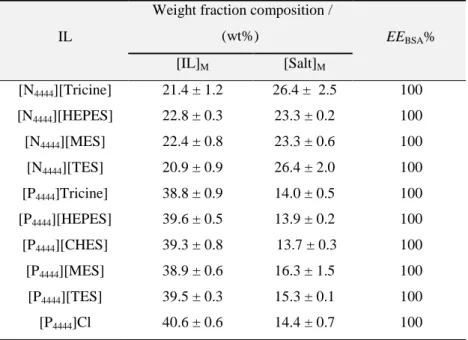
Table 2.3. Percentage extraction efficiencies of BSA, EEBSA%, and respective standard
deviations (σ) in the ABS composed of [N4444][GB] + K3C6H5O7 at 25 ºC and [P4444][GB]
+ K3C6H5O7 at 25 ºC and atmospheric pressure. Initial mixture compositions and
respective standard deviations (σ) are represented as [IL]M and [Salt]M. ... 48 Table 2.4. pH values of the coexisting phases ABS formed by ILs + K3C6H5O7. ... 49 Table 3.1. Identification of the systems able (), not able () or not tried ( ⃝) to form
two-phase systems with PPG with different molecular weights (400, 600, 1200) and the organic salt K3C6H5O7. ... 62 Table 3.2. Correlation parameters used to describe the experimental binodal data by Eq. 1
and respective standard deviations (σ) and correlation coefficients. ... 67
Table 3.3.. Data for the tie-lines (TLs) and tie-line lengths (TLLs). Initial mixture
compositions are represented as [Salt]M and [PPG]M whereas [Salt]Salt and [PPG]Salt are the
XVII
compositions and respective standard deviations (σ) are represented as [IL]M and
[PPG400]M. ... 70 Table 3.5. BSA stability test in 20% solution of IL conducted in HPLC. ... 75
XVIII
year of 2009 (dark grey) and the predicted rates for 2013 with a 95% prediction interval (light grey) for in men in the EU [11]. ... 5
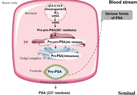
Figure 1.2. Schematic representation of PSA processing in epithelial cells of the prostate.
ER, endoplasmic reticulum. Adapted from Reference [46]. ... 14
Figure 1.3. Model of PSA biosynthesis in normal prostate epithelium versus cancer.
Adapted from Reference [48]. ... 15
Figure 1.4. Cartoon representation of the crystal structure of the Prostate Specific Antigen
(PDB ID: 1PFA). Catalytic Ser195, His57, Asp102 are highlighted in big ball and stick. Trp is highlighted in small ball and stick. Tyr is highlighted as sticks. Cys is highlighted as sticks [48]….. ... 16
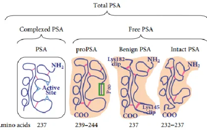
Figure 1.5. Schematic representation of the partitioning of free prostate-specific antigen
(fPSA) into various precursor isoforms of PSA (proPSA), benign PSA and inactive PSA (iPSA) in serum, with the respective amino acid number. cPSA = complexed prostate-specific antigen [62]. ... 20
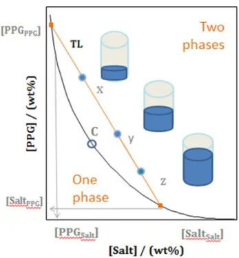
Figure 1.6. Representation of the phase diagram of an ABS. Binodal curve: (-); Critical
point: C; tie-line (TL); Mixture compositions at the biphasic region (X, Y and Z). Adapted from Reference [92]... 26
Figure 1.7. Chemical structures of the cations of nitrogen-based ILs. ... 29 Figure 1.8. BSA structure [122] ... 30 Figure 2.1. Chemical structures of the studied good buffers ionic liquids: (i) [Tricine]-; (ii) [TES]-, (iii) [CHES]-; (iv) [HEPES]-, (v) [MES]-; (vi) Cl-; (vii) [P4444]+; (viii) [N4444]+. .... 40 Figure 2.2. Ternary phase diagrams for the systems composed of IL + K3C6H5O7 + water
at 25 °C and atmospheric pressure in wt% (left) and in mol.kg-1 (right): () [P4444][Tricine], () [P4444][MES], () [P4444][HEPES], () [P4444][TES], ()
[P4444][CHES], and () [P4444]Cl [132]. ... 42 Figure 2.3. Phase Ternary phase diagrams for systems composed of IL + K3C6H5O7 +
XIX
Figure 2.4. Evaluation of the cation nature in the ternary phase diagrams composed of IL +
K3C6H5O7 + water at 25 °C and atmospheric pressure in wt% (left) and in mol.kg-1
(right): () [P4444][Tricine], () [P4444][MES], () [P4444][HEPES], () [P4444][TES],
() [P4444][CHES], (▲) [N4444][Tricine], (▲) [N4444][MES], (▲) [N4444][HEPES], (▲)
[N4444][TES], (▲) [N4444][CHES]. ... 44 Figure 2.5. Phase diagram for the ternary systems composed of GB-IL + K3C6H5O7 +
water, with the corresponding binodal fitting of data using Eq. 1 (-)... 45
Figure 2.6. Phase diagram for the quaternary system composed of K3C6H5O7 +
[P4444][TES] + Water at 25ºC and atmospheric pressure: binodal curve data (); TL data
(+); adjusted binodal data using Eq. 1 (-). ... 47
Figure 2.7. Visual appearance of the BSA extraction with an ABS formed by IL+ salt +
water…. ... 48
Figure 3.1. Synthesis of ionic liquids in an inert atmosphere. ... 57 Figure 3.2. Chemical structure of the studied ILs and PPG: i) [Ch][MES]; (ii)
[Ch][HEPES]; (iii) [Ch][Tricine]; (iv) [Ch][Bit]; (v) [Ch][DHCit]; (vi) [Ch][Gly]; (vii) [Ch]Cl (viii) [Ch][DHPhs], (ix) [Ch][Ac], (x) [Ch][Lac], (xi) [Ch][But], (xii) [Ch][Prop], (xiii) PPG. ... 60
Figure 3.3. Phase diagrams for systems composed of PPG 400 + IL + water at 25ºC and
atmospheric pressure in wt% (left) and in mol.kg-1 (right): () [Ch][Ac]; () [Ch][Gly]; () [Ch][lac]; () [Ch][Prop]; () [Ch][But]; () [Ch][DHCit]. ... 64
Figure 3.4. Phase diagrams represented in molality for the systems composed of PPG + IL
+ water in units wt% (left) and in mol.kg-1 (right): () [Ch]DHPhs]; () [Ch][Bit]; () [Ch]Cl. ... 65
Figure 3.5. Phase diagrams represented in molality for the systems composed of PPG + IL
+ water in units wt% (left) and in mol.kg-1 (right): () [Ch][Tricine]; () [Ch][HEPES]; () [Ch][MES]. ... 66
XX (-). 69
Figure 3.7.Size exclusion chromatograms of BSA in ternary system composed of PPG
400+ IL + water at 25 ºC. X-axis – Absorbance 280 (mV) a y-axis - Elution time (min). ..71
Figure 3.8. BSA extraction with ABS formed by PPG + IL + water with visible protein
precipitation at the interface. ... 72
Figure 3.9. Size exclusion chromatogram of BSA in PBS solution with 20% of [Ch][Ac].
Yellow line: BSA in PBS; Red line: BSA in 20% of [Ch][Ac]. ... 74
Figure 4.1. Different compositions along the same the same TL obtained by applying the
lever-arm rule. Salt + IL + water ABS (left), and PPG 400 + IL + water ( right). MP: mixture point; 5x, 7x, 10x, 12x, 20x, 40x corresponds to concentration factors. ... 81
Figure 4.2. Concentration factors (fc) of BSA (EBSA%) in the systems composed of
[Ch][Tricine] + PPG400 + water: fc=7 and f= 12; p1 and p2 represent the different
experiments. The filled line: experimental values; dashed green line: theoretical values. . 83
Figure 4.3. Concentration factors (fc) of BSA (EEBSA%) in the systems composed of
[N4444][Tricine] + salt + water: fc=2, 5, 10, 20 and 40; p1;p2;p3;p4;p5 represents the
different experiments. Filled lines: experimental values; dashed green line: theoretical values.. ... ……….84
Figure 5.1. Extraction efficiencies of PSA (EEPSA%) for the IL-rich phase in the systems
composed of K3C6H5O7 + [N4444][Tricine] + water at 25 (±1)º C and atmospheric pressure
XXI
σ – standard deviation;
Abs – absorbance (dimensionless);
AbsILPSA – absorbance PSA at the maximum wavelength in the IL-rich and in the salt-rich
AbsSaltPSA – absorbance PSA at the maximum wavelength in the salt-rich
Mw – molecular weight (g·mol-1);
R2 – correlation coefficient (dimensionless);
α – ratio between the top weight and the total weight of the mixture (dimensionless);
– concentration of ionic liquid (wt% or mol·kg-1
);
– concentration of ionic liquid in the ionic-liquid-rich phase (wt%); – concentration of ionic liquid in the salt-rich phase (wt%);
– concentration of ionic liquid in the initial mixture (wt%); – concentration of salt (wt% or mol·kg-1
);
– concentration of salt in the ionic-liquid-rich phase (wt%);
– concentration of salt in the salt-rich phase (wt%);
– concentration of salt in the initial mixture (wt%); – concentration of salt (wt% or mol·kg-1
); – concentration of PPG (wt% or mol·kg-1
);
– concentration of PPG in the ionic-liquid-rich phase (wt%);
– concentration of salt in the PPG-rich phase (wt%); – concentration of PPG in the initial mixture (wt%);
EEBSA% – percentage extraction efficiency of BSA (%);
EEPSA% – percentage extraction efficiency of PSA (%);
FCBSA – BSA concentration factor;
wIL – weight of the IL-rich phase;
XXII A2M – alpha-2-macroglobulin;
API – alpha-1-protease inhibitor ABS – aqueous biphasic systems; B – bottom phase;
BPH – benign prostate hyperplasia; BPSA – benign PSA;
BSA – bovine serum albumin C – critical point;
CaP – prostate cancer;
ELISA – enzyme-linked immunosorbent assay; DRE – digital rectal examination;
ER – endoplasmic reticulum;
ERSPC – european randomized study of screening for prostate cancer; FDA – Food and Drug Administration;
fPSA – free PSA; GB – Good’s buffers;
HEPES – N-cyclohexyl-2-aminoethanesulfonic acid; HPLC – High-performance liquid chromatography; iPSA – intact PSA;
K2CO3 –potassium carbonate;
K3C6H5O7 – potassium citrate;
K3PO4 – potassium phosphate;
LLE – liquid-liquid extraction;
MALDI-TOF MS – matrix-assisted laser desorption/ionization - time of flight mass spectrometry;
NNS – number needed to screen; NNT – number needed to treat; PEG – polyethylene glycol; PI –prediction interval; pI– isoelectric point;
XXIII PSA – prostate specific antigen;
proPSA – precursor isoforms of PSA; ROS – reactive oxygen species; T – top phase;
tPSA – total PSA; TL – tie-line;
TRIFA – immunochemiluminescent; TRUS – transrectal ultrasound; RNA – ribonucleic acid;
SDS-PAGE – sodium dodecyl sulfate-polyacrylamide gel electrophoresis; SPE – solid-phase extraction;
SPR – surface plasmon resonance technology;
[Ch][Ac] – (2-hydroxyethyl)trimethylammonium (cholinium) acetate; [Ch][But] – (2-hydroxyethyl)trimethylammonium (cholinium) butanoate; [Ch]Cl – (2-hydroxyethyl)trimethylammonium (cholinium) chloride;
[Ch][DHPhs] – (2-hydroxyethyl)trimethylammonium (cholinium) dihydrogen phosphate [Ch] [DHCit] – (2-hydroxyethyl)trimethylammonium (cholinium) dihydrogen citrate [Ch][Gly] – (2-hydroxyethyl)trimethylammonium (cholinium) glycolate;
[Ch][HEPES] – (2-hydroxyethyl)trimethylammonium (cholinium) 2-[4-(2-hydroxyethyl)piperazin-1-yl]ethanesulfonate;
[Ch][Lac] – (2-hydroxyethyl)trimethylammonium (cholinium) lactate;
[Ch][MES] – (2-hydroxyethyl)trimethylammonium (cholinium) 2-(N-morpholino)ethanesulfanate;
[Ch][Prop] – (2-hydroxyethyl)trimethylammonium (cholinium) propionate;
[Ch][Tricine] – (2-hydroxyethyl)trimethylammonium (cholinium) N-2(2-hydroxy-1,1-bis(hydroxymethyl)ethyl)glycinate;
[C8mim]Br –1-octyl-3-methylimidazolium bromide;
[C6mim]Cl – 1-hexyl-3-methylimidazolium chloride;
[P4444]Cl – tetrabutylphosphonium chloride;
XXIV
[N4444][MES] – tetrabutylammonium 2-(N-morpholino)ethanesulfonate;
[P4444][MES] – tetrabutylphosphonium 2-(N-morpholino)ethanesulfonate;
[N4444][HEPES] – tetrabutylammonium 2-[4-(2-hydroxyethyl)piperazin-1-yl]ethanesulfonate;
[P4444][HEPES] – tetrabutylphosphonium 2-[4-(2-hydroxyethyl)piperazin-1-yl]ethanesulfonate;
[N4444][TES] – tetrabutylammonium 2-[[1,3-dihydroxy-2-(hydroxymethyl)propan-2-yl]amino]
ethanesulfonate;
[P4444][TES] – tetrabutylphosphonium 2-[[1,3-dihydroxy-2-(hydroxymethyl)propan-2-yl]amino]
ethanesulfonate;
[N4444][Tricine] – tetrabutylammonium N-2(2-Hydroxy-1,1-bis(hydroxymethyl)ethyl)glycinate;
3
1.1 Scope and Objectives
Prostate cancer is a prevalent, worldwide concern among male adults [1]. At present, there is no curative therapy available for locally advanced or metastatic prostate disease, which makes its early detection a powerful way to increase the curative success rate [2]. A desired goal in the development and identification of prostate cancer is based on finding biomarkers by noninvasive assays to replace the currently used diagnostic techniques, like biopsy that it is still known as the diagnostic “gold standard”. However, this procedure has a high risk of adverse events, such as bleeding and sepsis, and it is associated with 15% to 20% of false-negative results [3]. On the other hand, the identification and quantification of biomarkers in human serum and urine can be seen as an alternative approach to the standard tissue biopsy procedure. For instance, prostate specific antigen (PSA) has been identified as the most reliable tumor marker for an early diagnostics of prostate cancer [4].
Commercial kits for the PSA quantification are now offered by several companies, but some of them require the identification and characterization of immunoassay-qualified antibodies and highly qualified technical operators [5]. Also, the investment on time and resources required to generate such immunoassays are considerable, what often obstruct it’s development on clinically specific chemistry laboratories [6]. Therefore, the main objective of this work consists on the development of an alternative platform for the extraction and concentration of PSA from human urine samples using a liquid-liquid extraction procedure. To this end, aqueous biphasic systems (ABS) formed by ionic liquids (ILs), an inorganic salt and water, as well as the combination of ILs and polymers will be investigated. Since commercially available cancer biomarkers are highly cost products and are distributed in small concentrations, it is necessary to carry out previous optimizations tests with cheaper and model compounds in order to achieve an improved ABS. Thus, bovine serum albumin (BSA), the most abundant protein in blood plasma of many species, was chosen as a model protein.
As a first approach, several experimental studies were attempted aiming at developing an optimized size-exclusion high-performance liquid chromatography (HPLC) quantification method for the model protein that can be applied further, only with small adjustments, on the PSA quantification. After defining the conditions for the quantification method, a large array of novel systems were investigated by combining potassium citrate (K3C6HPO4) with Good’s buffers ionic liquids (GB-ILs), i.e., ILs that can maintain the pH
4
of the aqueous solution, being this an important property when dealing with proteins. All the new ABS were characterized by their ternary phase diagrams, lines (TLs) and tie-line lengths (TLLs) at 25 ºC, followed by studies on their application in the extraction/concentration of BSA. All the GB-ILs were synthesized in this work and their synthesis and characterization are also described.
In a second stage, ABS formed by polypropylene glycol 400 (PPG 400) and cholinium-based ILs were further explored. In addition to the ternary phase diagrams, TLs and tie-line lengths (TLLs) initially ascertained, their extraction and concentration abilities were also evaluated. The stability of BSA in the ILs solutions was also evaluated.
Finally, it was addressed the real and prospective application of IL-based ABS for the possible concentration of cancer biomarkers from biological fluids, maintaining BSA as the model protein. In this context, two of the best systems, namely K3C6H5O7 +
[N4444][Tricine] and PPG 400 + [Ch][Tricine] were selected. The results revealed that the
system formed by K3C6H5O7 + [N4444][Tricine] is promising for the extraction process
(100% of extraction achieved in a single-step procedure) and further concentration that could be used in PSA clinical analysis in the near future. Aiming at supporting this statement the ABS formed by K3C6H5O7 and [N4444][Tricine] was used in an isolated
experiment with PSA and where it is confirmed the complete extraction of the cancer biomarker for the IL-rich phase by UV-spectroscopy.
5
1.2 Prostate Cancer Overview
1.2.1 Epidemiology and Risk Factors
Prostate cancer (CaP) is the third most common cancer-related cause of death in men, and resulted in more than 29,720 estimated deaths by the end of 2013 [1], a value that exceeds the expected number (28,170) of the year before [7]. In Europe, CaP is one of the most frequent solid neoplasms, with a predicted death rate of 10.52 cases per 100 000 men for 2013 (Figure 1.1) [8]. The vast majority (approximately 95%) of prostatic tumors are adenocarcinomas, whereas “adeno” refers to the glandular structure while carcinoma relates to the origin of cancer from the prostatic epithelium [9].
Figure 1.1. Bar-plots of standardized death rates per 100,000 individuals (men) for the year of 2009 (dark
grey) and the predicted rates for 2013 with a 95% prediction interval (light grey) for in men in the EU [11].
With the increasing interest from the medical community, several risk factors for prostate cancer have been identified, of which the most common include old age, race, inherent susceptibility and environmental factors [10]. More specifically, those at higher risk for developing prostate cancer include men over the age of 65, African-American individuals, and those who first-line relatives have/had CaP (the risk is at least doubled). In addition, factors such diet, sexual behaviour, alcohol consumption, exposure to ultraviolet radiation and occupational exposure have all been discussed as being of etiological importance [10].
Despite prostate cancer is a multifactorial disease, a lot of scientists affirm that the most significant risk factor is advanced age [11]. About 60% of all prostate cancer cases
6
are diagnosed in men with 65 years old or older, and 97% occur in men with 50 years old or older [12]. In addition, the incidence of prostate cancer increases more with age than any other type of cancer [10]. The relationship between CaP and advanced age likely reflects the interplay of environmental, physiological, and molecular influences with normal consequences of aging that presumably exacerbate the related effects. During aging, the progressive accumulation of DNA adducts and an increase in the DNA strand-break frequency in most tissues tend to occur [10]. It is believed that these age-related changes are caused by oxidative stress, which arises as a result of an imbalance in cellular prooxidant-antioxidant status [11]. Cellular oxidants, such as free radicals and reactive oxygen species (ROS), are produced during natural metabolic processes. ROS are highly reactive and potentially damaging to cells, because they directly damage macromolecules and organelle functions [10]. Damage to DNA by ROS results in single-strand and double-strand breaks, apurinic and apyrimidinic sites, ring-saturation of thymine derivatives, and adduct formation. Moreover, ROS can catalyze the oxidative modification of proteins, including enzymes involved in DNA repair. Together, these direct and indirect influences of ROS on DNA create an ideal environment for mutagenesis and tumor initiation [10].While the precise molecular consequences of aging involved in the development of prostate cancer have not been totally elucidated, several studies described gene expression changes associated with aging, particularly in prostatic stroma, and including genes involved in inflammation, cellular senescence and oxidative stress as the most possible causes [11].
At present, there is no curative therapy available once the disease spreads the limits of the organ. Since there are no effective therapeutic options for advanced prostate cancer, early detection of the tumor is pivotal and can increase the curative successful rate [2].
1.2.2 Diagnosis
Most cancer patients have a low-risk,clinically localized disease at diagnosis and can be treated effectively with surgery and radiation. Like most types of cancer, if cancer prostate is diagnosed in its early stages, there is a higher probability of an efficient treatment (Table 1.1). However, CaP survival is related to many factors, especially the extent of the tumor at the time of diagnosis. Approximately 10% to 20% of patients are diagnosed with locally advanced (affecting nearby tissues, such as the bladder or rectum)
7
or metastatic disease (affecting other areas in the body, usually the lymph nodes or bone). In general, the outcomes for such cases are less likely to be improved by therapy than those with lower volume or grade tumors [13]. The most common clinical symptoms are haematuria (presence of erythrocytes in the urine) and urinary obstruction, although they usually do not appear until the tumor has already invaded the nearby tissue or the lymph nodes. Cancer that spreads outside the gland may also result in lower extremity oedema from regional lymphatic obstruction or pain from bone metastasis [14]. Nevertheless, for most men, prostate cancer is of slow growing and does not result in clinical signs or symptoms during their lifetime. Consequently, the screening for CaP if of outmost importance, and requires that diagnostic tests must be performed in the absence of any symptoms or indications of disease, and which is often a major challenge [15].
Table 11. Year relative survival rate corresponding to the stage that prostate cancer was diagnosed [16].
Currently, prostate cancer diagnostic tests include digital rectal examination (DRE), prostate-specific antigen (PSA) blood test and transrectal ultrasound (TRUS) guided biopsy [15]. PSA test and DRE are used as primary screening tools in early detection of prostate cancer and TRUS-guided needle biopsies are techniques more directed to confirm diagnosis already proposed by PSA or DRE testing, or both [2]. Histopathological interpretation by Gleason score, a score based on the histological pattern of the tumor, is also applied in the diagnosis of CaP, being very useful, especially in stratifying patients into different risk groups [17]. This score represents a strong measure of how aggressive the prostate cancer is and can be used to determine the prognosis and type of therapy [17].
Although DRE and TRUS guided biopsies are widely employed by diagnosticians, they present some disadvantages. DRE has poor reliability, particularly in small tumors that have not reached the prostatic capsule [18]. TRUS guided biopsy is known for being a very invasive technique that carries out significant risks, such as subsequent infections. Two recent large cohort studies [3, 19] demonstrated that there is a significant
Stage 5-year relative survival rate
Local 100%
Regional 100%
8
hospitalization rate due to infections following biopsy, with a 3–4 fold increase above normal levels. In addition, these techniques exhibit limits on the ability to diagnose CaP because they do not permit the clinic to distinguish between benign prostate hyperplasia (BPH) and CaP [2]. Histopathological studies of prostate tissue can identify CaP in most cases, however, these methods also present some limitations. First of all, it is an invasive technique since it is necessary to perform a biopsy. Secondly, the Gleason’s grading scale
used by pathologists is at least semi-quantitative since it may be difficult to search every cell of every tissue slice. At last, there is a lack of concordance between the threshold of scoring between different pathologists [20]. In contrast, the quantification of PSA levels in biological fluids, especially in serum from a simple blood sample, is a less invasive screening tool, is easily performed, acceptable by the general population, accurate, and significantly affects the outcome of the disease. Consequently, PSA tests remain the most commonly used screening approach for prostate cancer [21].
1.3. Tumour Biomarkers
The introduction of biomarkers for disease diagnosis and management has revolutionized the practice of oncology. The National Cancer Institute defines a biomarker as “a biological molecule found in blood, other body fluids, or tissues that is a sign of a normal or abnormal process or of a condition or disease” [22]. Biomarkers are molecules whose detection or evaluation provides information about a disease beyond the standard clinical parameters that are gathered by the clinician. These can be proteins, metabolites, RNA transcripts, DNA, or epigenetic modifications of DNA, among other alterations. They can be detected in patient tissue samples, obtained either by biopsy or surgical resection, or noninvasively through the isolation of cells and/or molecules from bodily fluids, such as blood or urine. It’s now established that biomarkers can be valuable in the identification of the disease and its progression, identify high-risk individuals, and allow to monitor the individuals’ response to treatments. Furthermore, the identification of biomarkers can be carried out by non-invasive techniques and low-cost techniques [23] Therefore, the detection of tumour markers plays an important role in screening, diagnosis and to evaluate the prognosis of the diseases. Unlike most cancers, prostate cancer management has long used biomarkers. PSA has been identified as the most reliable tumor marker for the early diagnostics of prostate cancer [24].
9
1.4. Prostate Specific Antigen as a Cancer Prostate Biomarker
Prior to the 30s, there were no biochemical markers for the screening of prostate adenocarcinoma, and the diagnosis was only achieved by clinical history and physical examination. In 1938, Gutman observed, for the first time, that serum acid phosphatase (PAP), one of the major proteins secreted by prostate columnar epithelium secretory cells, appeared in high levels in patients with metastatic adenocarcinoma of the prostate [25]. However, since PAP is present in many normal blood components and organs and is highly concentrated in several non-prostatic malignancies, it exhibits lack in specificity for prostatic tissue and which makes it useless as a tumor marker [26]. In 1979, Wang et al. [27] reported on the purification of the PSA from prostate tissue. The authors showed that the antigen was present in normal and benign hyperplastic, as well as in malignant prostatic tissue and that it could not be detected in any other human tissue [27]. Since this discovery, PSA emerged as a potential prostate cancer biomarker. The first utility of PSA as a biomarker was in the monitoring of the disease status in patients with prostate cancer. After these findings, and because the PSA test is an inexpensive blood test that can be easily obtained, its application led to its increasing role as a screening modality. In 1994, the FDA approved PSA as a screening tool based on the seminal work of Catalona and colleagues [28] where they reported that the combination of the serum PSA measurement with levels ≥ 4.0 ng/mL, combined with other clinical findings, such as the results of DRE, improved the detection of prostate cancer.
1.4.1. Prostatic Cancer Screening
Prostate cancer diagnosis can be achieved by studying the prostate specific antigen levels in blood, where a high level could indicate the presence of cancer. In general, a PSA value of > 4.0 µg/L (and more recently > 2.5 µg/L) has been defined in the literature [29] as abnormal and it is frequently used as a cut-off, although for younger men a cut-off level of < 2.5 ng/mL is often used. For values between 4.0 and 10.0 µg/L, the grey zone, there exists a 22% to 27% likelihood of cancer, whereas values above 10 µg/L yield up to a 67% chance of prostate cancer [30]. Nevertheless, the PSA levels increase steadily with aging, and some urologists advocate the use of age-related “normal” PSA cut-points [31], rather than using the limit of > 4 µg/L as universal. Oesterling et al. [32] and Anderson et al. [33] noted that the upper limit of the PSA cutoff, achieving 95% of specificity, would increase
10
with age. Therefore, to maintain the equitable 95% specificity or an equivalent 5% false-positive rate across different ages, the PSA cutoff for recommending biopsy must necessarily increase with age. Based on cohorts of 537 patients with cancer-free status and benign prostatic hyperplasia, and 1,716 cancer-free, symptom-free, healthy volunteers who were between 40 and 79 years old, more general reference ranges were proposed as described in Table 1.2 [31].
Table 12. Values of reference range of PSA levels in serum for men with 40-79 years old [32, 33].
References
Age Group PSA Cutoff (ng/ml)
40–49 50–59 60–69 70–79
Oesterling et
al. [32] 2.5 3.5 4.5 6.5
Anderson et
al. [33] 1.5 2.5 4.5 7.5
In addition, it is already known that PSA levels in black men are higher compared to those in white men regardless of age, which also implies a careful interpretation of PSA levels [31]. Another fact that is important to have in mind is that the PSA level is a continuous parameter, so the higher the value, the more likely is the existence of CaP (Table 1.3) [29].
Table 13. Risk of prostate cancer in relation to PSA values in serum [29].
1.4.2. Prostate Cancer Stage and Grade
PSA levels strongly discriminate different cancer stages: they are higher in men with localized disease, and even higher in patients with metastatic [30]. Numerous subsequent studies have confirmed the original data from Stamey et al. [34], where they affirmed that a higher PSA level is associated with a large tumor volume [35], higher clinical stage [34],
PSA level (ng/mL) Risk of CaP (%)
0–0.5 6.6
0.6–1 10.1
1.1–2 17.0
2.1–3 23.9
11
pathological stage [20] and Gleason grade [20, 35]. Usually, the PSA levels in biological fluids are combined with the Gleason sum score. This score ranges from 2 to 10 and consists of 2 summed grade patterns that can vary from 1 (well differentiated) to 5 (poorly differentiated) [20]. The combination of these two techniques allows the risk stratification and clinical stage of men with localized CaP, and which include low risk men (PSA < 10 ng/mL and Gleason grade ≤ 6), intermediate risk men (PSA 10-20 ng/mL or Gleason grade = 7) and high risk men (PSA > 20ng/mL or Gleason grade 8-10). These risk stratifications then guide the treatment decisions and enable to predict disease characteristics and treatment outcomes.
1.4.3. Monitoring Therapy and Disease Recurrence
After the diagnosis of prostate cancer, the question regarding the best treatment for each patient arises. If there is an indication that the cancer is localized within the prostate and that the patient has a life expectancy larger than ten years, radical prostatectomy is the treatment of choice [36]. Many patients are cured of prostate cancer by this treatment and the frequency of performing this procedure has increased significantly to ∼40% of all prostate cancers, partially owing it to the PSA screening. PSA testing in post-prostatectomy patients is of most importance in deciding who has residual disease, who has relapsed (and when) and who can be considered cured. This has been the most widely accepted application of PSA testing in clinical practice [36]. After radical prostatectomy, PSA should decrease to undetectable concentrations because all the source tissue has been removed. The serum half-life of PSA has been estimated as 2.2-3.2 days [37]. PSA reaches a new steady state after a given number of half-lives subsequent to surgery, depending on the amount of prostatic tissue that was present. Non-negligible PSA and increasing PSA concentrations indicate either that the entire prostate was not excised or that PSA is being produced by metastases of the original tumor. Lange et al. [38] reported that patients with a PSA level < 0.4 µg/L, 3-6 months after radical prostatectomy, lead to recurrence 6 to 49 months after surgery (P < 0.0001).
The widespread use of the PSA test led to the prostate cancer being diagnosed at earlier stages [4]. The patients proportion diagnosed at local stages increased significantly and diagnoses of disseminated disease were drastically reduced [4]. Consequently, a decline in mortality rate has been observed which is also attributed to the possibility of
12
making a prompt treatment of the localized disease [4]. The European Randomized Study of Screening for Prostate Cancer (ERSPC) detected an improvement in cancer mortality rate in a PSA screened group [21]. More recent data from the Gottenberg arm of the ERSPC trial have emerged [39]. With a longer term 14-year follow-up period, in comparison with the 9-year follow-up data of the ERSPC trial, this study demonstrated a reduction in the number needed to screen (NNS) and the number needed to treat (NNT) to prevent 1 death by 5- and 4-fold, respectively [39]. Of note, the Gottenberg arm study had a shorter screening interval and lower PSA threshold required for initiation of further investigations [39]. When the data from the ERSPC trial are extrapolated in a longer period of follow-up, a similar reduction in NNS and NNT is also seen in comparison with the original 9-year follow-up data [40]. This indicates that significant benefits from treating PSA screened diseases are likely to occur over a decade after treatment. Support for PSA screening may arise as the longer-term follow-up data of these trials. Moreover, additional studies [41, 42] proved that PSA levels indicate the risk of prostate cancer, years or even decades, before diagnosis. All these studies [41, 42] show that men who will eventually develop prostate cancer have increased PSA levels years or decades before the cancer is diagnosed.
Despite the great utility of PSA for CaP detection, as a single test, it also has several limitations. While essentially organ-specific, PSA is not cancer-specific since raised levels may also indicate benign prostatic hyperplasia, prostatitis (inflammation of the prostate), or small tumors that do not prove to be fatal. Another issue with the PSA test is that false positives are common, and many men with elevated PSA levels do not have prostate cancer at all [23]. Furthermore, treatment of these slow-growing tumors is costly and often involves life changing surgeries that may not be necessary. These facts put in cause the viability of the routine PSA screening for prostate cancer [23]. Despite these drawbacks, routine PSA screening has been advocated as a mean to reduce disease-specific mortality by detecting early CaP, amenable to radical treatment with curative intent. Influential groups, such as the American Urological Association, the National Comprehensive Cancer Network, and the European Association of Urology support the importance of PSA screening in CaP [43].
In the last years, several studies were conducted with the finality of refining the PSA test, increasing its diagnostic accuracy by improving the specificity of PSA in the early
13
detection of CaP. They include the measurement of the PSA density of the transition zone and the quantification of free to total PSA ratio and PSA molecular forms such as free PSA (fpsa), precursor isoforms of PSA (proPSA) and benign PSA (BPSA) [18]. However, the determination of the PSA density implies a biopsy. On the other hand, the measurement of molecular forms of PSA seems to be the most promising diagnostic tools so far [44].
1.5. PSA Molecular Characteristics
PSA is a serine protease that belongs to the family of glandular kallikrein related peptidases, clustered in a locus that spans approximately 280 kb of chromosome 19q133–4 [45]. This protein is encoded by the prostate-specific gene kallikrein 3 (KLK3). PSA gene expression in the prostate is regulated by androgens; two functional androgen-response elements are present in the proximal promoter of the PSA gene [45]. PSA in blood exists in multiple forms: free or in complexes with various protease inhibitors; as proprotein or mature protein; and as intact or nicked [45]. These forms, and their implications for prostate cancer, will be discussed further below.
1.5.1 Biosynthesis
Transcription and expression of PSA are basically restricted to prostate epithelial cells and periurethral glands, and are dependent upon androgen mediation. PSA is translated as a proenzyme, pre-pro-PSA (261 amino acids), with 24 additional residues in the pre-region (17 residues), and which are the signal peptide and the propeptide (seven residues). The signal peptide directs the protein to the membrane of the endoplasmic reticulum (ER) [46]. In the ER, the prepeptide is removed and the resulting pro-PSA is transported within vesicles to the plasma membrane, where it is secreted into the lumina of prostate ducts (Figure 1.2). The protease or proteases responsible for the clipping of PSA remained unidentified for a long time [46]. It was suggested that PSA can be autocatalytic, but the cleavage sites observed are highly suggestive of a trypsin like enzyme. Moreover, it was demonstrated that this enzymatic activity belongs to hK2, the protein most closely related to PSA (~80% amino acid sequence identity), being responsible for the cleavage of propeptide to form the extracellular mature PSA (237 amino acids) [47]. The cleavage of the N-terminal seven amino acids from proPSA generates the active enzyme, which has five intrachain disulfide bonds, a single asparagine-linked oligosaccharide, and a weight of
14
33 kDa. This proPSA cleavage normally occurs between the arginine at position 7 and isoleucine at position 8, with the isoleucine becoming the N-terminus of the mature active protein [45]. The sugar-chain structure of PSA is determined by a series of processing reactions catalyzed by Golgi glycosidases and glycosyl transferases. In its final form, PSA is secreted into semen, at concentrations of 0.5–5 mg/mL, becoming one of the major proteins in seminal plasma. In comparison, it is found at lower concentrations in the serum [47].
Figure 12. Schematic representation of PSA processing in epithelial cells of the prostate. ER, endoplasmic
reticulum. Adapted from Reference [46].
Active PSA generated by hK2 can diffuse into the circulation, where it is rapidly bounded by protease inhibitors. This active PSA also undergoes proteolysis in the ER to generate inactive PSA which can enter the bloodstream (much smaller proportion than in semen) and circulates in an unbounded state (free PSA). In CaP, the loss of basal cells, the basement membrane and also the normal lumen architecture, results in a decrease in the ER processing of pro PSA to active PSA, and active PSA to inactive PSA, and with relative increases in bound PSA and proPSA in serum (Figure 1.3) [45]. While PSA is primarily produced by prostatic epithelial cells, PSA has also been detected in trace amounts in the paraurethral and perianal glands, endometrium, normal breast tissue, breast
Seminalfluid
Golgi complex
15
tumors, breast milk, female serum, adrenal neoplasms and renal cell carcinomas [48] However, these sites do not normally contribute to measurable levels of PSA in the circulation [48, 49].
Figure 13. Model of PSA biosynthesis in normal prostate epithelium versus cancer. Adapted from Reference
[48].
1.5.2. Structure
PSA is a glycoprotein with 28 kDa consisting of a single polypeptide chain of 237 amino acids and a single N-linked sugar-moiety [50]. This N-linked moiety comprises about 8% of the total mass of this protein. With regard to this moiety, a biantennary complex type with fucose linkages to the inner most N-acetylglucosamine (GlcNAc) was proposed, by NMR spectroscopy, as the major sugar-chain structure of PSA purified from seminal plasma [46]. It was also demonstrated that PSA is composed of 92% of peptides and 8% of carbohydrates, whereas the carbohydrate fraction is constituted by 4.84% of hexoses, 2.87% of hexosamines, and 0.25% of sialic acid [51]. The backbone contains a single carbohydrate unit attached at asparagine45 that increases the molecular weight by another 2-3 kDa and, in its mature form, exhibits five disulphide bonds [50].
In Figure 1.4, the 3D structure of PSA is displayed. The protein consists of two 6-strand antiparallel β-barrels and three α-helices. The catalytic site (Ser 195, His 57, and Asp 102) is conserved and localized in a cleft between two β-barrels [50]. The presence of
16
this characteristic His-Asp-Ser triad and a catalytic domain similar to those of other kallikrein-related peptidases is the reason for the categorization of PSA as a serine protease. The sequence also contains the GWG motif, which is a typical pattern present in many proteins with proteolytic activity. This motif contains Trp located in the β-barrel nearby the disulphide bond Cys 157-Cys [50]. Like for all serine proteases, the substrate and inhibitor recognition is mainly governed by the binding of the P1 residue to the S1 pocket of the enzyme [52].
Figure 14. Cartoon representation of the crystal structure of the Prostate Specific Antigen (PDB ID: 1PFA). Catalytic Ser195, His57, Asp102 are highlighted in big ball and stick. Trp is highlighted in small ball and
stick. Tyr is highlighted as sticks. Cys is highlighted as sticks [48].
1.5.3. Physiological Role
There is a great deal of interest and in determining the biochemical and physiological role of PSA. While most of the other kallikreins have trypsin-like proteolytic activity [52], PSA is considered a chymotrypsin-like protease. It shows similarities with chymotrypsin in the S1 specificity pocket of the catalytic site (presence of a serine at the bottom) and also preference for cleavage after the hydrophobic residues in the P1 position [52]. Despite these chymotrypsin similarities, PSA also displays enzymatic properties that differentiate it from chymotrypsin and other serine proteases. These differences suggest that there may be highly specific protein substrates for PSA that are not yet identified [53, 54]. Lilja et al. [55] revealed, in 1988, that the physiological function of PSA is the liquefying of seminal fluids, and this remains the most commonly accepted hypothesis until today. PSA liquefy
17
the seminal clot formed after ejaculation, in order to facilitate the transport of spermatozoa along the female reproductive tract. Gel structure dissolution occurs due to the PSA restricted chymotrypsinlike endoproteolytic activity, which allows the cleavage of semenogelin 1 and 2 (SEMG1, SEMG2), fibronectin, laminin and gelatin [55, 56]. Its activity is strongly inhibited by Zn2+, that is 100 times more abundant in semen than in serum [57].
Outside its physiological role, PSA may participate in the process of neoplastic growth and metastasis. PSA may influence, in particular, the tumoral growth of prostate cells through the degradation of the insulin like growth factor [58]. Other authors suggest that PSA performs the proteolysis of extracellular matrix proteins, such as fibronectin and laminin, and could also mediate invasion and the metastasis of CaP cells [49]. Further, PSA has been shown to stimulate the mitogenic activity of osteoblasts, possibly due to the activation of latent transforming growth factor-β and proteolytic modification of cell adhesion receptors [49].
1.5.4. Physicochemical Properties
PSA is a glycoprotein that has several isoenzymes, two intact and nicked forms of PSA, in the isoelectric point (pI) range of 6.5–8 [59]. The intact isoenzymes display high enzyme activity and, on the other hand, nicked forms display very low or no enzymatic activity [60]. It seems that the differences in the sialic acid content are the origin on the existence of these multiple isoforms. Each PSA molecular form contains six immunoreactive, antibody binding sites (epitopes) [61]. Serum levels of substances are dependent on their production and distribution, but also on their rate of elimination. Free PSA is quickly eliminated from blood, with a terminal half-life of ~14 hours [49]. For total PSA (tPSA) measured in the conventional PSA tests, the terminal half-life is about 2.2-3.2 days [51]. In Table 1.4 some important biochemical characteristics of PSA are summarized.
18
Table 14. Biochemical characteristics of PSA [59, 60] [49, 51, 61].
Biochemical characteristics of PSA Molecular Mass (kDa) 28–33
Number of epitopes 6 Class Glycoprotein Subunit Monomer pI 6.5–8 Half-life fPSA ~ 14 hours tPSA 2.2-3.2 days 1.5.5. Molecular Derivatives
It was initially assumed that the PSA measured in serum was the natural 28 kDa form of the protein containing 237 amino acids [51]. Only later it was discovered that PSA in serum appears in two different forms, either as complexed to other proteins (inhibitors) or as a free unbound form, “complexed PSA” and “free PSA”, respectively [47]. Both complexed PSA and free PSA are combined to give a measure of tPSA conventionally used in the diagnostic of prostate cancer. While PSA isolated from seminal plasma is composed mainly of the catalytically active free form, in serum, the majority of PSA is complexed with endogenous inhibitors [45]. The most recognized inhibitors are alpha-1-antichymotrypsin (ACT), alpha-2-macroglobulin (A2M) and alpha-1-protease inhibitor (API). These protease inhibitors prevent the potential damage of the protease activity of PSA [62]. ACT covalently bound to PSA is the most common form and has a molecular weight of 80-90 kDa. The formation of this complex was described by the esterification of Ser-189 of PSA with 358 of ACT. This leads to the cleavage of ACT between Leu-358 and Ser-359, as indicated by Sodium Dodecyl Sulfate -polyacrylamide gel electrophoresis (SDS-PAGE) and N-terminal sequence analysis [63]. Matrix-assisted laser desorption/ionization - time of flight mass spectrometry (MALDI-TOF MS) revealed molecular weights of 80.8 kDa for PSA-ACT, 28.3 kDa for PSA, and 56.9 kDa for ACT [64].
Several researchers have found that as the percentage of fPSA decreases, the probability of having cancer increases, suggesting that this form could possibly make an improved diagnostic [44]. The most convincing results came from a careful meta-analysis of 66 studies, which clearly verified a better diagnostic performance (that is, a higher
19
prostate cancer detection rate) for fPSA than for tPSA [65]. It was also established that the calculation of the percentage of free PSA in relation to tPSA (the free PSA ratio), allows a modest but significant improvement, in the discrimination of prostate cancer from benign disease such as benign prostatic hyperplasia. This combination seems to increase in patients with borderline or intermediate PSA values (4-10 ng/mL) and negative DREs [49].
1.5.5.1. Free PSA
Of the various modifications of serum PSA used to improve its specificity in detecting CaP, the concept of free to total PSA ratio (f/t PSA) is the most widely used approach [35]. It has been shown that the analysis of free PSA reduces the number of negative biopsies by 25% [66]. Free PSA is generally lower in prostate cancer, and ranges from 1% to 10%, when compared to the benign prostate enlargement [66]. This patterns is mostly due to the reduced exposure of PSA to abundant proteolytic enzymes in seminal fluid [66]. Since fPSA provides better cancer discrimination, several investigations have focused on further discriminating fPSA into different molecular forms [66, 67]. fPSA in serum is now known to be composed of at least three distinct forms of inactive PSA, and as shown in Figure 5. The first PSA form may contain a number of minor variants, but appears to be largely composed of intact PSA that is similar to native, active PSA, except for structural or conformational changes that have rendered it to be enzymatically inactive. A second form of PSA, called BPSA, is an internally cleaved or degraded form of PSA that is more highly associated with Benign prostatic hyperplasia (BPH) [68]. BPSA is identical to native mature PSA, and also contains 237 amino acids, but has 2 internal peptide bond cleavages at Lys182 and Lys145. The last has been identified as the proenzyme or precursor forms of PSA (proPSA), and is the form mostly associated to cancer[69]. ProPSA in serum and tissues was found to be comprised of several truncated proPSA forms (Figure 1.5), containing from 1 to 5 amino acids in the pro leader peptide instead of the native 7 amino acids. The native proPSA (otherwise known as [-7]proPSA) exists in a truncated form containing a seven amino acid N-terminal pro-leader peptide (APLILSR). Partial cleavage of [-7]proPSA results in the shortening of the seven amino acid pro-leader peptide to form [-5]proPSA (LILSR), [-4]proPSA (ILSR) and [-2]proPSA (SR) [62]. Immunohistochemical studies have shown that BPSA is expressed preferentially in the transitional zone of the prostate and is associated with pathological BPH, while proPSAs
20
are expressed almost exclusively in the peripheral zone of the prostate where most prostate cancers are known to emerge [62]. The truncated proPSA form contains pro leader peptides of 2 amino acids, and where [-2]proPSA is of particular interest [45]. One potential explanation for the enrichment of truncated proPSA forms is that these truncated forms are more resistant to activation than the intact proPSA. The truncated proPSA forms are therefore more stable. Subsequent studies on prostate tissues already proved that these PSA forms are highly enriched in prostate tumors and are a more specific prostate cancer marker [69]. Furthermore, amino acid sequencing showed that the proPSA is mainly comprised of a truncated form of [-2]proPSA rather than the usual seven amino acids ([-7]proPSA) [62].
Figure 15. Schematic representation of the partitioning of free prostate-specific antigen (fPSA) into various
precursor isoforms of PSA (proPSA), benign PSA and inactive PSA (iPSA) in serum, with the respective amino acid number. cPSA = complexed prostate-specific antigen [62].
The proPSA forms are especially useful in the 2.5–4 ng/mL PSA range, while the other PSA forms show little diagnostic utility [70]. A current systematic review and meta-analysis demonstrated that the percentage of [-2]proPSA in relation to fPSA ([-2]proPSA/fPSA × 100) has greater accuracy than tPSA or fPSA in detecting CaP, and also that [-2]proPSA might be helpful in preferentially identifying those patients who might harbor a more aggressive form of the disease [71]. Based on this, Beckman Coulter (Coulter ACCESS® immunoassay system) introduced a measure of prostate health index
21
(phi) (64). This value is calculated from a combination of tPSA, free PSA and a measure of a truncated PSA isoform, [-2]proPSA [62].
In healthy adult males aged ≤ 50 years-old, the concentrations are 106
-fold higher in seminal fluid than in blood, in which the median PSA level is ~0.6 ng/mL [49]. In seminal fluid, PSA predominantly occurs in a free and active single-chain form, and only a minor proportion (≤5%) exists as inactive (due to internal cleavages) [56, 72]. The occurrence of this inactive form is due to a formation of covalent complexes with SERPINA5 (protein C inhibitor), which is abundantly released from the seminal vesicles [73]. In contrast, the majority of PSA that enters in blood is intact, non-catalytic and the predominant proportion is covalently linked to ACT [45]. PSA levels in blood span a ~105-fold range, from <0.1 to 104 ng/mL, with levels above 102 ng/mL found almost exclusively in men with advanced prostate cancer [49]. The increased blood levels of PSA in men with cancer cannot be explained by increased PSA expression; during the development and progression of prostate cancer, PSA expression may actually decrease slightly. The increased blood PSA levels must instead be caused by increased release of PSA into blood, consequently to the already mention rupture of prostate gland architecture, characteristic of CaP [49].
1.5.6. Stability of Sample Storage of Total and Free PSA
Clinical results for quantifying serum PSA are not usually urgently requested, and hence, samples are commonly stored and assayed in bundles or in batches for cost-efficiency [74]. PSA species are both pH- and temperature-labile, which implies a special care in sample handling and storage, since it might influence PSA stability in serum. Changes in immunoreactivity or alterations in the binding affinity with the specific antibody during the sample store are the major drawbacks [75]. Several investigators have reported the stability of tPSA in serum samples [65, 75]. It was already confirmed that tPSA is sufficiently stable at room temperature for 24h, after serum separation and storage in a refrigerator at 4°C for up to 1 week, or in a domestic freezer at -20°C for 1 month (76). In 2008, Reed et al. [75] have gone further when demonstrating that tPSA measured in serum exhibits a high correlation with the ones measured in the same serum after 7 years of storage at -80 ºC. However, this is not valid for fPSA since it is the most thermolabile isoform. In general, it is recommended to prepare samples for analysis within 8 h after venipuncture, with the storage of samples at 4 ºC when analyzed on the day of collection,
![Figure 2.1. Chemical structures of the studied good buffers ionic liquids: (i) [Tricine] - ; (ii) [TES] - , (iii) [CHES] - ; (iv) [HEPES] - , (v) [MES] - ; (vi) Cl - ; (vii) [P 4444 ] + ; (viii) [N 4444 ] +](https://thumb-eu.123doks.com/thumbv2/123dok_br/15791539.1078203/63.892.273.680.262.631/figure-chemical-structures-studied-buffers-liquids-tricine-hepes.webp)
![Figure 2.2. Ternary phase diagrams for the systems composed of IL + K 3 C 6 H 5 O 7 + water at 25 °C and atmospheric pressure in wt% (left) and in mol.kg-1 (right): () [P 4444 ][Tricine], () [P 4444 ][MES], ()](https://thumb-eu.123doks.com/thumbv2/123dok_br/15791539.1078203/65.892.138.770.439.732/figure-ternary-diagrams-systems-composed-atmospheric-pressure-tricine.webp)
![Figure 2.3. Phase Ternary phase diagrams for systems composed of IL + K 3 C 6 H 5 O 7 + water at 25 °C and atmospheric pressure in wt% (left) and in mol.kg-1 (right): (▲) [P 4444 ][Tricine], (▲) [N 4444 ][MES], (▲)](https://thumb-eu.123doks.com/thumbv2/123dok_br/15791539.1078203/66.892.135.765.135.425/figure-ternary-diagrams-systems-composed-atmospheric-pressure-tricine.webp)
![Table 2.2. Data for the tie-lines (TLs) and tie-line lengths (TLLs). Initial mixture compositions are represented as [Salt] M and [IL] M whereas [Salt] Salt and [IL] Salt are the composition of IL and salt at the IL-rich](https://thumb-eu.123doks.com/thumbv2/123dok_br/15791539.1078203/69.892.126.793.198.854/table-data-lengths-initial-mixture-compositions-represented-composition.webp)
![Figure 3.3. Phase diagrams for systems composed of PPG 400 + IL + water at 25ºC and atmospheric pressure in wt% (left) and in mol.kg-1 (right): () [Ch][Ac]; () [Ch][Gly]; () [Ch][lac]; () [Ch][Prop];](https://thumb-eu.123doks.com/thumbv2/123dok_br/15791539.1078203/87.892.145.792.153.443/figure-phase-diagrams-systems-composed-water-atmospheric-pressure.webp)
![Figure 3.4. Phase diagrams represented in molality for the systems composed of PPG + IL + water in units wt% (left) and in mol.kg-1 (right): () [Ch]DHPhs]; () [Ch][Bit]; () [Ch]Cl](https://thumb-eu.123doks.com/thumbv2/123dok_br/15791539.1078203/88.892.144.799.155.454/figure-phase-diagrams-represented-molality-systems-composed-dhphs.webp)