Current Medicinal Chemistry, 2013, 20, 3049-3068 3049
Development of Plasmodium falciparum Protease Inhibitors in the Past
Decade (2002–2012)
B. Pérez
1, C. Teixeira
*,1,2, J.R.B. Gomes
2and P. Gomes
11
Centro de Investigação em Química da Universidade do Porto, Departamento de Química e Bioquímica, Faculdade de Ciências, Universidade do Porto, R. do Campo Alegre, 687, P-4169-007 Porto, Portugal; 2CICECO, Departamento de Química, Universidade de Aveiro, Campus Universitário de Santiago, 3810-193 Aveiro, Portugal
Abstract: New drug targets for the development of antimalarial drugs have emerged after the unveiling of the
Plasmo-dium falciparum genome in 2002. Potential antimalarial drug targets can be broadly classified into three categories
ac-cording to their function in the parasite’s life cycle: (i) biosynthesis, (ii) membrane transport and signaling, and (iii) he-moglobin catabolism. The latter plays a key role, as inhibition of hehe-moglobin degradation impairs maturation of blood-stage malaria parasites, ultimately leading to remission or even cure of the most severe blood-stage of the infection. Intraerythro-cytic Plasmodia parasites have limited capacity to biosynthesize amino acids which are vital for their growth. Therefore, the parasites obtain those essential amino acids via degradation of host cell hemoglobin, making this a crucial process for parasite survival. Several plasmodial proteases are involved in hemoglobin catabolism, among which plasmepsins and fal-cipains are well-known examples. Hence, development of P. falciparum protease inhibitors is a promising approach to an-timalarial chemotherapy, as highlighted by the present review which is focused on the Medicinal Chemistry research ef-fort recorded in the past decade in this particular field.
Keywords: Plasmodium falciparum, hemoglobin catabolism, plasmepsin, falcipain, falcilysin, aminopeptidase, proteases, an-timalarial.
1. INTRODUCTION
Malaria is one of the most threatening infectious diseases that mainly affects the world’s poorest countries in tropical areas [1]. Approximately 40% of the world population lives at risk of this disease and this constitutes a large burden on the health and economic development of low-incomes coun-tries [1-3]. Although the World Malaria Report 2011 showed that there has been significant and durable progress in bat-tling this disease, emergence of parasite resistance to antima-larial medicines remains a threat to this continued stride to-ward the reduction of malaria cases in the world. There are five species of Plasmodium which cause malaria in human. Those species are P. ovale, P. malariae, P. knowlesi [4, 5],
P. vivax and P. falciparum (Pf), the latter being the deadliest
[6, 7]. Malaria is transmitted by a female infected Anopheles mosquito that harbors Pf sporozoites in its salivary glands; by biting a human, the mosquito injects those sporozoites into the blood-stream, and they are then carried to the liver to develop into merozoites, which subsequently are released into the blood stream invading host red blood cells and mul-tiplying asexually as trophozoites. Some erythrocytic para-sites develop into male and female gametocytes, the only forms able to infect other mosquitoes and there reproduce sexually, permitting completion of the life cycle [2].
*Address correspondence to this author at the Centro de Investigação em Química da Universidade do Porto, Departamento de Química e Bioquími-ca, Faculdade de Ciências, Universidade do Porto, R. do Campo Alegre, 687, P-4169-007 Porto, Portugal; Tel: +351 234 401 423; Fax: +351 234 401 470; E-mail: [email protected]
Currently, there are drugs targeting different stages in the malaria life cycle such as chloroquine 1, artemisinin 2, and primaquine 3 (Fig. 1). However, the increasing spread of parasite strains resistant to currently used antimalarials has put enormous pressure on public health systems to introduce new treatments [8]. Given that the hope for a long-lasting vaccine against malaria is yet an unmet goal, it appears that control of the disease has to rely mostly on chemotherapy in the foreseeable future [9].
N O HN NH2 N Cl HN N O O O O O H H 1 2 3
Fig. (1). Structure of known antimalarials: chloroquine (1), artemis-inin (2), and primaquine (3).
Since the unveiling of the Pf genome, a decade ago, new targets for development of antimalarial drugs have arisen [10]. Antimalarial targets can be broadly classified into three 1875-533X/13 $58.00+.00 © 2013 Bentham Science Publishers
categories according to their function in the parasite’s life cycle: (i) biosynthesis, (ii) membrane transport and signal-ing, and (iii) hemoglobin catabolism. To the first target cate-gory belong Pf enzymes in charge of generating nutrients required for malaria parasite growth. For instance, there are different biosynthesis pathways today used as targets for the discovery of antimalarial drugs such as the folate biosynthe-sis pathway [11-13]. The second target category includes pathways which mediate the uptake of nutrients into cells and the generation and maintenance of transmembrane elec-trochemical gradients, for instance, the plasmodial surface anion channel (PSAC) [14-16]. Other examples which fall within this target category are enzymes whose substrates are involved in intracellular signal transduction, for instance, farnesyltransferase [17-19]. Finally, targets within the third target category play an essential role in the development of intraerythrocytic malaria parasites. Since the digestion of hemoglobin is presumably an essential catabolic function performed by the blood stage parasites, the proteases partici-pating in this pathway have been proposed as targets for the development of novel antimalarial drugs [20-22]. This re-view will mainly focus on such proteases and their inhibitors developed in the past decade.
2. HEMOGLOBIN CATABOLISM
During the intraerythrocytic development of the asexual stages of Pf, the parasite inhabits a parasitophorous vacuole (PV) formed inside the red blood cell (RBC); host cell he-moglobin is endocytosed by the PV and transported to an acidic compartment known as the parasite’s food vacuole (FV). Within the FV, proteases degrade most of the host he-moglobin to free amino acids that can then be incorporated into newly synthesized proteins [23] and used to modulate the osmotic status of the host cell [24, 25]. In addition, re-moval of hemoglobin also frees up space within the RBC for parasite replication [26]. Early in the degradative pathway, free heme is released and oxidized from the ferrous (Fe2+) state to the ferric (Fe3+) hematin. Both heme and hematin are potentially toxic to the parasite [27]. To counter this, the parasite has evolved a detoxification system resulting in the formation of the hemozoin pigment, an inert crystalline -hematin polymer [28, 29]. Antimalarial 4-amino-quinolines, such as chloroquine (1, Fig. 1), appear to function by disrupt-ing this sequestration, leaddisrupt-ing to an accumulation of toxic heme products [30].
Studies suggest that the proteases in charge of hemoglo-bin degradation are essential for the parasite growth since the parasite has limited ability for de novo biosynthesis of the amino acids needed for its own proteins. It has been shown that Pf can also obtain amino acids exogenously, as parasites can survive in media supplemented only with isoleucine, an amino acid not contained in hemoglobin; yet, in the absence of an exogenous amino acids source, the parasite can solely rely on hemoglobin degradation to obtain most of the re-quired amino acids [31]. Moreover, it has been clearly shown that Pf utilizes hemoglobin as an amino acid source for pro-tein synthesis, as amino acids from hemoglobin degradation have been detected in parasite’s proteins, and in vitro ex-periments where hemoglobin digestion has been impaired
revealed that parasite development and morphology were significantly affected [32].
Extensive study of this protease cascade has resulted in the model outlined in (Fig. 2) for hemoglobin degradation. There is evidence that the degradative enzymes function in a semi-ordered pathway [30], with aspartyl proteases, being the first to participate in this proteolytic pathway by making the initial cleavage in intact hemoglobin. Subsequent degra-dations of globin into small peptides chains are carried by cysteine proteases, metalloprotease falcilysin (FLN), and dipeptidyl amino peptidase 1 (DPAP1) [33]. Following, the small peptides are transported to the parasite cytoplasm where they are terminally degraded to amino acids by exopeptidases (Fig. 2) [33].
From the above, it is understandable that proteases in-volved in this catabolic pathway have become of substantial interest for the malaria research community. Certain inhibi-tors for these enzymes have been tested in in vitro experi-ments and found to block Pf growth and its expansion to other erythrocytes [35-37]. Therefore, the search for potent inhibitors of these proteases has become a new strategy for developing new antimalarial drugs.
Fig. (2). Proteolytic cascade in hemoglobin degradation by Pf para-sites. For a more detailed explanation refer to: http://priweb.cc.huji. ac.il/malaria/FramHemoglobindigest.html and see ref [34].
2.1. Aspartyl Proteases (Plasmepsins)
Pf contains at least 10 aspartyl proteases, known as
plas-mepsins: plasmepsins I, II, and IV–X, and histidine aspartic protease (HAP). The precise role of each of the plasmepsins in the parasite metabolism is not clear. To date, the most extensively studied plasmepsins are plasmepsin I (Plm I; EC number: 3.4.23.38), plasmepsin II (Plm II; EC number: 3.4.23.39), plasmepsin IV (Plm IV; EC number: 3.4.23.B14) and HAP. These aspartyl proteases are expressed during the erythrocytic stage of the parasite [38], have all been shown to be directly involved in the process of hemoglobin degra-dation [38, 39], and have all been characterized structurally [40]. High levels of sequence homology (60%-70%) [41] are observed between Plm I, II, IV, and HAP, which also lies in a cluster on the same gene [39]. Compared to Plm II, the binding site regions of Plm I, IV, and HAP show 84%, 68%, and 39% identity, respectively [42].
Plm I and Plm II catalyze the initial step in the breakdown of hemoglobin by the parasite by making the first cleavage of hemoglobin between Phe33 and Leu34 of the -chain to gen-erate globin and free heme [38, 39]. In addition to its partici-pation in hemoglobin degradation, the interest in Plm IV is also motivated by the fact that it is the only plasmepsin located in the FV of Pf which has orthologs in the other Plasmodium species infecting human [41]. HAP is unique in the sense that it has a histidine in the place of the first canonical aspartic acid. Whether this results in an aspartic- or serine-protease-like mechanism has been subject of discussion [43]. Computa-tional predictions indicate that only the aspartic acid (Asp214) is directly involved in catalysis, while the histidine residue (His34) provides critical stabilization along the cleavage [44]. Recent knock-out studies revealed that single, double, and even triple knock-outs of FV plasmepsins are viable, suggest-ing that multiple plasmepsins must be targeted to produce anti-parasitic effects [45]. This also indicates that there is a high number and functional redundancy of plasmepsins in the he-moglobin pathway. However, it has been shown that Plm in-hibitors significantly attenuated parasitemia both in culture and in animal models [46].
As the crystal structure of Plm II was the first one to be publicly available, most research focused on the develop-ment of Plm II inhibitors. Plm II contains 329 amino acids and its binding cleft, also known as the catalytic dyad, is constituted by Asp34 and Asp214, which are bridged by a water molecule [47]. Hydrolysis of the peptide bond between hemoglobin’s Phe33 and Leu34 takes place once Asp214 abstracts one of the protons from the water molecule, and generates two peptide chains (Fig. 3) [35, 47]. The cleavage mechanism concludes when the peptide products leave the active site and the water bridge between Asp34 and Asp214 is regenerated.
Plm I and II have a 73% sequence homology suggesting that both enzymes can be inhibited by related molecules. Therefore, Plm I and II have become the main targets of most plasmepsin inhibitors developed as potential antimalar-ial drugs. Yet, these plasmepsins show high structural simi-larity with human cathepsin D, which makes selectivity an important factor to take into account when designing suitable inhibitors.
Well known HIV-1 aspartyl protease inhibitors, such as ritonavir, indinavir, nelfinavir, lopinavir, saquinavir,
ata-zanavir and amprenavir, were also found to inhibit Pf aspar-tyl proteases and the development of parasites at pharmaco-logically relevant concentrations [48]. All of these inhibitors undergo non-covalent interactions with the proteases and share key structural structures for the inhibition of aspartyl proteases, which is a hydroxyl or hydroxyl-like moiety that coordinates to the catalytic dyad and mimics the transition state for peptide bond hydrolysis. The (S)-hydroxyl group displaces the water molecule from the catalytic site and forms a hydrogen bond with Asp [49, 50]. Like these HIV-1 aspartyl protease inhibitors, most Plm I and II inhibitors mimic the tetrahedral intermediate formed during the aspar-tyl protease catalysis. There are several transition state ana-logue cores used for the design of Plm inhibitors [35, 51-65], but the most important include the statine core [54, 56, 57, 64], the reversed-statine core [53, 54], or a hydroxyethy-lamine motif (Fig. 4) [56, 61, 62].
Studies using encoded combinatory libraries based on the statine core structure 4 (Fig. 5) allowed determining that "-branched side chains are preferred in P2 and hydrophobic side chains as phenyl or isobutyl in P1. In addition, P2 and P3 substituents impart selectivity in Plm II inhibitors (Fig. 5) [66, 67]. One advantage of this type of inhibitors is that they don’t cross inhibit other proteases such as serine-, cysteine-, or metallo-proteases [68].
Recently reported statine-based inhibitors include the one found by Bosisio and co-workers [64]. They coupled a series of statine-based inhibitors with primaquine 3 and found low nanomolar inhibitors of Plm II with IC50 between 0.59 to 400 nM and low micromolar activity in vitro against Pf, the best of which (5) is shown in (Fig. 6). A direct correlation be-tween the compounds’ activity against Plm II and the in vitro parasite growth suggested that the main mechanism of these inhibitors was Plm II inhibition and consequently, the diges-tion of hemoglobin that is essential for Pf survival as stated before. Compound with the linker derived from a succinic acid was the least active of the series which is in agreement with the finding suggesting that an aromatic substituent is preferred in P3 for Plm II inhibition [66]. The introduction of an aromatic ring as a linker in general structure 6 increased the activity as expected and compounds with a naphthyl were more active against Plm II. Still, there was not significant increase of the resulting derivatives against parasite growth. Compound 5 was considerably more active against the en-zyme and parasite growth with IC50 0.59 nM on in vitro
in-33Phe HN O Leu34 H O H 34Asp O OH Asp214 O O 33Phe HN Leu34 34Asp O O Asp214 HO O 33Phe H2N O Leu34 34Asp O OH Asp214 O O OH O OH H Scissile bond
hibition of Plm II, and IC50of 0.4 M on in vitro inhibition of growth of Pf D10 [64]. Additional studies were done to ver-ify the dual-action of compound 5. The products of the hy-drolysis of Lys-Leu peptide bond in compound 5 were as-sessed as inhibitors of the parasite development and Plm II activity. Even though such products were both active against parasite growth in the micromolar range, compound’s activ-ity against the parasite development was found to be mainly due to Plm II inhibition. H N O OH P1 -branched side chain Hydrophobic side chain
Extended and large aromatic side chain
N H 2P O 3P Impact on selectivity '1P O NH 4
Fig. (5). General structure requirements for Plm I and Plm II inhibi-tors based on the statine core.
Samuelsson and co-workers reported [54] a series of statine-reversed core inhibitors of Plm I and Plm II. The best inhibitor 7 of the series (Fig. 7) exhibits Ki values of 250 nM and 1.4 M for Plm I and II, respectively. To increase affin-ity to both plasmepsins a benzyl group was added to the N-terminus to interact with the S1’ pocket of the proteases. Furthermore, a hydrazine moiety was inserted to allow flexi-bility to the benzyl substituent in the N-terminus to better fit
in the S1’ pocket. All the compounds were tested against Plm I and II activity. From the inhibition results it can be inferred that the N-benzyl derivatives were almost inactive whereas the aza-benzyl derivatives promote the inhibition of both plasmepsins. Also, the presence of the carboxybenzyl group in the P2’ position seems to increase the activity of the compounds. Samuelsson and co-workers [54] also reported that the basic piperidine- and pyridine-substituents are pre-sent in the majority of the potent inhibitors, for instance, see compound 7.
In 2003, Nöteberg and co-workers [61] reported a series of compounds, including a basic hydroxyethylamine transi-tion state isostere, designed and synthesized as inhibitors of Plm I and Plm II. These compounds were designed on the basis of previous findings from Ellman’s group, who deter-mined that large substituents were suitable for P1’ position [69]. In comparison to Ellman’s compounds, Nöteberg’s had the nature of the peptidic inhibitor minimized using a single prime side amino acid residue bearing a biphenyl side chain. This yielded compounds which were highly selective for plasmepsins over cathepsin D, the most active of which (8) is displayed in (Fig. 8).
In the same year, the same group also synthesized and screened compound libraries based on the general structure 9, and found inhibitors with Ki values in low nanomolar range that targeted the malaria proteases Plm I and Plm II, again with high selectivity versus cathepsin D (Fig. 9) [62]. This study allowed not only to find very active and selective inhibitors, like 10 (Fig. 9), as well as to demonstrate that
Fig. (4). Transition-state mimicking groups in peptidomimetic plasmepsin inhibitors: reduced amine [52, 59], statine [54, 56, 57, 64], droxypropylamine [51], reversed-statine [53, 54], dihydroxyethylene (C- and N-duplicated) [35, 55, 63], norstatine [58, 60, 65], and hy-droxyethylamine [56, 61, 62].
very diverse side chains in P1’ and P3 positions are suitable for Plm I and II inhibition.
N N H O N Bn NHCbz OpBrBn OH 7
Fig. (7). The most active Plm I and Plm II inhibitor, based on the statine-reversed core, designed by Samuelsson and co-workers [54]. Bn=Benzyl; Cbz=Benzyloxycarbonyl. N H H N N O O OH H N NH2 O 8
Fig. (8). The most active Plm I and Plm II inhibitor of the biphenyl series developed by Nöteberg and co-workers: Ki(Plm I)= 115 nM;
Ki(Plm II)=121 nM [61].
Most of the reports on Plm II inhibitors describe non co-valent interactions with the aspartyl proteases. However, there are a few examples of irreversible inhibitors in the lit-erature [70-72]. Woster and co-workers [71] studied the syn-thesis and screening of Plm II inhibitors and found that three compounds produced irreversible inactivation of the enzyme with IC50 values in the low nanomolar range. They hypothe-sized that an (S)-hydroxyl substituent moiety bearing a latent electrophile should act as an irreversible inhibitor of Plm II. For instance, Asp214 or an adjacent basic amino acid could abstract an acidic proton from the inactive electrophile
re-sulting in the generation of an ,"-unsaturated system (11), ketenimine (12), or an allene (13 and 14) in the catalytic site (Fig. 10). R1 NH H N O O OH H N NH2 O R2 9 N H H N O O OH H N NH2 O 10 O O
Fig. (9). Core structure 9 of the library of potential Plm I and Plm II inhibitors studied by Nöteberg’s team. Compound 10 was the most active and selective inhibitor, with Ki(Plm I)=12 nM; Ki(Plm II)=110nM; and Ki(Cathepsin D)=3300 nM [62].
N H H N O O OH N H N O R H N O NHR' O R=CH2Cl; R'=H (11) R=CH2CN; R'=H (12) R=CH2CCH; R'=H (13) R=H; R'=CH2CCH (14)
Fig. (10). Structures of irreversible Plm II studied by Woster’s group [71]. N MeO HN N H O NH2 H N O N H O O N H O O H N N H OH O 5 N MeO HN N H O NH2 H N O N H O R N H O O H N N H OH O 6
Fig. (6). General structure of statine-based inhibitors coupled with primaquine (6) designed by Bosisio and coworkers [64]. Compound 5 corresponds to the most active inhibitor of this series.
Woster’s group found compounds 11, 13, and 14 to be active in nanomolar range between 20-350 nM. Interestingly, compound 14 showed the greatest selectivity for Plm II over cathepsin D despite the fact that the latent electrophile was not adjacent to the (S)-hydroxyl moiety. Compound 11 was a poor inhibitor of the parasite activity in vitro in the blood stage, contrary to compounds 13 and 14 which displayed IC50 values of 7.7 and 9.2 "M, respectively, in the infected erythrocyte assay. Still, the mechanism through which com-pounds 11, 13, and 14 exert their activity against the enzyme is unknown. Thus, additional studies are required in order to explain the specific amino acid residues which participate in the covalent bond formation.
In general, the aforementioned peptidomimetic inhibitors exhibit low nanomolar activity in vitro against plasmepsins, however their in vitro activity against blood stage parasites usually drops significantly. In other words, effective inhibi-tion of plasmepsins was found to correlate poorly with para-site killing in culture [73]. This frequent effect might be at-tributed either to compounds possessing unfavorable phar-macokinetic properties, e.g. poor cell permeability, or to the fact that not all four plasmepsins are inhibited equally well.
Although most Plm I and II inhibitors are peptidomimetic compounds that mimic the tetrahedral intermediate formed during the aspartyl protease catalysis, some nonpeptidic molecules have generally been more successful than the pep-tidomimetic inhibitors in demonstrating high activities in cell culture [74]. This may be the result of more appropriate structural profiles for membrane permeability. A number of nonpeptide plasmepsin inhibitors displaying low nanomolar range against parasite growth in vitro (Fig. 11) have been reported [75].
Via a high-throughput screening of the Roche compound library, substituted piperidines were identified and further developed as inhibitors of the aspartyl protease renin [76]. Subsequent structural optimization of the initial lead struc-ture, found to inhibit Plm I and Plm II with an IC50 value of about 1 "M for both variants, resulted in compounds, such as compound 15 (Fig. 11), exhibiting nanomolar activity in cellular assays [77].
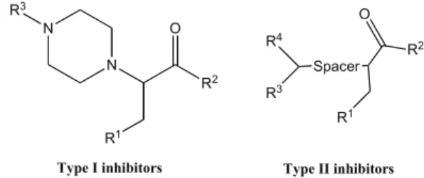
Another study, reported by Boss and co-workers [78], of high-throughput screening of a commercial library using fluorescence resonance energy transfer (FRET) assays to measure the Plm II activity, identified low M inhibitors such as type I and type II (Fig. 12) as promising compound classes. Further optimization of the compounds was able to
increase the activity of type I and type II inhibitors by 250-fold and a factor of 60, respectively. Such improvements led to compound 16 (Fig. 11) and 17 (Fig. 13), respectively [77, 78]. The binding modes of the 4-aminopiperidines (com-pound 16) were elucidated by X-ray crystallography (PDB-code: 2BJU) [79], which certainly will facilitate the subse-quent structure-based drug design of this promising class of compounds.
For type II inhibitors, replacement of the n-pentyl chain by shorter ones, as well as the insertion of a heteroatom in the chain, leads to a decrease on the activity of the inhibitors against Plm II. Still, little is known about the consequences on the activity of type II inhibitors upon alterations of R3 and R4 substituents. Boss and co-workers [78] demonstrated that hydrophilicity and polarity in the bis-heteroaryl moiety could be added to improved activity against the parasite growth. Their studies also showed that the aryl-amide unit and the aryl-amine are beneficial for global activity. Compound 17, an aryl–amine derivative shown in (Fig. 13), was found to be one of the most promising inhibitors against Plm II and para-site growth of the series of compounds considered, having IC50 values of 374 nM against Plm II and 273 nM against the parasite development.
In addition, other non peptidic inhibitors of plasmepsins include the achiral oligoamines reported by Diederich and co-workers [80]. They developed an initial library of 11 compounds and tested them against six different aspartyl proteases. Several hits were identified and some of them were found to be also selective. They demonstrated that the sulfonamides acceptor groups are preferred over the carbonyl moiety because they allow additional degrees of freedom. Polymethylene links confer flexibility to the oligoamine de-rivatives, which requires a high degree of pre-organization from the molecules before binding to the protein. One of the most active inhibitors of this series against Plm II, compound 18 shown in (Fig. 14), exhibited Ki value of 7.0 M.
Noteworthy, several of the peptidomimetic and nonpep-tide plasmepsin inhibitors have been assessed against both chloroquine-resistant and chloroquine-sensitive strains of Pf with similar potencies against both strains, indicating no cross-resistance between chloroquine and plasmepsin inhibi-tors [64, 75].
2.2. Cysteine Proteases (Falcipains)
After aspartyl proteases carry out the first cleavage of the hemoglobin into two peptide chains, falcipains (FPs) come to
N H O O O O OH N O N OH N 15 16
play their role to transform those peptides into smaller ones. FPs are papain-like cysteine proteases and the best character-ized Cys proteases of the malaria parasite, in which a Cys catalyzes protein hydrolysis via nucleophilic attack to the carbonyl carbon of a susceptible bond (Fig. 15). Analysis of the Pf genome sequence suggested the existence of four FPs: FP1, FP2 (EC number: 3.4.22.B69), FP2’, and FP3 [81].
N R1 N O R2 R4 R3 Spacer R2 O R1 R3
Type I inhibitors Type II inhibitors
Fig. (12). General structure of type I and type tertiary amines as inhibitors of Plm II [78]. N O N 17 N
Fig. (13). Compound 17, one of the most active aryl-amine deriva-tives against Plm II and parasite development with IC50 values of 374 nM and 273 nM, respectively [78]. S N N H N S O O O O 18
Fig. (14). Compound 18, one of the most active oligoamine deriva-tives against Plm II with Ki value of 7 #M, reported by Diederich and co-workers [80].
FP1 is distantly related to the other FPs in terms of se-quence (<40% amino acid identity) and its exact physiologi-cal role has yet to be elucidated. Some studies suggested that FP1 could be important in oocyst production during parasite development in the mosquito midgut [82] and also could help in the invasion of the host cell by Pf. However, disrup-tion of the FP1 gene does not disturb the parasite growth in the blood stage of the malaria life cycle [83]. Furthermore, a study showed that FP1 knockout parasites developed nor-mally in erythrocytes, suggesting that this protease alone is neither required for parasite invasion nor for intracellular development within erythrocytes [84]. FP2, FP2’ and FP3 are closely related and appear to be the key hemoglobinases in the acidic FV. FP2 is the most studied and FP2’ is thought
to arise by gene duplication, presenting 93% a.a. similarity with FP2 [85]. N N His H Asn NH2 O NHR R O Cys S N N His H Asn NH2 O NHR R O S Cys N N His Asn NH2 O R Cys S O H +H2O -NH2R N N His Asn NH2 O R OH O Cys S H NHR
Fig. (15). Mechanism of peptide cleavage mediated by falcipains, Cys proteases of Pf.
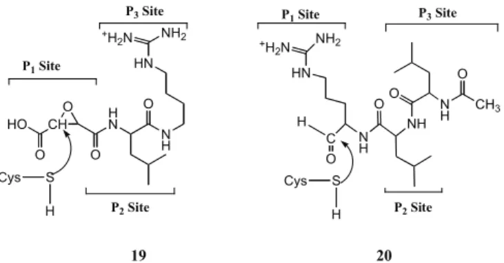
FP2 and FP3 are single polypeptide chains presenting a high similarity in sequence (68% identity) and sharing simi-lar sized prodomains. Their catalytic site has Cys and His residues (Fig. 16), whose side chains form a thio-late/imidazolium ion pair, and has also an Asn, responsible for correct orientation of the ion pair [81]. FP2 and FP3 con-tribute equally to hemoglobin degradation and both require reducing environment and acidic pH for optimal activity. Interestingly, the disruption of FP2 gene revealed that the loss of this enzyme alone is not sufficient to cause parasite lethality, thus suggesting the participation of additional cys-teine proteases for parasite invasion and growth in human erythrocytes [86]. In contrast, the FP3 gene could not be dis-rupted, revealing that FP3 is essential to erythrocytic para-sites and therefore indicating that efforts to develop cysteine protease inhibitors as antimalarial drugs should probably be focused on FP2 and FP3 [86]. The structural similarities of FP2 and FP3 can be a reason for these proteases performing similar functions in the FV. One difference between FP2 and FP3 is that FP3 showed to be far less active than FP2 against peptide substrates. However, FP3 is more active and stable at acid pH (as in the parasite’s FV) than FP2 [87]. Also, it has been estimated that the concentration of FP2 in trophozoites is 1.8 times that of FP3. Still, FP3 processes globin two times faster than FP2 does [88]. The structures of FP2 and FP3 forming complexes with known inhibitors of other Cys proteases have been reported: the FP2 crystal structure was reported in a complex with inhibitor E64 19 (PDB code=3BPF) [83] and the crystal structure of FP3 in a com-plex with Leupeptin 20 (PDB code=3BPM) [83] (Fig. 16) and with a vinyl sulfone (PDB code=3BWK) [89]. Both FP2
and FP3 showed to have a preference for substrates with a hydrophobic residue, especially leucine, at P2 position.
Although both FP2 and FP3 contribute almost equally to the digestion of hemoglobin, FP3 is usually less amenable to inhibition by peptidyl-based small molecules [83]; conse-quently, there is a variety of FP2 inhibitors in the literature [86]. In general, FP inhibitors can be classified in peptidyl-based inhibitors (covalent irreversible and reversible inhibi-tors) [83, 90, 91], including peptidomimetic compounds [92, 93], and nonpeptidic small inhibitors [94-96].
CH HO O O O H N N H HN NH2 +H 2N O NH2 +H 2N HN C N H H O NH OO N H O CH3 P2 Site P1 Site
P3 Site P1 Site P3 Site
P2 Site Cys S H Cys S H 19 20
Fig. (16). Structures of known Cys protease inhibitors E64 (19) and Leupeptin (20).
2.2.1. Peptidyl-Based Inhibitors 2.2.1.1. Irreversible Inhibitors
Most of the falcipain inhibitors identified so far are pep-tidyl-based molecules [86]. Given that Cys proteases have their enzymatic roles dependent on a catalytic Cys residue, an electrophilic warhead is necessary to inhibit these prote-ases. Among such warheads, prone to bind in a irreversible way to FPs, like fluoromethyl ketones [97], epoxysuccinyl [98] or azirine [98] derivatives, the most used are Michael acceptors that irreversibly inactivate Cys proteases via alky-lation of the side chain thiolate of the Cys residue (Fig. 17) [37]. These Cys protease inhibitors include an unsaturated conjugated system such as vinyl sulfone or !,$-unsaturated carbonyls [99, 100]. N H R R1 R H Cys S H His N H R R1 R H Cys S H His N H R R1 R H Cys S His
Fig. (17). Schematic view of irreversible alkylation of a Cys via Michael addition.
Since Rosenthal and co-workers [100] reported in 1996 that vinyl sulfone inhibitors of FP2 blocked the development
of Pf in culture and presented antimalarial activity in vivo, there have been several efforts to synthesize Cys protease inhibitors. The best known attempt was done by the same research team in 2003 [101], when they reported the struc-ture-activity relationships (SAR) for a series of peptidyl vi-nyl sulfones, vivi-nyl sulfone esters, and vivi-nyl sulfonamides as inhibitors of FP2 and FP3. In that study, they demonstrated that the SAR for the two proteases are similar and found multiple compounds to be potent inhibitors of both falci-pains, especially amongst the vinyl sulfones (21, Fig. 18).
Studies of Rosenthal’s group showed that the SAR of FP2 and FP3 are similar and several compounds are potent inhibitors of falcipains and parasite growth. Also, the SAR for the inhibition of the proteases differed notably for those of inhibition of parasite development. In the SAR regarding the proteases, Rosenthal’s team found that inhibitors with the dipeptide core Leucine-homoPhenylalanine (Leu–hPhe) in P2-P1 provided potent inhibition for FP2, FP3 and for devel-opment of malaria parasites in vitro, and that potency was imparted by alterations of amino (P3)- and carboxy (P1’)-terminal substituents of vinyl sulfone. Still, substrates with Leu at P2 showed to be less active against FP3 than against FP2. Compounds with the core sequence Phe-hPhe exhibited modest FP2 inhibitory activity and changes at position P3 in these molecules had relatively low impact on activity. Com-pounds with the dipeptide sequence Phe-O-(phenyl)Ser had inhibitory activities with IC50 against FP2 from 56 to 290 nM. The same group then tested 30 compounds with the core sequence Phe-hPhe, which differed at the P1’ and P3 posi-tion. Vinyl sulfonamides and vinyl sulfones were seen to be less active against FP2 than the vinyl sulfonate esters. Still, inhibitory activity against parasite growth was higher for vinyl sulfones. Finally, these authors also found a pattern of activity against the parasite cultures, depending on the sub-stitution in the aryl ring of the vinyl sulfonate esters: pres-ence of the electron-donating methoxy group led to an in-crease of activity over the unsubstituted ring, whereas the last one showed to be more active than the one bearing an electron-withdrawing fluorine [101]. More recently, a com-putational 3D-QSAR study on these three vinyl sulfone, sul-fonamide and sulfonate ester families has identified critical regions where any change in the steric, electrostatic, and hydrophobic fields of the molecules may affect the inhibitory activity [102]. For instance, bulky groups at R2 position (Fig. 18) tend to decrease biological activity while electropositive groups at R2 backbone, R1 side-chain and near the SO2-R1’ group are preferred to improve FP2 inhibitory activity. Also, compounds that orient any hydrophobic part of R3 group towards Tyr78 and Leu84 would exhibit enhanced FP2 in-hibitory activity. Additionally, they also postulated that the general rank order of FP2 inhibitory activity, sulfonate esters > sulfonamide > sulfones, observed when comparing com-pounds that only differ on R1’ substituent could be related to the flexibility of this substituent.
Though peptidyl-based inhibitors like those above inhibit enzymatic activity of FPs at very low nanomolar range, their utility as therapeutic agents may be limited for their suscep-tibility to protease degradation and their poor absorption through cell membranes, so peptidomimetic scaffolds have been developed to bypass this problem [86]. A common strategy to avoid the therapeutic limitations of peptide-based
inhibitors is to lock a defined conformation of the peptide into a rigid scaffold. There are several examples of pepti-domimetic inhibitors of FP2 reported in the literature [92, 93, 103-106]. O’Neill and co-workers designed and synthesized novel 2-pyridone peptidomimetic FP2/3 inhibitors with chemical structures shown in (Fig. 19) [107]. All of these molecules displayed antimalarial activity in the micromolar range, being 22 the most active inhibitor of the series with an IC50 (3D7 Pf) value of 5.7 $M, followed by the ,"-unsaturated methyl ester 23 with an IC50 (3D7 Pf) value of 27.9 $M. Compound 24 was less active than their counter-parts bearing the unnatural segment of hPhe residue. How-ever, only 24 expressed activity against FP2/3, whereas the low solubility of 22 prevented its evaluation as a potential FP inhibitor. N R' N Cbz O R= C2H4Ph; R'= SO2Ph (22) R= C2H4Ph; R'= COOMe (23) R= CH2Ph; R'= SO2Ph (24) R H
Fig. (19). FP2/3 peptidomimetic inhibitors studied by O’ Neill and co-workers [107]. Cbz=Benzyloxycarbonyl.
Other peptidomimetics bearing Michael-acceptor moie-ties for irreversible FP inhibition have been explored as an-timalarials. For instance, Moreira and co-workers have de-veloped vinyl sultams [108], aza vinyl sulfones [109], squaric acid [110] and 3-methylene substituted indolinone derivatives [111]; only some of the latter compounds were able to inhibit Pf parasites in vitro at IC50 as low as 140 nM, the remaining presenting IC50 values in the micromolar range. In general, none of these compounds was able to ex-hibit potent FP inex-hibition activity.
Peptidomimetic nitriles have also been reported in the lit-erature as potential FP inhibitors. Nitriles are known to in-hibit cysteine proteases by the formation of a covalent thio-imidate adduct resulting from the nucleophilic attack of the active site Cys residue [112]. Using a rational structure-based molecular modeling focusing on optimal occupancy of the main apolar FP2 pockets to obtain potent and particularly
selective inhibitors, Diederich and co-workers [103] were able to design nitrile inhibitors of FP2 action, and showed that the ideal occupation of the selectivity-determining S2 pocket and the balanced electrophilicity of the nitrile group seem to be essential to achieve activity and selectivity. The 3D modeling studies specifically revealed that 1-aminocyclohexanecarboxylic acid should be the central core of the nitrile derivative to address the active site of FP2. The cyclohexyl motif should orientate the other substituents in the molecules to promote their interactions with the S2 and S3 pockets. They synthesized the nitriles derivatives and assessed their biological activity against FP2. Accordingly, the S2 pocket is accessible for different substituents of 5- or 6-membered aromatic systems such as pyridin-3-yl, 4-methoxyphenyl, 2-4-methoxyphenyl, 3,4-di4-methoxyphenyl, furan-2-yl, and thiophen-2-yl. Also, loss of activity was ob-served when the 2-aminoisobutyric acid substituted the 1-aminocyclohexanecarboxylic acid as a center core. Most of the derivatives exhibited high selectivity against FPs versus cathepsin B and L, as well as against the serine protease -chymotrypsin, making them interesting targets for further optimization studies toward the discovery of new potent an-timalarials. Compound 25, one of the most active inhibitors of the series against FP2 action is displayed in (Fig. 20).
N C N H O H N O O 25
Fig. (20). Compound 25, one of the best inhibitors designed by Diederich and co-workers [103] with a Ki value against FP2 of 1.2 $M.
Most recent examples of peptidyl-based structures as po-tential FP inhibitors include those reported by Gomes and co-workers [105]. They designed and synthesized a series of cinnamic acid/4-aminoquinoline derivatives as potential in-hibitors of falcipain action and found the unsubstituted cin-namic acid derivative 26 (Fig. 21) to inhibit not only FP2 action but also the parasite growth in vitro in the micromolar range. The retro enantio dipeptide used to link the 4-aminoquinoline and the respective cinnamic acid was dem-onstrated to provide antimalarial activity. Interestingly, using the natural counterparts of the amino acids in the linker H N N H R3 S R1' O O O O R1 R2 P3 Site P1 Site P2 Site R1: homoPhe, O-(phenyl)Ser R2: Phe, Leu R1' substitutents: O N H O
Phenyl Vinyl Sulfone Vinyl Sulfonamide Vinyl Sulfonate Ester P1'Site
R
R
R 21
dipeptide was detrimental to compound inhibitory activity against FP2 and also resulted in a decrease in the antiplas-modial activity. Furthermore, some of the compounds from the series exhibited activity against hemozoin biocrystalliza-tion and FP2 making them good leads toward the develop-ment of potential dual action antimalarials.
N HN (R) HN (R) N H O O O (E) Cl 26
Fig. (21). The most active FP2 inhibitor among the new series pro-posed by Gomes and co-workers [105].
All the above examples demonstrate that, with the excep-tion of vinyl sulfones unveiled by Rosenthal’s group, com-pounds able to irreversibly inhibit FP and display nanomolar activities against Pf have been hard to find. Hence, several groups have developed novel structures where the vinyl sul-fone moiety is conserved in an attempt to ensure FP irre-versible inhibition. An example of such strategy is that of Zappalà’s team [92, 93]. These authors synthesized a series of Cys protease inhibitors based on the 1,4-benzodiazepine scaffold, which is known to act as a good mimetic of $-turns [113], the structural motif postulated as the biological active form of the D-Ser-Gly peptide [114]. Furthermore, benzodi-azepines are also known to enhance oral bioavailabilitity and also to increase stability toward premature proteolytic degra-dation by enzymes. In this connection, Zappalà and co-workers [93] focused their attention on the synthesis of two different series both including a vinyl sulfone in the P1’site (Fig. 22). One series contained hPhe in P1, a residue known to increase the inhibitors potency against FPs, and the second series included Gly in P1 to assess the relevance of the amino acid side chain for enzyme recognition. All of the vinyl sulfone inhibitors displayed activity against the parasite higher than 9.1 #M. As expected, compounds with hPhe residue in P1 site were generally better inhibitors of parasite development than compounds containing a Gly. Compounds 27a and 27b (Fig. 22) presented good FP2 inhibitory activi-ties and also the highest antiplasmodial activiactivi-ties of the cor-responding series [93]. In addition, the compounds were tested against cathepsin B and L and found to be weak inhibi-tors of these two enzymes, which can be considered a promis-ing result regardpromis-ing their selectivity toward parasite versus host Cys proteases. Thus, these compounds can be considered as leads for the development of new antimalarials.
Following, Zappalà’s group reported a series of com-pounds synthesized based on the same 1,4-benzodiazepine scaffold with the aim of improving the pharmacological properties of this type of irreversible inhibitors [92]. In that work, they described a highly potent and selective derivative with a vinyl ester warhead 28 (Fig. 23), which exhibited the highest potency with an activity two times higher than that of the standard E-64 (ksecond 1586000 M-1 min-1) and the highest
enzymatic affinity (Ki =17 nM) among the series. Although 28 displayed a Ki 17 nM, it did not result as active in vivo, supposedly, due to the difficulty for the derivative to cross the biological membranes of the parasites and reach FP2.
H N O O N N O HN O Ph F3C Cl SO2R' R 27a, R=CH2CH2Ph R'=C2H5 27b, R=CH2CH2Ph R'=Phe
Fig. (22). FP2 inhibitors, designed by Zappalà’s team, combining a benzodiazepine core and a vinyl sulfone warhead [93].
H N O O N N O HN O Ph F3C Cl R' R 28, R=H R'=COOMe
Fig. (23). Most potent compound of the series 28 reported by Zap-palà and co-workers [92].
The unmatched relevance of the dipeptidyl vinyl sulfone warhead for FP inhibition has been also recognized in re-search works where this moiety has been combined with other antimalarial scaffolds, e.g., artemisinin, to produce multi-target antimalarial molecules 29 (Fig. 24) [115].
O O O O O N H O HN S R3 O O O R2 R1 R1 = CH2Ph, R2= H, R3 = Ph, R1 = CH2Ph, R2 = CH2Ph, R3= Ph, R1 = CH2CH2Ph, R2 = CH2Ph, R3 = Ph, R1 = CH2CH2Ph, R2 = CH2CHMe2, R3= Ph, R1 = CH2CH2Ph, R2 = CH2Ph, R3=Me, R1 = CH2CH2Ph, R2 = CH2CHMe2, R3=Me, 29
Fig. (24). Artemisinin-dipeptidyl vinyl sulfone hybrids 29 devel-oped by Moreira’s team [115].
2.2.1.2. Reversible Inhibitors
An example of reversible inhibitors was reported by Zappalà’s team [104]. These authors synthesized a series of
Cys protease inhibitors based on the 1,4-benzodiazepine scaffold linked to a C-terminal aspartyl aldehyde building block that would inhibit the enzyme by forming a covalent reversible bond. All of the designed benzodiazepine deriva-tives presented activity against FP2 with IC50 values between 8 and 26 ZM. The derivative of compound 30 obtained by introducing a phenyl group at R position (Fig. 25) was the least potent inhibitor of the series with an IC50 of 21.54 "M. The introduction of an withdrawing or electro-donating group to the para position of the phenyl ring in the general structure 30 (Fig. 25) increased the activity against the enzyme. An enhancement of the activity against FP2 was also observed by the insertion of a methyl or a trifluoro-methyl group to the p-chlorophenyl ring (c.f. compound 31 in Fig. 25). To evaluate the selectivity of the compounds, the derivatives were tested against a panel of active recombinant human caspases (i.e., caspases 1-9) and the molecules dis-played inhibitory activity up to 50 "M.
H N O O N N O HN O O O HO Ph F3C Cl 31 HN O O N N O HN O O O HO Ph R 30
Fig. (25). General structure 30 for the design of benzodiazepine inhibitors with a P1-aspartyl aldehyde moiety and compound 31, one of the best inhibitors of the series [104].
Other examples of reversible inhibitors include the ones reported by O’Neill and co-workers [90]. They studied ex-amples of FP2 and FP3 inhibitors that do not have an ,"-unsaturated system included in the structure and inhibit FPs in a reversible manner. They were able to synthesize two molecules, namely, compounds 32 and 33 in (Fig. 26), bear-ing an aldehyde moiety which displayed activity against FP2 and FP3 in the low nanomolar range, and also expressed activity versus the 3D7 strain of Pf. Both molecules con-tained residues Leu and homoPhe, the key amino acids pre-viously identified by Rosenthal and co-workers [90], and were found to possess a good enzyme fit according to dock-ing studies.
Other peptidyl reversible inhibitors of falcipains include peptidyl aldehyde and -ketoamide derivatives developed by Rosenthal and co-workers [91]. They evaluated FP2 inhibi-tion and antimalarial activity of the series and found most of the compounds to inhibit FP2 activity in nanomolar range. Due to the high sequence similarities between FP2 and FP3, they decided to test the compounds also against FP3 and
activity against both falcipains resulted to be similar, sug-gesting that a single small specific protease inhibitor could be enough to inhibit both falcipains and consequently, inhibit hemoglobin degradation leading to parasite death. As seen previously in other works, the compounds which displayed higher activity against the protease had a Leu in P2 site con-trary to those who had a Phe in P2. Inhibition of FP2 was always accompanied by swollen and darkly stained parasitic FV, an experimental finding that agrees with blocking of hemoglobin degradation [116]. Since new ideal antimalarials should be active against all Pf strains, Rosenthal and co-workers decided to test the three best inhibitors of the series against other five Pf strains. They found the three com-pounds to inhibit more or less equally each strain and that there was no evidence that the activity against multidrug-resistant strains differed from the activity against broadly sensitive strains. The morpholino-carbonyl-leucine-homophe-nylalanine aldehyde 34 (Fig. 27) exhibited excel-lent activity against FP2 and parasite development. Rosen-thal’s team assessed the activity of the P. vinckei-mice and the compound 34 showed antimalarial activity when it was administered intraperitonially but this activity was fairly low. They inferred that compound 34 activity could be mainly due to the short life of the compound. Thus, due to the poor pharmacokinetic of the compound to provide consistent lev-els in blood and to improve drug delivery, mice were dosed by a subcutaneous infusion pump continuously. However, the antimalarial activity continued to be modest and the mice eventually died by the infection.
N H H N R O H O R=Cbz (32); Mu (33)
Fig. (26). FP2/3 reversible inhibitors developed by O’Neill and co-workers [90]. Mu=Morpholine urea; Cbz=Benzyloxycarbonyl.
O N O H N O H O 34
Fig. (27). Most active peptidyl reversible inhibitor 34 developed by Rosenthal and co-workers [91].
2.2.2. Non-Peptidic Inhibitors
In an effort to develop non-peptidic FP2 inhibitors, Zap-palà’s group reported a series of 1-aryl-6,7-disubstituted-2H-isoquinolin-3-ones which were synthesized and tested against FP2, as well as against cultured Pf strain FCBR para-sites. Most compounds presented activity against the enzyme though there were not selective. The most potent compound of the series 35 (Fig. 28) displayed a Ki=2.3 "M against FP2 emphasizing the importance of hydrophobic and bulky groups at position 6 and 7 of the isoquinoline scaffold [117]. Further work is required in order to improve the selectivity of these compounds because they resulted to be slightly more active against human cathepsins than FP2.
N BnO BnO O NH2 OMe 35
Fig. (28). Most potent compound 35 of the series reported by Zap-palà and co-workers [117].
Several other non-peptidic small inhibitors of FPs have been found [118-121], some of them through computer-based drug design and virtual screening studies, for instance, compounds 36-39 (Fig. 29) [94-96, 120]. Recently, Avery and co-workers [120] identified a total of 28 non-peptidic low micromolar inhibitors of FP2 and FP3 and elaborated SAR for each series. Some of the complex trends in the SAR could be explained by the energetical changes associated with the displacement of water molecules upon ligand bind-ing, and other could be related with poor chemical reactivity of the reactive centers of these compounds. The motivation to use water energetical changes was due to the fact that the SAR could not be explained by none of the following de-scriptors: hydrogen bonds, van der Waals, and electrostatics interactions, or by ligand strain, or even by using traditional computational approaches such as molecular mechan-ics/generalized Born surface area (MM-GBSA), docking scores, and molecular mechanics interaction energies. Some of the molecules identified in this study also inhibited para-site growth in culture.
Some hybrid drugs, capable of targeting two processes in the malaria life cycle where one of the targets is FP2 tion, can fall into the category of non-peptidic small inhibi-tors [122, 123]. For instance, in 2010, Chibale’s group iden-tified chalcone-chloroquine hybrid 40 as a promising antima-larial (Fig. 30) [122, 123]. Compound 40 and related hybrid structures were synthesized and showed to be active against
Pf. The molecules were tested as inhibitors of both FP and
hemozoin formation, and some of them were found to be active in the micromolar range against FP. However, com-pound 40 and related structures did not show correlation between FP inhibition and parasite growth in culture. Still, there was consistent correlation between in vitro antimalarial
potency and inhibition of hemozoin formation, suggesting that this could be the primary mechanism of their antimalar-ial activity. H N N OH O N N H HO O O H N S S H N O O O O O S N H S H2N O N N S O O 36 37 38 S H N O N N O HO O 39
Fig. (29). Non-peptidic FP inhibitors developed by Avery and co-workers [120]. O O N N N N Cl OMe OMe MeO 40
Fig. (30). Hybrid drug 40 designed to include FP inhibition ( ,"-unsaturated system as Michael acceptor) in its mode of antimalarial action [122, 123].
Other non-peptidic inhibitors include structural analogs of triazole (Fig. 31) and benzothiazole (Fig. 32) cores show-ing moderate inhibition of falcipains, reported by Avery and co-workers [124]. In the triazole series, in an effort to opti-mize the activity of compound 41 against FP2, only few tria-zoles with general structure 42 were found to display activity against FP2 but none was more active than the parent
com-pound. The low activity of these derivatives might be as-cribed to the unsuitable or shorter hydrophobic R groups used for this family as compared to the parent compound 41 that had an IC50 value of 2.2 "M. Indeed, molecules with bulkier R groups were the ones exhibiting modest activity against FP2. N O N H O N N N R 42 N O N H O N N N N 41 NH O F
Fig. (31). General structure used to design triazoles by Avery’s team [124]. N S S O H N O NH3Cl N S S O H N O NH3 N S S O H N S O O O F3C 43 44 45 Cl
Fig. (32). Benzothiazole based inhibitors as Plm II designed by Avery and co-workers [124].
In the benzothiazole series, compounds 43 and 44 were predicted by docking studies to interact with the polar resi-dues of the S2 pockets of FP2 and FP3; these compounds inhibited both FP2 (IC50 between 12.22-12.75 "M) and FP3 (IC50 between 13.77 and 14.94 "M), but also displayed activ-ity against homologous mammalian cysteine proteases lack-ing polar residues, suggestlack-ing their low selectivity. Only compounds 44 and 45 inhibited the growth of W2 strain of
Pf, with IC50 values of 2.08 "M and 4.65 "M, respectively.
Other non-peptidic FP inhibitors include heteroarylni-triles. For instance, Fiandor and co-workers [118] explored the pyrimidinenitrile scaffold for FP inhibition: optimization studies led to the discovery of 2-cyano-5-chloropyrimidines and 2-cyano-5-bromopyrimidines as promising inhibitors of the enzymes. Although some compounds inhibited falcipains in the subnanomolar and low nanomolar ranges, they only inhibited parasite growth in the micromolar range. However, introduction of a protonable amine resulting in analogs such as compound 46 (Fig. 33) led to an increase in antiplasmo-dial activity. N N C N Br N N O N N 46
Fig. (33). One of the best 2-cyano-5-bromopyrimidines, 46, devel-oped by Fiandor’s team: FP2 IC50=0.5 nM, FP3 IC50=3 nM and Pf W2 IC50=1 nM [118].
Organometallic reversible FP inhibitors have also been proposed, namely, a series of gold derivatives reported by Messori and co-workers [125]. They found gold complexes capable of pronounced and reversible inhibition of FP2 in the micromolar range. Still, they were unable to establish a cor-relation between enzyme inhibition and antiplasmodial activ-ity. Compound 47 (Fig. 34) displayed the lowest Ki (1.4 "M) of the series with an IC50 of 5.11 "M against parasite growth.
N N O Au Cl Cl [PF6] 47
Fig. (34). Gold derivative 47, with micromolar activity against parasite development [125].
More recently, Gomes and co-workers [105] developed non-peptidic inhibitors of FP2 including a cinnamoyl moiety. They synthesized a series of cinnamic acid/4-amino quino-lines conjugates 48 (Fig. 35) with FP2 inhibitory activity in the micromolar range. By analyzing the binding modes of 48 docked to FP2, those authors observed that most of the com-pounds present the cinnamoyl group at the S2 cavity and having the vinyl group relatively close to the active residue of the cysteine protease. However, none of these inhibitors displayed antiplasmodial activity, probably due to low per-meation into the infected red blood cell or into the parasito-phorous vacuole inside it.
The combination of the inhibitory activity results regard-ing all falcipain inhibitors reported so far with the recent availability of crystal structures of both FP2 and FP3, will
strongly help and support all the medicinal chemists cur-rently involved in the discovery of potent falcipain inhibitors as promising molecules to combat malaria disease.
N HN Cl O NO2 48
Fig. (35). Best inhibitor of the cinnamic acid derivatives reported by Gomes’ team [105].
2.3. Falcilysin
The zinc metalloprotease falcilysin (FLN) is another pro-tease that participates in the hemoglobin degradation proc-ess, specifically, in the hydrolysis of short globin peptides produced by FP-mediated proteolysis of globin [75, 126]. FLN is highly active at acidic pH, which is consistent with its role in the FV, but it is also active at neutral pH, although with a different substrate specificity. At neutral and acidic pH this protease prefers to cleave sites in which P1’ residues are bulky hydrophobic amino acids and has strong prefer-ence for methionine at P3’; however, in what concerns P2’ residues, FLN favors hydrophobic residues at pH 5.2, whereas at pH 7.2 it has stronger preference for Arg. Finally, at P4’ and P5’, FLN prefers acidic residues at acidic pH but is less selective at neutral pH. Studies by Goldberg and co-workers [127] revealed that FLN is not only present in the FV but also in association with vesicular structures else-where in the parasitophorous vacuole, suggesting an ex-panded role for this protease in Pf biology. Despite some chelators, such as 1,10-phenanthroline [128] presented some activity toward this protease, no specific inhibitor was found so far [127-129].
2.4. Dipeptidyl Amino Peptidase 1
Once FLN converts short globin polypeptides into oli-gopeptides consisting of 5–10 amino acids, dipeptidyl amino peptidase 1 (DPAP1), a lysosomal exopeptidase, sequentially cleaves oligopeptide substrates from their N terminus into dipeptides [87, 130]. Attempts to disrupt the DPAP1 gene have been unsuccessful, which suggests that the enzyme makes an important contribution to hemoglobin catabolism during the intraerythrocytic cycle [131]. While DPAP1 is one of the three related DPAP enzymes encoded in the para-site genome, current evidence suggests that only DPAP1 resides in the food vacuole [130]. DPAP1 is also potentially a good drug target because the closest human homologue is cathepsin C, a protease that is not essential in mammals [130, 132, 133]. DPAP1 differs from other endopeptidases in that they recognize the N-terminal free amine of substrate proteins and cleave N-terminal dipeptides [134]. Structur-ally, its active site is relatively small compared to that of other endopeptidases and its substrate specificity is mainly dictated by the N-terminal residue P2 [134]. This smaller active site is more amenable to binding of bioactive
mole-cules with low molecular weight and should be more suitable for computational docking and in-silico design of inhibitors.
Although DPAP1 is a fairly new target, some inhibitors of this protease have been already developed: Bogyo and co-workers [134] screened several non-peptidic scaffolds from a large library of small compounds that was initially designed to target cysteine cathepsins in mammals, and found stable covalent DPAP1 inhibitors 49 (Fig. 36) which kill Pf at low nanomolar concentrations.
Bogyo findings demonstrated that inhibition of DPAP1 by small compounds results in an immature trophozoite and, consequently, parasite death. This strongly suggests that de-velopment of DPAP1 inhibitors may be a key strategy to-ward malaria chemotherapy.
NH2 N N N O O F F F F 49
Fig. (36). Best DPAP1 inhibitor found by Bogyo’s team [134].
2.5. Exoaminopeptidases
Pf contains nine exoaminopeptidases [135], four of
which are methionine aminopeptidases (MetAPs). MetAPs catalyze the removal of N-terminal methionine during pro-tein synthesis [129, 136] and their inhibition can impair Pf growth in culture [24, 129, 137, 138]. The remaning five Pf exoaminopeptidases include an aspartic aminopeptidase, a prolyl aminopeptidase, a post-prolyl aminopeptidase, a leucine aminopeptidase, and an alanine aminopeptidase. These five aminopeptidases are considered to be potential targets for inhibition of hemoglobin degradation. Neutral alanine and leucine metallo-aminopeptidases [139], involved in the generation of free amino acids from the previously generated dipeptides, are the pivotal Pf exoaminopeptidases. Bestatin 50 (Fig. 37) has been found to inhibit both the activ-ity of these enzymes and malaria parasite growth in vitro [36]. H N OH O NH2 OH O 50
Fig. (37). Bestatin 50, a reported inhibitor of Pf Leu and Ala amin-opeptidases [36].
2.5.1. M1 Alanine Aminopeptidase
PfA-M1 (EC number: 3.4.11.2) is a plasmodial zinc
aminopeptidase that belongs to the M1-family which dis-plays its activity at pH 7.4 and has a broad substrate spec-trum [140, 141]. PfA-M1 is found in both trophozoites and
schizonts, being involved in hemoglobin degradation and erythrocyte reinvasion [142]. Therefore, design and synthesis of potential inhibitors of the PfA-M1 activity can lead to the discovery of new antimalarials [141, 143, 144]. For example, Deprez-Poulain’s group first identified three non peptidic and non-selective inhibitors of this protease, and further op-timized their results, and found the desired specificity against mammalian neutral aminopeptidases (APN), the host M1 family prototype [141]. The compounds were based on malonic hydroxamic template 51 (Fig. 38) and the influence of the malonic substituents on the activity against PfA-M1 was analyzed: compounds bearing a benzyl or a m-phenozy-benzyl group in R1 presented higher activity compared to those with an isobutyl substituent in that position. Binding of hydroxamate to the Zn2+ is expected to be the main anchor-ing point of the potential inhibitor. Therefore, substituents on R1 position should discriminate those molecules which will bind to the enzyme from those which will not. Compounds bearing a benzyl amine in R2 position, when presenting a fluorine atom in the para position or a methyl group on the
ortho position, expressed strong increase on activity. To
re-move the chiral carbon, the same authors synthesized unsatu-rated analogues of 51, and found that a Z-configuration 52 led to increased activity, whereas an E-configuration had the opposite effect. Additionally, they found that cyclization of 52 to produce 53 was detrimental for enzyme inhibitory ac-tivity. R2 H N R1 O O OH H N HN O O OH 51 52 F H N N O O OH 53 F
Fig. (38). PfAM-1 inhibitors developed by Deprez-Poulain’s group [141].
In addition, Deprez-Poulain’s group has also developed quinoline inhibitors of PfA-M1 [145]. They discovered that the presence of a Zn-chelating group is essential for the ac-tivity. They also found that the hydroxamate group is more efficient than a carboxylic acid group. The three PfA-M1 inhibitors are also inhibitors of the parasite growth and the quinoline moiety allows these compounds to also inhibit heme biocrystallization. The best inhibitor 54 (Fig. 39) of the synthesized compounds exhibited IC50 values of 0.854 M and 0.317 M against PfA-M1 or parasite growth, respec-tively.
Recently the X-ray structures of PfA-M1 in the apo form, in complex with bestatin (50, Fig. 37), and with a phosphine-based inhibitor, hPheP(CH2)Phe (55, Fig. 40), originally designed for the cytosol leucine aminopeptidase [146], were determined by Dalton and co-workers (PDB codes: 3EBG, 3EBH, 3EBI) [143]. The availability of these crystal struc-tures will trigger the explanation of different affinities pre-sented by different inhibitors allowing the early phase of inhibitors optimization. N N HN N Cl N O O HN OH 54
Fig. (39). The best quinoline-based inhibitor of PfA-M1developed by Deprez-Poulain’s group [145].
2.5.2. M17 Leucine-Aminopeptidase
There is an overexpression of M17 leucine aminopepti-dases (EC number: EC 3.4.11.1) in the malaria parasite; ac-tually, the expression level of M17-family is ~18-fold higher than the M1-family [147, 148]. Leucine aminopeptidase (LAP) is an exopeptidase that has a preference for substrates which contain leucine or alanine in the N-terminus. Plasmo-dial LAP (PfLAP) exhibits its optimal activity at pH 7.2 and is inactive below pH 6, which is consistent with no activity in the FV. Apart from bestatin 50, other inhibitors of M17 leucine aminopeptidases have been reported [149, 150]. For instance, Gardiner and co-workers reported in 2007 a series of phosphinate dipeptide analogues as potential PfLAP in-hibitors [150]. By screening different phosphinate analogues of dipeptides against functionally recombinant PfLAP, those authors were able to identify four compounds as potent in-hibitors of the parasite PfLAP activity and from those four molecules, compounds 55, 56 displayed in (Fig. 40) pre-sented the highest inhibitory effect against PfLAP. These two compounds resulted to be even more active than bestatin 50 against PfLAP and also showed activity against malaria parasite growth in culture in the low micromolar range. Docking of compound 55 against the enzyme showed that an amino group and two oxygen atoms are able to interact with the two zinc metal ions in the active site of PfLAP. In addi-tion, the hydrophobic S1 in PfLAP can accommodate the hPhe of ligand 55. The affinity of compound 56 to PfLAP was significantly similar to that of 55 because the additional hydroxyl group from the Tyr residue is solvent exposed and does not promote additional interactions with the enzyme. Molecular docking studies showed that the phosphinic moie-ties in both 55 and 56 are able to form unique contacts with the metals ions and residue Lys386 of the enzyme’s active site. These interactions do not occur with the corresponding fragment in bestatin and, therefore, can be accounted for the high binding affinity of 55 and 56 over bestatin 50 for
![Fig. (6). General structure of statine-based inhibitors coupled with primaquine (6) designed by Bosisio and coworkers [64]](https://thumb-eu.123doks.com/thumbv2/123dok_br/15724071.1070804/5.892.144.414.546.631/general-structure-statine-inhibitors-primaquine-designed-bosisio-coworkers.webp)
![Fig. (20). Compound 25, one of the best inhibitors designed by Diederich and co-workers [103] with a K i value against FP2 of 1.2](https://thumb-eu.123doks.com/thumbv2/123dok_br/15724071.1070804/9.892.247.715.155.330/fig-compound-best-inhibitors-designed-diederich-workers-value.webp)
![Fig. (29). Non-peptidic FP inhibitors developed by Avery and co- co-workers [120]. OONNNNClOMe OMe MeO40](https://thumb-eu.123doks.com/thumbv2/123dok_br/15724071.1070804/12.892.500.815.221.699/fig-non-peptidic-inhibitors-developed-avery-workers-oonnnnclome.webp)
![Fig. (31). General structure used to design triazoles by Avery’s team [124]](https://thumb-eu.123doks.com/thumbv2/123dok_br/15724071.1070804/13.892.100.464.274.428/fig-general-structure-used-design-triazoles-avery-team.webp)
