303
lipoprotein associated with culprit vessel
in ST-elevation myocardial infarction
– a promising marker?
[28]
PatRíCia naPoleão, Mafalda selas, aleXandRa toste, antónia tuRkMan, valeska andReozzi, ana MaRia viegas-CResPo,
teResa PinheiRo, Rui CRuz feRReiRa
Centro de Biologia Ambiental, universidade de Lisboa, Lisboa, Portugal Serviço de Cardiologia, Hospital de Santa Marta, Lisboa, Portugal Centro de Estatística e Aplicações, universidade de Lisboa, Lisboa, Portugal
Laboratório de Feixe de Iões, Instituto Tecnológico e Nuclear, Sacavém & Centro de Física Nuclear, universidade de Lisboa, Portugal Rev Port Cardiol 2009; 28 (3): 303-308
Recebido para publicação: Setembro de 2008 • Aceite para publicação: Janeiro de 2009 Received for publication: September 2008 • Accepted for publication: January 2009
RESUMO
Variações longitudinais de lipoproteínas de baixa-densidade oxidadas associadas à artéria “culpada” no enfarte do miocárdio com elevação ST – um marcador promissor?
Objectivo: Estudaram-se as variações de
lipoproteínas de baixa densidade oxidadas (oxLDL) na fase aguda do enfarte do miocárdio e ao longo do período de recuperação, e ainda explorar as relações deste marcador com a gravidade da doença coronária. Foram avaliados 50 doentes com enfarte do miocárdio com elevação de ST nas primeiras 24h de evolução (grupo AMI). Para efeitos de comparação estabeleceu-se um grupo de 25 voluntários saudáveis (grupo referência). Os doentes do grupo AMI foram seguidos imediatamente após o enfarte do miocárdio (nas primeiras 24h de evolução, antes da angiografia e administração de inibidores das glicoproteínas IIb/IIIa), e ainda ao longo do período de recuperação, 48h e 40 dias após a intervenção. As concentrações de oxLDL foram medidas por ELISA, e verificou-se que estavam significativamente elevadas nos doentes com AMI na fase aguda (Dia 0), decrescendo progressivamente ao longo do período de recuperação. Verificou-se ainda que as concentrações de oxLDL estão
ABSTRACT
The aim of the present study was to investigate variations in oxidized LDL (oxLDL) at the onset of acute myocardial infarction (AMI) and over the recovery period, exploring their relationship with coronary disease severity. A follow-up of 50 AMI patients was evaluated against 25 healthy volunteers (reference group). The AMI patients were evaluated at three time points: at admission before the administration of IIb/IIIa inhibitors and angioplasty, and two and 40 days after intervention. Plasma oxLDL concentrations were measured by ELISA. oxLDL was found to be significantly higher in AMI patients in the acute phase relative to reference levels, decreasing progressively over the recovery period. The results also demonstrated that oxLDL levels were decreased in patients with the left circumflex artery (LCX) as culprit vessel compared to the left anterior descending coronary (LAD) or right coronary artery (RCA). The results highlight a significant increase in oxLDL concentration related to coronary artery disease severity, as conditions such as LCX lesions are usually associated with a favorable prognosis, contrasting with LAD-associated conditions that can compromise large areas of myocardium. The results thus suggest that oxLDL may constitute a promising marker in assessment of AMI evolution.
Several studies have shown a correlation between plasma levels of oxLDL products and the presence and/or severity of coronary
artery disease (CAD)(3-5). However, the clinical
importance of this marker remains to be established, both at the onset of acute episodes and at different stages of disease evolution, as does its possible prognostic value.
The objective of this study was to investigate serial changes in oxLDL in the plasma of patients with coronary artery disease immediately after acute myocardial infarction and during the recovery period, and to explore the relation of this marker with disease severity and extent.
METHODS Study groups
A total of 75 subjects (16 women and 59 men) were recruited at the Cardiology Department of Santa Marta Hospital, Lisbon. Among them, 50 patients with acute myocardial infarction constituted the myocardial infarction (AMI) group. The subjects in this group all had ST-elevation myocardial infarction (ST-element changes and creatine kinase >3 times normal) in the first 24 hours of evolution and underwent
INTRODUCTION
C
oronary plaque disruption, with consequentplatelet aggregation and thrombosis, is the most important mechanism by which athero - s clerosis leads to acute ischemic syndromes such as acute myocardial infarction (AMI).
The oxidative hypothesis of atherosclerosis recognizes oxidative stress and subsequent mo d - i fication of low-density lipoprotein (LDL) as a central paradigm. The transport of native and oxidized LDL (oxLDL) across the endothelium into the artery wall is therefore an important
initiating event(1).
Human studies have confirmed that oxLDL and oxidized lipid by-products are present
within atherosclerotic plaques(2). The oxidation of
LDL has been associated with atherothrombosis, as it promotes apoptosis of endothelial cells and macrophages and upsets the anticoagulant balance in endothelium, which in turn stimulates tissue factor production and promotes platelet adhesion, facilitating plaque destabilization and
erosion(2). oxLDL therefore plays an important role
in initiating and maintaining the inflammatory
process in atherosclerotic lesions(2), and levels of
oxLDL in blood appear to be associated with this process.
Key words Oxidized low-density lipoprotein; Acute coronary syndromes; Longitudinal study; Coronary disease severity; Culprit vessel.
diminuídas ao longo do período de estudo nos doentes cujo vaso culpado foi a artéria coronária circunflexa relativamente à artéria coronária direita ou ascendente. Os resultados apontam para um aumento importante das concentrações de oxLDL relacionado com a gravidade da doença coronária, uma vez que condições clínicas como lesões culpadas na artéria coronária circunflexa estão usualmente associadas a prognósticos mais favoráveis, por contraste com condições associadas a lesões na coronária direita, as quais provocam danos em áreas mais vastas do miocárdio. Desta forma, os resultados sugerem que as concentrações de oxLDL poderão constituir um marcador promissor na avaliação da evolução do enfarte agudo do miocárdio.
Palavra-chave
Lipoproteínas de baixa-densidade oxidadas; Síndromes coronárias agudas; Estudo longitudinal; Gravidade de doença coronária; Artéria culpada.
305 Statistical analysis
Data were summarized as mean and standard error (SE) for continuous variables and as proportions for categorical variables. Non-continuous variables were analyzed using a 2x2 table and Fisher’s exact test. Differences between healthy subjects and AMI patients at each time point were compared using a general linear model ANOVA.
Plasma oxLDL concentrations were studied in AMI patients by modeling them as a response variable over time, since observations on the same individual are inter-correlated. A logarithmic transformation of oxLDL values was applied. The time variable was categorized as 1, 2 and 3 for the three consecutive samplings (Days 0, 2 and 40), and hence changes in oxLDL per time unit should be interpreted as changes between these consecutive samplings. A linear mixed effects model was used to fit oxLDL variations over time and the association with biochemical and disease severity indicators and medication data. The model takes into account that for each patient, measures at the three different time points are associated. The model intercept gives the mean oxLDL level in the acute phase and estimates the differences in mean slopes between study time points.
Values of p<0.05 were considered statistically significant. The calculations were performed using SPSS (version 10.0) and R (version 2.5) software.
RESULTS
Characteristics of the study groups
The demographic characteristics and baseline clinical data are listed in Table I. Since coronary disease risk factors and comorbidities were ex-clusion criteria in the REF group, subjects en-rolled inevitably differed from patients (Table I). Prior to the myocardial infarction only 12 patients were not taking any kind of medication. After admission, hospital medication in-cluded aspirin (75%), beta-blockers (11%), ACE inhibitors (61%), statins (36%) and platelet inhibitors (68%). During follow-up all patients took platelet inhibitors (clopidogrel, 71%; aspirin, 75%) in addition to the above medication.
primary percutaneous transluminal coronary angioplasty as reperfusion therapy.
A reference group (REF) of 25 healthy non-smoking volunteers with no previous history of coronary disease or chest discomfort was also established to help with interpretation of baseline lipid oxidation. These volunteers did not undergo coronary angiography.
Subjects aged over 90 years or with significant comorbidities including peripheral artery di-sease or carotid artery didi-sease, known history of malignancy or infectious diseases, chronic renal insufficiency or previous AMI in the last five years, were excluded.
Detailed hospital data were recorded, in-cluding age; gender; coronary risk factors (smok-ing, diabetes, hypertension, hyperlipidemia, personal and fa mily history of CAD); and levels of C-reactive protein (CRP), troponin T and N-terminal pro-B-type natriuretic peptide (NT-proBNP). Pre-event medication, in-hospital and follow-up therapy were also recorded.
Informed consent was obtained for all subjects enrolled, and the study was approved by the local Ethical Committee.
Study protocol and sampling
A longitudinal follow-up of all patients in the AMI group was carried out on three different occasions. For all AMI patients blood samples were obtained from the right femoral artery before the administration of IIb/IIIa inhibitors and angioplasty (Day 0). Additionally, blood samples were obtained by venous puncture two (Day 2) and 40 (Day 40) days after the initial angioplasty intervention.
Blood samples were drawn into pyrogen-free blood collection tubes with EDTA and immediately centrifuged at 2500 rpm for 10 minutes. The plasma-EDTA was collected after centrifugation and placed on ice. Aliquots were stored in liquid nitrogen until analysis (no longer than 6 months).
Oxidized LDL (oxLDL) concentrations were determined using a commercial ELISA (Mer-codia, uppsala, Sweden). Each sample was assayed in duplicate; the intra-assay variation among the duplicates for all samples was <10%. oxLDL concentrations were expressed in u/l.
variations in mean slopes between time points studied.
Table II. Biochemical data and inflammatory markers in the two study groups.
REF
(n=25) (n=50)AMI
Day 0 Day 2 Day 40
Lipid profile TC (mg/dl) 192±6 199±7 b 169±8 b 152±6 b LDL-C (mg/dl) 123±6 133±6 b 103±6 b 89±5 b HDL-C (mg/dl) 54±2 40±2 b 37±3 b 37±2 b TG (mg/dl) 74±5 113±9 b 104±6 b 131±14 b
oxLDL (u/l) 57±5 92±6 a 81±6 a,c 57±5 c,d
Inflammatory marker CRP(mg/dl) 0.37±0.03 0.71±0.08 a 3.9±0.6 a,c 0.43±0.05c,d Others Glucose (mg/dl) 90±2 177±10 b 137±8 b 118±7 b Albumin (g/dl) 3.9±0.1 3.4±0.1a 3.2±0.1 a 3.9±0.1 Troponin T (ng/ml) 0.01±0.0 2.25±0.72 a 4.15±0.83 a,c 0.05±0.03c,d N-BNP (pg/ml) 38±6 663±128 a 1843±266 a,c 781±103 a,c,d CRP: C-reactive protein; HDL-C: high-density lipoprotein cholesterol; LDL-C: low-density lipoprotein cholesterol; N-BNP: N-terminal pro-B-type natriuretic peptide; TC: total cholesterol; TG: triglycerides. Data are expressed as mean ± SE. a: p<0.05 vs. REF group; b: not compared (see text); c: p<0.05 vs. AMI group at day 0; d: p<0.05 vs. AMI group at day 2.
To evaluate the possible influence of drug intake on oxLDL concentrations, the 12 AMI patients who were not taking any kind of pre-event medication were compared to the remaining patients. oxLDL levels were not statistically different (p=0.245) between the two groups. To further evaluate the possible influence of drug therapy on serial changes in oxLDL, pre-event, in-hospital and follow-up medication data were categorized and added to the linear mixed effects models as covariables. None of the drugs administered before or after admission significantly influenced changes in oxLDL concentrations over time (data not shown).
Associations with coronary artery disease severity
The linear mixed effect model used to describe mean oxLDL decrease over time was also tested including number of diseased vessels, culprit vessel and biochemical indicators such as CRP, troponin T and NT-pro BNP as covariables. No associations were found between serial changes
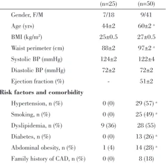
Table 1. Baseline clinical characteristics of the subjects enrolled REF (n=25) AMI (n=50) Gender, F/M 7/18 9/41 Age (yrs) 44±2 60±2 a BMI (kg/m2) 25±0.5 27±0.5 Waist perimeter (cm) 88±2 97±2 a Systolic BP (mmHg) 124±2 122±4 Diastolic BP (mmHg) 72±2 72±2 Ejection fraction (%) - 51±2
Risk factors and comorbidity
Hypertension, n (%) 0 (0) 29 (57) a
Smoking, n (%) 0 (0) 25 (49) a
Dyslipidemia, n (%) 9 (36) 28 (55)
Diabetes, n (%) 0 (0) 13 (26) a
Abdominal obesity, n (%) 1 (4) 14 (28) a
Family history of CAD, n (%) 0 (0) 8 (18) BMI: body mass index; BP: blood pressure. Values are expressed as mean ± SE, except otherwise indicated. a p<0.05 vs. REF reference group. AMI: acute myocardial infaction group.
Forty-seven percent of AMI patients had multivessel disease. The culprit vessel was the left anterior descending coronary artery (LAD) in 25 patients, the right coronary artery (RCA) in 17, and the left circumflex artery (LCX) in only 8 patients.
Lipid and glucose data obtained for the two study groups could not be directly compared as fasting blood tests were only performed for REF individuals (Table II). Maximum values of CRP (an acute phase inflammatory indicator), NT-proBNP and troponin T (markers of ventricular failure and myocardial damage) were observed at Day 2.
Since the assumption behind longitudinal analysis is that missing data are missing completely at random, the missing information was examined in detail. Patients who died (n=3) or were lost to follow-up (n=10) did not significantly differ from other patients in terms of age, risk factors, comorbidity or clinical characteristics (data not shown).
Longitudinal variations in plasma oxLDL
At the onset of myocardial infarction (Day 0) plasma oxLDL concentrations were markedly increased compared to the values obtained for the reference group (Table II). The levels decreased significantly over time, reaching values similar to the REF group 40 days after infarction (Table
307
are usually associated with a favorable prognosis, contrasting with LAD-associated conditions that can compromise large areas of myocardium.
Although it is widely accepted that oxLDL exists in blood, the mechanisms and kinetics governing oxLDL in the circulation remain
unclear(4), especially the question of where
oxidative modification of LDL takes place(6).
Several authors have proposed that LDL oxidation may not preferentially occur in
blood(3-4). The plasma oxLDL associated with
CAD may derive from the arterial wall by
back-diffusion(3) or oxidative modification of LDL by
leukocytes at sites of plaque rupture(1,6). The high
levels of oxLDL at AMI onset (Day 0) observed in the present study support the hypothesis that an increase in oxLDL levels is associated with
plaque instability, as previously suggested(2,5,7-8).
The inflammatory process associated with plaque rupture and ischemia/reperfusion injury promotes the generation of excessive pro-oxidants, disrupting the antioxidant/oxidant
balance in blood(7). The gradual reduction in
oxLDL concentrations with clinical stabilization observed in this study could be partially due to changes in the redox balance, through either an increase in the activity of antioxidant systems or a reduction in oxidant levels, as proposed by
Nakuro et al.(7).
There are few studies reporting oxLDL measurements at the onset of AMI or a few days
afterwards(5,7-8). Longitudinal data for oxLDL in
AMI is limited(9), but overall a good agreement
was found between data sets at discrete time points obtained in this work with similar disease conditions reported by other authors. Enhanced oxLDL levels have been reported in CAD patients in different clinical situations such as myocardial infarction, stable and unstable
angina, or transplantation(2-3,5,10).
To the best of our knowledge this is the first study reporting an association between circulating oxLDL decreases and the LCX culprit vessel in AMI patients. The very high levels of oxLDL in patients with LAD or RCA as culprit vessel and the low levels of oxLDL in LCX patients suggest that oxLDL may be associated with CAD severity and possibly with lesion characteristics that depend on the infarct-related artery. The above findings are consistent with the hypothesis that oxLDL has an important role in the atherosclerotic process, both as an inflammatory mediator and in oxLDL and the biochemical markers studied
(p=0.204, p=0.092 and p=0.114, respectively) or the number of diseased vessels (p=0.973).
However, examining serial variations in oxLDL by culprit vessel showed that patients with LCX as culprit had the lowest oxLDL levels (Figure 1). Although a general tendency for oxLDL to decrease with time was observed in all cases, low oxLDL levels were consistently associated with patients having LCX as culprit vessel (p=0.046) compared to those with LAD or RCA. The model significance was correlated particularly strongly with oxLDL concentrations at Day 0 (Figure 1).
Figure 1. Influence of the culprit vessel (LAD – squares; RCA – circles; and LCX - triangles) on oxLDL concentrations (mean, SE) from myocardial infarction onset (Day 0) until Day 40. Significances (p<0.05) vs. LAD (a) and RCA (b) at
each time point are indicated.
DISCUSSION
The present study demonstrates that plasma oxLDL levels were markedly elevated at MI onset before therapeutic intervention and decreased shortly afterwards (Day 2), continuing to fall 40 days later. The decline in oxLDL concentrations with time was significant and these serial changes were influenced by culprit lesion location. AMI patients with the LCX artery as culprit vessel had the lowest oxLDL concentrations, particularly at MI onset, unlike patients in whom the cause of infarction was the LAD or RCA. Thus, these results suggest that the increase in oxLDL concentration is related to CAD severity, as conditions such as LCX lesions
ACKNOWLEDGEMENTS
The work was financially supported by Fun-dação para a Ciência e Tecnologia (SFRHI/ BD/18822/2004) and by Liga dos Amigos do Hospital de Santa Marta.
Address for reprints: Pedido de separatas para: PatRíCia naPoleão
Laboratório de Feixes de Iões Instituto Tecnológico e Nuclear Estrada Nacional 10 2685–953 Sacavém, Portugal Tel: +351219946250 Tlm: 351919673550 Fax: +351219941525 e-mail: [email protected] as a product of imbalance between pro- and
anti-oxidant systems. Plaque composition and activity have been associated with enhanced inflammatory indices and different lesion
characteristics in the three coronary arteries(11).
Supplementary studies, using different imaging techniques for lesion assessment, are therefore essential to verify this hypothesis. New imaging approaches with histological characterization based on backscattered ultrasound may bring new insights in lesion characteristics and complexity. This will be of the utmost importance to identify the degree of dependence of circulating oxLDL levels on culprit vessel and lesion type and thereby validate its usefulness as a marker of plaque instability.
A major limitation of this study is the limited number of subjects enrolled so far in some subgroups. Large-scale studies involving more patients may be needed to support the results obtained. Nevertheless, serial changes in oxLDL values in AMI patients and their association with the infarct-related artery suggest that this marker may be useful to broaden understanding of plaque instability and to improve evaluation of coronary artery disease severity.
REFERENCES
1. Madamanchi NR, Vendrov A, Runge MS. Oxidative stress and vascular disease. Arterioscler Thromb Vasc Biol 2005;25:29-38. 2. Stocker R, Keaney JF. Role of Oxidative Modifications in Atherosclerosis. Physiol Rev 2004;84:1381-478.
3. Holvoet P, Vanhaecke J, Janssens S, Werf FV, Collen D. Oxidized LDL and malondialdehyde-modified LDL in patients with acute coronary syndromes and stable coronary artery disease oxidized. Circulation 1998;98:1487-94.
4. Toshima S, Hasegawa A, Kurabayashi M et al. Circulating oxidized low density lipoprotein levels: a biochemical risk marker for coronary heart disease. Arterioscler Thromb Vasc Biol 2000;20:2243-7.
5. Inoue T, yaguchiI, uchida T et al. Clinical significance of the antibody against oxidized low-density lipoprotein in acute myocardial infarction. Cardiology 2002;98:13-7.
6. Wen y, Leake DS. Low Density Lipoprotein undergoes Oxidation Within Lysosomes in Cells. Circ Res. 2007;100:1337-43.
7. Naruko T, ueda M, Ehara S et al. Persistent high levels of plasma oxidized low-density lipoprotein after acute myocardial infarction predict stent restenosis. Arterioscler Thromb Vasc Biol 2006;26:877-83.
8. Johnston N, Jernberg T, Lagerqvist B, Siegbahn A, Wallentin L. Oxidized low-density lipoprotein as a predictor of outcome in patients with unstable coronary artery disease. Int J Cardiol 2006;113:167-73.
9. Inami S, Okamatsu K, Takano M et al. Effects of statins on circulating oxidized low-density lipoprotein in patients with hypercholesterolemia. Jpn Heart J 2004;45:969-75.
10. Nordin Fredrikson G, Hedblad B, Berglund, Nilsson J. Plasma oxidized LDL: a predictor for acute myocardial infarction? J Internal Med 2003;253:425-9.
metalloproteinases, and cell death in human carotid plaques: impli cations for plaque stabilization. Circulation 2001;103:926-33.
11. Leon M, Klauss V, Stone G at al. The right coronary artery has a different plaque component profile when compared to the other two epicardial arteries: IVuS Report from the Global VH-IVuS Registry. Am J Cardiol 2006; Abstract TCT246.