Effect of the pH in the formation of β-galactosidase
1
microparticles produced by a spray-drying process
2 3
Berta N. Estevinhoa, *, Irena Ramosa,b, Fernando Rochaa 4
5
a – LEPABE, Departamento de Engenharia Química, Faculdade de Engenharia da 6
Universidade do Porto, Rua Dr. Roberto Frias, 4200-465 Porto, Portugal 7
b – Departamento de Engenharia Química, Universidade Federal Rural do Rio de Janeiro, 8 Brasil 9 10 11
Abstract
12The objective of this work was to investigate the influence of pH in the 13
microencapsulation process, using a modified chitosan to microencapsulate the enzyme 14
β-galactosidase, by a spray-drying technique. Structural analysis of the surface of the 15
particles was performed by Scanning Electron Microscopy (SEM), showing that the 16
obtained microparticles have an average diameter smaller than 3.5 μm and in general a 17
regular shape. The activity of the enzyme was studied by spectrophotometric methods 18
using the substrate O-Nitrophenyl-β,D-galactopyranoside (ONPG). The parameters of 19
Michaelis-Menten were calculated. The value of Km decreases with the decrease of the 20
pH, which can be associated to an increase of the affinity between the enzyme and 21
*Corresponding author: Tel: +351225081678 Fax: +351225081449; e-mail:
This article was published in International Journal of Biological Macromolecules, 78, 238-242, 2015
substrate to smaller pH´s. The highest value of the parameter Vmax, representing the 22
maximum reaction rate at a given enzyme concentration, was obtained at pH 6. 23
24
Keywords:
Microencapsulation, modified chitosan, β-galactosidase, spray-drying. 25Introduction
26
The challenge of this work was to evaluate the effect of the pH on the β-galactosidase 27
microencapsulation process, by a spray drying technique. We propose the use of a 28
modified chitosan to encapsulate the enzyme β-galactosidase, which has a very important 29
role in health and industry [1–6]. 30
Many people experienced gastrointestinal disorders including abdominal distention, 31
cramps, flatulence, and/or watery stools after the ingestion of milk or milk products, 32
caused by a β-galactosidase deficient. Most people with this problem are not able to digest 33
lactose well, they are discouraged from consuming milk, and by this way may lose a 34
major source of calcium and high-quality proteins from their diets. For these lactose-35
intolerant people, hydrolyzed-lactose milk, cultured dairy products and sweet acidophilus 36
milk that include microbial organisms producing β-galactosidase have been 37
recommended as milk substitutes [7]. However some questions have been reported by 38
some authors about these options. For example the hydrolyzed-lactose milk for lactase-39
deficient subjects has a sweeter taste than whole milk [7,8]. 40
Encapsulation of β-galactosidase can be a solution. Microencapsulation can provide a 41
physical barrier between the core compound and other components of the product. For 42
example, the microencapsulation in liposomes, which can segregate β-galactosidase from 43
lactose in milk under storage conditions. In this strategy of microencapsulation, lipid 44
vesicles are carriers for the β-galactosidase enzymes, protecting them [7–9]. In such a 45
lipid vesicle assisted lactose hydrolysis process, the entrapped enzyme is added to milk 46
and is released into the stomach by the presence of bile salts, allowing an ‘in situ’ 47
degradation of lactose [7,8]. 48
An important factor in the microencapsulation process is the choice of an encapsulating 49
agent, which is very important for the encapsulation efficiency and microcapsule stability. 50
Chitosan is a widely used biopolymer [10,11]. Chitosan has interesting intrinsic 51
properties, such as biocompatibility, biodegradability and also anticholesterolemic, 52
hypocholesterolemic, antimicrobial, and antioxidant [12]. Modified Chitosan has been 53
used for different microencapsulation processes considering the advantages of being 54
soluble at neutral pH [13,14] but insoluble at acid pH. In order to develop this kind of 55
chitosans, many attempts have been made to modify the molecular structure of chitosan, 56
and thereby improve or control its properties [15–17]. 57
Also the methodology and the experimental conditions will influence the type of 58
microparticles that will be obtained. In this study, a spray-drying technique was used. 59
Spray drying is a relatively low cost technology, rapid, reproducible, allowing easy scale-60
up, when compared with other microencapsulation techniques, justifying the preference 61
in industrial terms [18–21]. The process is flexible, offering substantial variation in 62
microencapsulation matrix, is adaptable to commonly used processing equipment and 63
produces particles of good quality. Spray drying production costs are lower than those 64
associated with most other methods of encapsulation [22]. 65
Using in the β-galactosidase microencapsulation process, a spray drying technique, we 66
will simplify the process of obtaining β-galactosidase microencapsulated formulation, 67
increasing the possibility of human application. Studies with β-galactosidase 68
microencapsulated by a spray drying technique have already been developed by the 69
authors, which optimized the spray drying methodology applicate to β-galactosidase and 70
the selection of the encapsulating agent, in previous works [23,24] however is necessary 71
to clarify how the pH of the immobilization can affect the activity of the enzyme. This 72
study will focus in this question how the pH can affect the size, morphology of the β-73
galactosidase microparticles and activity of the enzyme. 74
Experimental
76 77
Reagents
78
Water soluble chitosan (pharmaceutical grade water soluble chitosan) was obtained from 79
China Eastar Group (Dong Chen) Co., Ltd ((Batch no. SH20091010). Water soluble 80
chitosan was produced by carboxylation and had a deacetylation degree of 96.5% and a 81
viscosity (1%, 25 ºC) of 5 mPa.s. 82
β-galactosidase enzyme (Escherichia coli) from Calbiochem (Cat 345,788 ; EC number: 83
3.2.1.23) with a specific activity of 955 U mg-1 protein and BSA (bovine serum albumin) 84
were purchased from Sigma Aldrich (A7906-100g) . The enzyme substrate O-nitrophenyl 85
β, D-galactopyranoside (ONPG) was purchased from Merck (ref 8.41747.0001). 86
87
Experimental conditions – Spray-drying process
88
The same type of procedure (methodology and operational conditions) was followed for 89
all the types of microparticles prepared. All the solutions were prepared with deionised 90
water at room temperature. Water soluble chitosan 1% (w/v) solutions were prepared with 91
different pH (5.2, 6, 7. 8 and 9), after 2 hours agitation at 1200 rpm. The pH of the chitosan 92
solution was adjusted with hydrochloric acid for different pH values. 93
A solution with a concentration of enzyme (0.1 mg mL-1) was prepared from stock 94
solution in phosphate buffer 0.08 M at pH 7.7. To the enzyme stock solution BSA was 95
added to obtain a final concentration of 1 mg BSA mL-1. BSA is used to stabilize some 96
enzymes and to prevent adhesion of the enzyme to reaction tubes, pipet tips, and other 97
vessels. 98
The solution containing the enzyme (5 mL) was added and mixed with the chitosan 99
aqueous solution (25 mL) at constant agitation speed of 1200 rpm, during 10 min at room 100
temperature. 101
The five prepared chitosan-enzyme solutions (with different pH) were spray-dried using 102
a spray-dryer BÜCHI B-290 advanced (Flawil, Switzerland) with a standard 0.5 mm 103
nozzle. The spray-drying conditions, solution and air flow rates, air pressure and inlet 104
temperature were set at 4 mL min-1 (15%), 32 m3 h-1 (80%), 6.5 bar and 115 ºC, 105
respectively. The outlet temperature, a consequence of the other experimental conditions 106
and of the solution properties, was around 58 ºC. 107
108
Scanning electron microscopy characterization
109
Structural analysis of the surface of the particles was performed by Scanning Electron 110
Microscopy (SEM) (Fei Quanta 400 FEG ESEM/EDAX Pegasus X4M). The surface 111
structure of the particles was observed by SEM after sample preparation by pulverization 112
of gold in a Jeol JFC 100 apparatus at Centro de Materiais da Universidade do Porto 113 (CEMUP). 114 115
β-galactosidase activity
116The activity of the β-galactosidase was measured according to the methodology described 117
by Switzer and Garrity [25]. The enzyme activity was evaluated, based on absorbance 118
values, by UV-visible spectrophotometry (UV-1700 - PharmaSpec - SHIMADZU) at 420 119
nm and at room temperature. 120
The enzyme activity was tested with the substrate ONPG. A stock solution of ONPG was 121
prepared with a concentration of 2.25 mmol L-1. Then, the enzyme was exposed to 122
different ONPG concentrations (0.225, 0.198, 0.180, 0.135, 0.090, 0.068, 0.045 and 0.018 123
mmol L-1). 124
The enzymatic reaction started by adding the enzyme solution (either in the free form, or 125
in the microencapsulated form) to the cuvette containing the buffer solution and the 126
substrate ONPG. The reaction volume was kept constant in all the experiments and equal 127
to 2.5 mL. The cuvette was stirred for 20 s. The formation of an orange coloured product 128
[O-nitrophenol (ONP)] that absorbs at 420 nm allowed the monitoring of the enzymatic 129
reaction. The value of the absorbance was recorded at time intervals of 30 s. The enzyme 130
concentration, in the microencapsulated enzyme assays, was estimated by mass balance 131
and corresponds to the same value used in the free enzyme assays (enzyme concentration 132
0.001 mg mL-1). 133
134
Determination of β-galactosidase kinetic parameters
135
For an enzyme concentration of 0.001 mg mL-1, several concentrations of ONPG have
136
been tested between 0.018 and 0.225 mmol L-1. For each β-galactosidase reaction curve
137
the initial velocity was calculated, according to the methodology described by Switzer 138
and Garrity [25]. A linear regression method, Lineweaver-Burk method, was performed 139
to determine the Michaelis-Menten parameters. 140
141 142 143
Results and Discussion
144
The spray drying methodology and the operation conditions were optimized based on 145
preliminary studies [23,24,26]. The product yield (quantity of powder recovered reported 146
to the quantity of raw materials) was in average 30%. This value is low. The authors have 147
already reported yields ranging between 30% and 50% for the microencapsulation of β-148
galactosidase with different biopolymers [23,24]. Erdinc & Neufeld (2011) referred that 149
at inlet temperatures of 150 and 175°C, higher moisture removal and product yield was 150
observed. The final moisture content of the particles was around 6%, and the product 151
yield was 35% [27]. In this work the inlet temperature is lower (115 ºC) increasing the 152
probability of obtaining low product yields. At low temperatures the deposition of 153
particles on the cylinder or/and on the cyclone wall was observed, leading to a lower 154
product yield. On the other hand the particles formed are very small, and the efficiency 155
of the cyclone to separate small particles decreases, some of them being aspirated with 156
the air leaving the spray dryer. The sample volume was small (30 ml) implying also higher 157
relative losses. 158
The prepared microparticles were characterized and the enzymatic activity was evaluated. 159
Structural analysis of the surface of the particles was performed by SEM (Figure 1), 160
showing that the obtained microparticles have an average diameter smaller than 3.5 μm, 161
and in general a smooth surface and a regular shape. The diameter of the particles was 162
confirmed by laser granulometry using a Coulter Counter-LS 230 Particle Size Analyser 163
(Miami, USA). For all the assays, microparticles with an average size (differential volume 164
distribution) around 3.4 – 3.5 μm with a variation coefficient of distribution around 55% 165
were obtained. For a differential number distribution the average size of the 166
microparticles is around 0.10 – 0.11 μm with a variation coefficient of the distribution 167
around 100%. It was not observed any significant difference in the size of the 168
microparticles obtained with different pH. On the other hand, the increase of the pH of 169
the modified chitosan solution allowed the formation of microparticles more defined, and 170
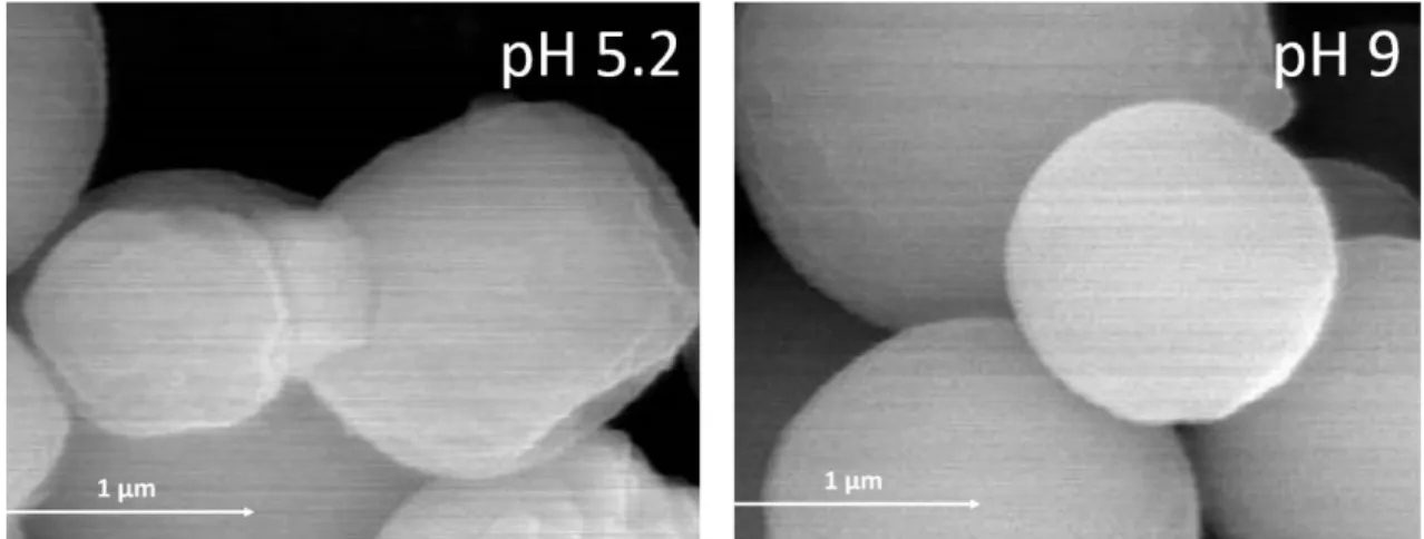
with a more regular shape (Figure 2). Further, differences in the dispersibility of the 171
microparticles in water due to pH were not observed. 172
With the present work we also intend to compare the behaviour of the enzyme β-173
galactosidase when microencapsulated with different solutions of modified chitosan with 174
different pH. The success of the β-galactosidase microencapsulation depends on various 175
factors such as pH, ionic strength, surface and protein properties such as isoelectric point 176
of the protein and history of dependence of protein-adsorption kinetics [28]. Since the 177
activity of β-galactosidase may be significantly reduced or lost during the 178
microencapsulation process, the selection of the encapsulating agent and the pH are very 179
important. The pH is an important factor that significantly influences encapsulation 180
efficiency [29]. β-galactosidase works in a relatively broad pH range: enzymes from fungi 181
act between pH 2.5–5.4, yeast and bacterial enzymes act between pH 6.0–7.0. Depending 182
on the natural source where lactose is present, pH values range between ~ 3.5 or 5.6 of 183
acid whey to 6.5 of milk. [28]. The isoelectric point of β-galactosidase is around 4.6 [30]. 184
Chitosan is a positive polymer in acidic solutions, and its positive potential decreases with 185
increasing solution pH [29]. In experimental works with α-galactosidase (isoelectric 186
point also 4.6), the positive potential of α-galactosidase decreased when the pH was 187
increased from 3.0 to 4.5, after which the repellent force between chitosan and α-188
galactosidase weakened. [29] 189
In this work a modified chitosan was tested, which is a less positive polymer in acid 190
solutions than the normal chitosan; on the other hand β-galactosidase from a bacterial 191
source was used, these enzymes acting better between pH 6.0–7.0. So, our kinetic results 192
will be obtained for a combination of factors. 193
For the free enzyme, in a previous study [23], the highest value of the enzyme activity 194
was obtained at pH 6.8, which is in agreement with results obtained by other authors [31]. 195
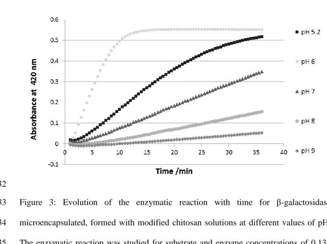
In Figure 3, the evolution of the enzymatic reaction with time for the microencapsulated 196
enzyme formed was observed. The highest velocity was reached when the enzyme was 197
microencapsulated at pH 6. With the increase of the pH for values higher than 6, the 198
velocity decreases and the same happened when the pH decreases for values lower than 199
6. So the optimal pH to do the microencapsulation of the β-galactosidase with this 200
modified chitosan is around pH 6. 201
This can be explained by the fact that the enzyme β-galactosidase has two active-site 202
carboxyl groups that can exist as –COO− (as nucleophile) and –COOH (as proton donor) 203
simultaneously at neutral pH [32] but also it depends on the amount of carboxyl and 204
amino group in chitosan. For example, some groups of chitosan can be charged more 205
positively by the effect of the decrease of the pH and can establish interactions with some 206
groups of the enzyme charged more negatively. The interactions between enzyme and 207
chitosan can change the conformation of the enzyme and/or can make difficult the access 208
to the active center of the enzyme, by this way the activity of the enzyme will decrease. 209
So, different pH will influence the structure of the enzyme and of the encapsulating agent 210
and the type of interactions between them and as referred before, the pH of the solution 211
will affect the strength of the interaction between chitosan and β-galactosidase. 212
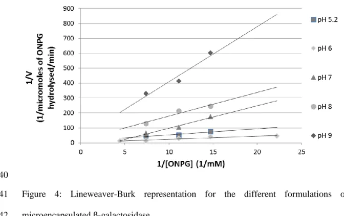
For each β-galactosidase reaction curve, the initial velocity was calculated, and the 213
Lineweaver-Burk linearization was performed to determine the Michaelis-Menten 214
parameters (Figure 4). 215
The Michaelis-Menten parameters were determined for the microencapsulated 216
formulations with different pH and are presented in Table 1. The values related to the free 217
enzyme have already been determined by the authors [26]. 218
The parameter Vmax, representing the maximum reaction rate at a given enzyme 219
concentration, decreased its value after microencapsulation process thus confirming what 220
has been observed by other authors [26,33]. Some active centres are likely to be blocked 221
after microencapsulation, which reduces the reaction rate, causing the decrease of the 222
maximum reaction velocity. The highest value of the Vmax was obtained from the 223
microencapsulated β-galactosidase formulation obtained at pH 6, being more than four 224
times higher than the Vmax obtained from the formulations produced with different pH 225
(Figure 5). However, this value is smaller than the Vmax obtained with the free enzyme 226
[26]. 227
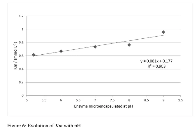
The parameter Km was associated to the affinity between the enzyme and the substrate. 228
A smaller value of Km indicated a greater affinity between the enzyme and substrate, and 229
it means that the reaction rate reaches Vmax faster. The value of Km increased in these 230
assays of microencapsulation assays with pH of the microencapsulation formulation, this 231
means that the affinity between the enzyme and the substrate decreased (Figure 6). A 232
linear correlation between the value of Km and the pH of the β-galactosidase 233
microencapsulation solution was obtained. 234
The β-galactosidase immobilization on chitosan was studied by Carrara and Rubiolo [34]. 235
These authors obtained chitosan beads of 2.2 mm diameter, bigger than the microparticles 236
that we obtained in this work. The higher activity value of the immobilized enzyme 237
compared with those of the free β-galactosidase is only 10.7% of the free enzyme values. 238
In our study, for a microencapsulation process at pH 6, the enzyme keeps 55% of the 239
activity of the free enzyme. A different pH provoked a decrease in the activity of the 240
enzyme. 241
After six months storage at controlled ambient conditions (4ºC), a small decrease in 242
enzyme activity was observed, as described in a previous work [23], and no significant 243
differences in the appearance, color, and particle size distribution were identified. 244
Comparing the results obtained in this study with the previous ones [23,24,26], we can 245
conclude that the selection of the pH for the immobilization (microencapsulation) of the 246
enzyme is so important as the selection of the encapsulating agent or the selection of the 247
operational conditions of the spray dryer for the optimization of the β-galactosidase 248 activity. 249 250
Conclusion
251The main objective of this work was to study the influence of pH in the β-galactosidase 252
microencapsulation process, with a modified chitosan through a spray-drying process. 253
β-galactosidase microparticles with an average diameter smaller than 3.5 μm and in 254
general a regular shape were obtained. 255
The parameters of Michaelis-Menten were calculated for all the β-galactosidase 256
formulations. The value of Km decreases with the decrease of the pH, which can be related 257
to an increase of the affinity between the enzyme and substrate to smaller pH´s. 258
The highest value of the Vmax was obtained for the microencapsulated β-galactosidase 259
formulation obtained at pH 6, being more than four times higher than the Vmax obtained 260
for the formulations produced with different pH. However this value is smaller than the 261
Vmax obtained with the free enzyme. For a microencapsulation process at pH 6, the
262
enzyme keeps 55% of the activity of the free enzyme. 263
264
Acknowledgments
The authors thank Fundação para a Ciência e a Tecnologia (FCT) for the Post-doctoral 266
grant SFRH/BPD/73865/2010 (Berta N. Estevinho) and Programa Ciência sem Fronteiras 267
(Brasil) for the scholarship of Irena Ramos. 268
269
References
270
[1] D.S. Wentworth, D. Skonberg, W.D. Darrel, A. Ghanem, Application of chitosan‐ 271
entrapped β-galactosidase in a packed-bed reactor system, J. Appl. Polym. Sci. 91 272
(2004) 1294–1299. 273
[2] C.R. Carrara, E.J. Mammarella, A.C. Rubiolo, Prediction of the fixed-bed reactor 274
behaviour using dispersion and plug-flow models with different kinetics for 275
immobilised enzyme, Chem. Eng. J. 92 (2003) 123–129. doi:10.1016/S1385-276
8947(02)00129-8. 277
[3] K. Makowski, A. Białkowska, M. Szczesna-Antczak, H. Kalinowska, J. Kur, H. 278
Cieśliński, et al., Immobilized preparation of cold-adapted and halotolerant 279
Antarctic β-galactosidase as a highly stable catalyst in lactose hydrolysis., FEMS 280
Microbiol. Ecol. 59 (2007) 535–542. doi:10.1111/j.1574-6941.2006.00208.x. 281
[4] S.A. Ansari, Q. Husain, Lactose hydrolysis from milk/whey in batch and 282
continuous processes by concanavalin A-Celite 545 immobilized Aspergillus 283
oryzae β galactosidase, Food Bioprod. Process. 90 (2012) 351–359. 284
doi:10.1016/j.fbp.2011.07.003. 285
[5] A.K. Singh, K. Singh, Study on Hydrolysis of Lactose in Whey by use of 286
Immobilized Enzyme Technology for Production of Instant Energy Drink, Adv. J. 287
Food Sci. Technol. 4(2). 4 (2012) 84–90. 288
[6] Z. Grosová, M. Rosenberg, M. Rebroš, Perspectives and Applications of 289
Immobilised β-Galactosidase in Food Industry - a Review, Czech J. Food Sci. 26 290
(2008) 1–14. 291
[7] C.K. Kim, H.S. Chung, M.K. Lee, L.N. Choi, M.H. Kim, Development of dried 292
liposomes containing beta-galactosidase for the digestion of lactose in milk., Int. 293
J. Pharm. 183 (1999) 185–93. 294
[8] J.M. Rodríguez-Nogales, A.D. López, A novel approach to develop β-295
galactosidase entrapped in liposomes in order to prevent an immediate hydrolysis 296
of lactose in milk, Int. Dairy J. 16 (2006) 354–360. 297
[9] J.M. Rodriguez-Nogales, A. Delgadillo, Stability and catalytic kinetics of 298
microencapsulated β-galactosidase in liposomes prepared by the dehydration– 299
rehydration method, J. Mol. Catal. B Enzym. 33 (2005) 15–21. 300
doi:10.1016/j.molcatb.2005.01.003. 301
[10] B.N. Estevinho, A. Ferraz, L. Santos, F. Rocha, A. Alves, Uncertainty in the 302
determination of glucose and sucrose in solutions with chitosan by enzymatic 303
methods, J. Braz. Chem. Soc. 24 (2013) 931–938. 304
doi:http://dx.doi.org/10.5935/0103-5053.20130119 J. 305
[11] B.N. Estevinho, a Ferraz, F. Rocha, a Alves, L. Santos, Interference of chitosan in 306
glucose analysis by high-performance liquid chromatography with evaporative 307
light scattering detection., Anal. Bioanal. Chem. 391 (2008) 1183–8. 308
doi:10.1007/s00216-008-1832-3. 309
[12] I. Aranaz, M. Mengíbar, R. Harris, I. Paños, B. Miralles, N. Acosta, et al., 310
Functional characterization of chitin and chitosan, Curr. Chem. Biol. 3 (2009) 203– 311
230. 312
[13] B.M.A.N. Estevinho, F.A.N. Rocha, L.M.D.S. Santos, M.A.C. Alves, Using water-313
soluble chitosan for flavour microencapsulation in food industry., J. 314
Microencapsul. 30 (2013) 571–579. doi:10.3109/02652048.2013.764939. 315
[14] B.N. Estevinho, F. Rocha, L. Santos, A. Alves, Microencapsulation with chitosan 316
by spray drying for industry applications – A review, Trends Food Sci. Technol. 317
31 (2013) 138–155. doi:10.1016/j.tifs.2013.04.001. 318
[15] H. Sashiwa, N. Kawasaki, A. Nakayama, Chemical modification of chitosan. 14:1 319
Synthesis of water-soluble chitosan derivatives by simple acetylation., 320
Biomacromolecules. 3 (2002) 1126–1128. 321
[16] H. Zhang, S. Wu, Y. Tao, L. Zang, Z. Su, Preparation and Characterization of 322
Water-Soluble Chitosan Nanoparticles as Protein Delivery System, J. Nanomater. 323
2010 (2010) 1–5. doi:10.1155/2010/898910. 324
[17] Y.C. Chung, C.F. Tsai, C.F. Li, Preparation and characterization of water-soluble 325
chitosan produced by Maillard reaction, Fish. Sci. 72 (2006) 1096–1103. 326
doi:10.1111/j.1444-2906.2006.01261.x. 327
[18] J. Pu, J.D. Bankston, S. Sathivel, Developing microencapsulated flaxseed oil 328
containing shrimp (Litopenaeus setiferus) astaxanthin using a pilot scale spray 329
dryer, Biosyst. Eng. 108 (2011) 121–132. 330
doi:10.1016/j.biosystemseng.2010.11.005. 331
[19] A.L.R. Rattes, W.P. Oliveira, Spray drying conditions and encapsulating 332
composition effects on formation and properties of sodium diclofenac 333
microparticles, Powder Technol. 171 (2007) 7–14. 334
doi:10.1016/j.powtec.2006.09.007. 335
[20] N. Schafroth, C. Arpagaus, U.Y. Jadhav, S. Makne, D. Douroumis, Nano and 336
Microparticle Engineering of Water Insoluble Drugs Using a Novel Spray–Drying 337
Process, Colloids Surfaces B Biointerfaces. 90 (2011) 8–15. 338
doi:10.1016/j.colsurfb.2011.09.038. 339
[21] P. de Vos, M.M. Faas, M. Spasojevic, J. Sikkema, Encapsulation for preservation 340
of functionality and targeted delivery of bioactive food components, Int. Dairy J. 341
20 (2010) 292–302. doi:10.1016/j.idairyj.2009.11.008. 342
[22] K.G.H. Desai, H.J. Park, Recent Developments in Microencapsulation of Food 343
Ingredients, Dry. Technol. 23 (2005) 1361–1394. 344
[23] B.N. Estevinho, A.M. Damas, P. Martins, F. Rocha, The Influence of 345
Microencapsulation with a Modified Chitosan (Water Soluble) on β-galactosidase 346
Activity, Dry. Technol. 32 (2014) 1575–1586. 347
doi:10.1080/07373937.2014.909843. 348
[24] B.N. Estevinho, A.M. Damas, P. Martins, F. Rocha, Microencapsulation of β-349
galactosidase with different biopolymers by a spray-drying process, Food Res. Int. 350
64 (2014) 134–140. doi:10.1016/j.foodres.2014.05.057. 351
[25] R. Switzer, L. Garrity, Experimental Biochemistry, third edit, Freeman, 1999. 352
[26] B.N. Estevinho, A.M. Damas, P. Martins, F. Rocha, Study of the Inhibition Effect 353
on the Microencapsulated Enzyme β-galactosidase, Environ. Eng. Manag. J. 11 354
(2012) 1923–1930. 355
[27] B. Erdinc, R.J. Neufeld, Protein micro and nanoencapsulation within glycol-356
chitosan/Ca2+/alginate matrix by spray drying., Drug Dev. Ind. Pharm. 37 (2011) 357
619–627. doi:10.3109/03639045.2010.533681. 358
[28] Q. Husain, β galactosidases and their potential applications: a review, Crit. Rev. 359
Biotechnol. 30 (2010) 41–62. doi:10.3109/07388550903330497. 360
[29] Y. Liu, Y. Sun, Y. Li, S. Xu, J. Tang, J. Ding, et al., Preparation and 361
characterization of α-galactosidase-loaded chitosan nanoparticles for use in foods, 362
Carbohydr. Polym. 83 (2011) 1162–1168. doi:10.1016/j.carbpol.2010.09.050. 363
[30] P.S. Panesar, S. Kumari, R. Panesar, Potential Applications of Immobilized β-364
Galactosidase in Food Processing Industries., Enzyme Res. 2010 (2010) 1–16. 365
doi:10.4061/2010/473137. 366
[31] E. Jurado, F. Camacho, G. Luzón, J.M. Vicaria, Kinetic models of activity for β-367
galactosidases: influence of pH, ionic concentration and temperature, Enzyme 368
Microb. Technol. 34 (2004) 33–40. doi:10.1016/j.enzmictec.2003.07.004. 369
[32] Q.Z.K. Zhou, X.D. Chen, Effects of temperature and pH on the catalytic activity 370
of the immobilized β-galactosidase from Kluyveromyces lactis, Biochem. Eng. J. 371
9 (2001) 33–40. 372
[33] T. Haider, Q. Husain, Hydrolysis of milk/whey lactose by β galactosidase: A 373
comparative study of stirred batch process and packed bed reactor prepared with 374
calcium alginate entrapped enzyme, Chem. Eng. Process. 48 (2009) 576–580. 375
doi:10.1016/j.cep.2008.02.007. 376
[34] C.R. Carrara, A.C. Rubiolo, Immobilization of β-Galactosidase on Chitosan, 377
Biotechnol. Prog. 10 (1994) 220–224. doi:10.1021/bp00026a012. 378 379 380 381 TABLE CAPTIONS 382 383
Table 1: Michaelis-Menten parameters (Km and Vmax), for the different assays with free 384
and microencapsulated β-galactosidase. 385
386 387 388
FIGURE CAPTIONS
389 390
Figure 1: SEM images of β-galactosidase microparticles prepared at different pH´s (5.2, 391
6, 7, 8 and 9). Magnification = 12000 times, beam intensity (HV) = 15kV, distance 392
between the sample and the lens (WD) = 15 mm. 393
394
Figure 2: Surface and shape of the β-galactosidase microparticles prepared at pH´s 5.2 and 395
9. 396 397
Figure 3: Evolution of the enzymatic reaction with time for β-galactosidase 398
microencapsulated, formed with modified chitosan solutions at different values of pH. 399
The enzymatic reaction was studied for substrate and enzyme concentrations of 0.135 400
mmol L-1 and 0.001 mg mL-1, respectively, based on absorbance values, by UV-visible 401
spectrophotometry at 420 nm and at room temperature. 402
403
Figure 4: Lineweaver-Burk representation for the different formulations of 404
microencapsulated β-galactosidase. 405
406
Figure 5: Evolution of the Vmax with pH. 407
408
Figure 6: Evolution of Km with pH. 409
410 411
Table 1: Michaelis-Menten parameters (Km and Vmax), for the different assays with free 412
and microencapsulated β-galactosidase. 413
414 415
416 417
TOC GRAPHIC
419
420
Figure 1: SEM images of β-galactosidase microparticles prepared at different pH´s (5.2, 421
6, 7, 8 and 9). Magnification = 12000 times, beam intensity (HV) = 15kV, distance 422
between the sample and the lens (WD) = 15 mm. 423
424 425 426
427
Figure 2: Surface and shape of the β-galactosidase microparticles prepared at pH´s 5.2 and 428
9. 429 430
431
432
Figure 3: Evolution of the enzymatic reaction with time for β-galactosidase 433
microencapsulated, formed with modified chitosan solutions at different values of pH. 434
The enzymatic reaction was studied for substrate and enzyme concentrations of 0.135 435
mmol L-1 and 0.001 mg mL-1, respectively, based on absorbance values, by UV-visible 436
spectrophotometry at 420 nm and at room temperature. 437
439
440
Figure 4: Lineweaver-Burk representation for the different formulations of 441
microencapsulated β-galactosidase. 442
444
Figure 5: Evolution of the Vmax with pH. 445
447
448
Figure 6: Evolution of Km with pH. 449


