R E S E A R C H A R T I C L E
Open Access
Burkholderia pseudomallei transcriptional
adaptation in macrophages
Sylvia Chieng
1, Laura Carreto
2and Sheila Nathan
1*Abstract
Background: Burkholderia pseudomallei is a facultative intracellular pathogen of phagocytic and non-phagocytic cells. How the bacterium interacts with host macrophage cells is still not well understood and is critical to appreciate the strategies used by this bacterium to survive and how intracellular survival leads to disease manifestation.
Results: Here we report the expression profile of intracellular B. pseudomallei following infection of human macrophage-like U937 cells. During intracellular growth over the 6 h infection period, approximately 22 % of the B. pseudomallei genome showed significant transcriptional adaptation. B. pseudomallei adapted rapidly to the
intracellular environment by down-regulating numerous genes involved in metabolism, cell envelope, motility, replication, amino acid and ion transport system and regulatory function pathways. Reduced expression in catabolic and housekeeping genes suggested lower energy requirement and growth arrest during macrophage infection, while expression of genes encoding anaerobic metabolism functions were up regulated. However, whilst the type VI secretion system was up regulated, expression of many known virulence factors was not significantly modulated over the 6 hours of infection.
Conclusions: The transcriptome profile described here provides the first comprehensive view of how B. pseudomallei survives within host cells and will help identify potential virulence factors and proteins that are important for the survival and growth of B. pseudomallei within human cells.
Keywords: Burkholderia pseudomallei, Macrophage, Transcriptome analysis Background
Burkholderia pseudomallei causes melioidosis, a disease with considerable human mortality and morbidity in the tropics [1]. Clinical presentation of melioidosis varies from rapidly fatal septicemia and acute pneumonia, chronic or localized abscess formation to skin and soft tissue infections that progress rapidly to systemic infec-tion [2,3]. Treatment of melioidosis involves long peri-ods of antibiotic administration which are vital to eliminate B. pseudomallei and to prevent relapse [4,5].
Several features of melioidosis suggest that B. pseudo-mallei is a facultative intracellular bacterium. These in-clude long incubation periods of up to 62 years and the tendency to relapse [3,6]. B. pseudomallei has the ability to survive and proliferate within phagocytic and
non-phagocytic cells [7,8], and free-living amoeba [9] for months or years. The exact mechanism of invasion is still unknown, but it requires the rearrangement of host actin cytoskeleton and the involvement of BopE, an ef-fector protein of the type III secretion system (T3SS-3) [10]. After cellular uptake, B. pseudomallei escapes from endocytic vacuoles and once in the cytoplasm, induces host cell fusion and enters neighbouring cells by forming actin tails and membrane protrusions [11,12]. Vacuole escape and intracellular survival requires a functional T3SS-3, as mutants of T3SS-3 display delayed vacuolar escape, reduced actin formation and reduced capacity to survive intracellularly and spread to neighbouring cells [13,14]. It is possible that intracellular survival and inter-cellular spread may provide B. pseudomallei protection from host defences.
Recently, the type VI secretion cluster tss-5 (T6SS-1) was shown to contribute to intracellular growth of B.
pseudomallei [15]. The expression of this secretion
* Correspondence:[email protected]
1
School of Biosciences and Biotechnology, Faculty of Science and Technology, Universiti Kebangsaan Malaysia, Bangi 43600, Malaysia Full list of author information is available at the end of the article
© 2012 Chieng et al.; licensee BioMed Central Ltd. This is an Open Access article distributed under the terms of the Creative Commons Attribution License (http://creativecommons.org/licenses/by/2.0), which permits unrestricted use, distribution, and reproduction in any medium, provided the original work is properly cited.
system is dependent on the sensor regulators BprC and VirA-VirG (VirAG). The type VI secretion system is also thought to play a major role during bacterial transition from the phagosome to the cytosol [15]. Moreover, T6SS is also implicated in other important bacterial pathogens as the key virulence factor and is involved in translocation of effector proteins into eukaryotic cells [16,17]. Using in vivo expression technology, Shalom et al. [18] found that the B. pseudomallei tss-5 gene cluster was induced inside murine macrophages. Fur-thermore, Pilatz and colleagues demonstrated that a
B. pseudomallei tss-5 transposon mutant displayed
reduced ability to form plaques on PtK2 epithelial cell monolayers, indicating the requirement of tss-5 in cell-to-cell spread [19]. This mutant was also highly attenu-ated in mice with reduced bacterial load in the spleen, liver and lung at 48 h post-infection [19].
Previous studies have examined the detailed cellular responses of B. pseudomallei within eukaryotic cells. However, the overall picture of the mechanisms involved in adaptability to the intracellular lifestyle is still unclear. The availability of the complete genome sequence for B.
pseudomallei [20] enabled us to design a whole genome
DNA microarray to identify B. pseudomallei genes regu-lated during infection of macrophages. These data pro-vide an insight into genes involved in survival and adaptation of the bacteria within macrophage cells as well as a new understanding of the biology of host-pathogen interaction during melioidosis.
Results
Infection model and bacterial RNA isolation
Human macrophage-like U937 cells were chosen as the infection model as it has previously been used exten-sively to study B. pseudomallei interaction with host cells [7,8]. In this study, bacterial replication within macrophages was observed with a maximum CFU obtained at 6 h post-infection. The calculated doubling time for intracellular B. pseudomallei is about 6.4 h, in-dicating a slower growth rate compared to the control bacteria in RPMI (doubling time of ~ 1.9 h). The number of viable bacteria decreased consistently after 6 h post-infection and a 2-log reduction in viable cells was noted with prolonged incubation of up to 24 h. A similar de-cline in bacterial numbers following prolonged incuba-tion was previously reported in RAW264.7 cells [7,21]. Viability of infected U937 cells decreased over time and was drastically reduced by 6 h post-infection, suggesting a cytotoxic effect of B. pseudomallei on the host cells (Figure 1).
An ultrastructural study of infected U937 cells showed that the nuclei, mitochondria and vacuoles were swollen whilst the nuclear material appeared diluted and the cell membrane totally disrupted (Figure 2A). All these
Figure 1 B. pseudomallei intracellular growth and viability of infected U937. Intracellular survival of B. pseudomallei was monitored over a 24 h period, along with U937 cell viability after B. pseudomallei infection. The relative amount of bacteria recovered (log10scale) was plotted in grey bars while the relative % of viable
infected cells was indicated as a solid line. Vertical lines represent the standard deviation (SD) obtained from three independent experiments conducted in triplicate (n = 9).
Figure 2 Transmission electron micrograph of intracellular B. pseudomallei within U937 cells. (A) B. pseudomallei-infected U937 cells demonstrating oncotic-like morphology and disruption of cell membrane. ×7000 (B) Internalised B. pseudomallei within membrane-bound phagosome (arrowed) were observed at early stage of intracellular infection. ×8000 (C) Loss of phagosome membrane was clearly visible and free bacteria were seen in the cell cytoplasm (arrowed). ×8000 (D) A dividing bacillus in a phagosome with near-intact membrane (arrowed). Some membrane dissolution can be seen close to the ends of the bacterial cell. ×8000.
changes indicate that infected U937 cells displayed a phenotype similar to oncotic cells as previously observed in other cells infected with B. pseudomallei [21]. Loss of phagosome membrane in B. pseudomallei infected macrophage cells was established as early as 15 min post-incubation similar to that previously reported [22]. At 2 h post-infection, loss of phagosome membrane is
evident and bacilli were seen in the cytoplasm
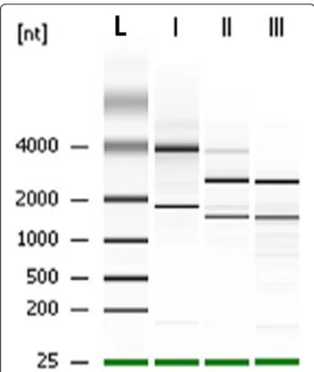
(Figure 2C). Moreover, replication of intracellular bac-teria was seen in the cytoplasm of host cells (Figure 2D). An infection period of 1 to 6 h was selected to study changes in the expression profile as more than 85 % of the infected U937 cells were still viable during this period (Figure 1). This would ensure maximum recovery of intracellular B. pseudomallei from viable cells to ob-tain sufficient intact bacterial RNA. We adopted the dif-ferential lysis method with saponin to prevent bacterial cell lysis and easily recover bacterial RNA. This strategy enriches the bacterial mRNA several thousand-fold [23]. Treatment with saponin specifically lyses eukaryotic cells without affecting bacterial viability [24,25] and in our study, the use of 0.1 % to 4 % saponin was not detrimen-tal to B. pseudomallei (data not shown). Differential cen-trifugation was combined with this method to efficiently remove host cellular debris. Based on the RNA integrity number (RIN) and the 16 S/23 S rRNA subunit ratio, the electrophoretic profiles of the total RNA extracted from intracellular bacteria showed no evidence of deg-radation and only minor host RNA contamination was detectable (Figure 3).
The global gene expression profile
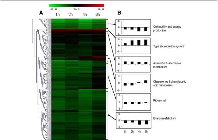
Temporal gene expression profiles obtained from intra-cellular B. pseudomallei were compared with the tran-scriptome of control bacteria grown in cell culture medium. Statistical analysis (p< 0.01) combined with a 2-fold variation cut off indicated that 2,797 genes were differentially expressed at 1 h, 2,755 at 2 h, 2,776 at 4 h and 1,918 genes at 6 h. Hierarchical clustering of gene expression levels revealed similar patterns of gene regu-lation over the period of infection (Figure 4A). The ma-jority of the genes with altered expression were down-regulated throughout the infection period compared to bacteria grown in control medium. Expression levels of these genes continued to decrease up to 4 h before in-creasing to levels lower or similar to control bacteria at 6 h. It appears that the adaptation of B. pseudomallei within U937 cells is rapid and most of the changes oc-curred as early as 1 h post-infection (Figure 5). With time, the number of significantly deregulated genes grad-ually decreased, suggesting that B. pseudomallei had
be-come adapted to the intracellular environment.
Functional classification of intracellularly modulated bacterial genes at each time point showed that most of
these genes encoded core functions such as metabolism, cell envelope, regulatory functions, transport and bind-ing. Many genes encoding proteins with unknown func-tion or hypothetical proteins were also modulated during infection (Figure 6).
Of the 5,721 probes represented on our microarrays, 1,284 transcripts representing 22.4 % of the B.
pseudo-mallei ORFeome, were differentially expressed during
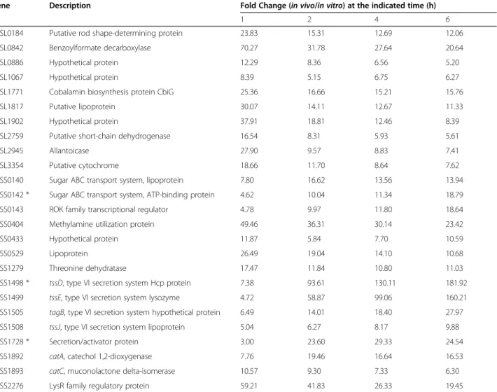
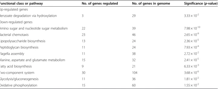
the infection time course. Of these, 25 genes demon-strated increased expression (Table 1) while 1,259 genes were down-regulated (Additional file 1). We performed gene function enrichment analysis on these genes to fur-ther categorize them into biological functions. Based on the statistical analysis of KEGG biochemical pathways, the number of genes involved in cellular functions such as biosynthesis of flagella and capsule, energy metabol-ism and regulatory systems was significantly higher dur-ing intracellular infection (Table 2).
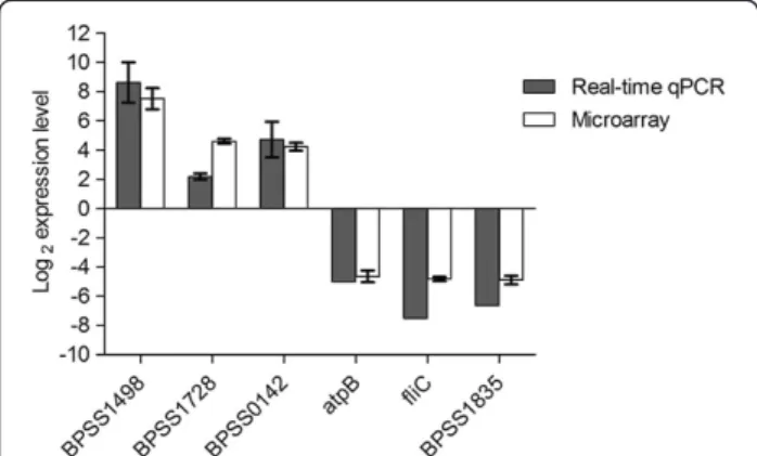
As an independent measure of differential gene ex-pression, we examined the relative expression of six up-or down-regulated genes selected from different func-tional categories by real-time qPCR (Table 1 and Add-itional file 1) on the same samples as those used for microarray analysis. Changes in expression were verified by real-time qPCR with a correlation of >0.95 to the microarray data (Figure 7). The strong correlation
Figure 3 Size chromatographic separation of RNAs. Total RNA were extracted from (I) unifected U937 cells, (II) B. pseudomallei harvested from infected U937 cells and (III) B. pseudomallei grown in vitro. Lane L represents RNA ladder (kb).
observed verified the efficiency and robustness of the designed microarray for high throughput screening of the B. pseudomallei transcriptome.
Due to the large number of significantly differentiated genes modulated during the infection, only data related
to genes that have some functional information are shown and discussed below. The identified genes are dis-cussed according to functional categories.
Intracellular metabolism and ion transport
In host-pathogen interactions, sufficient nutrition levels are necessary for the successful survival of the pathogen. Once intracellular, the bacteria must make metabolic adjustments to adapt to changes in nutrient availability. We noted a robust shutdown in the expression of B.
pseudomallei genes involved in metabolism. Genes
involved in glycolysis and oxidative phosphorylation were consistently down-regulated in intracellular B. pseudomallei. In addition, most genes involved in energy metabolism such as ATP synthase and NADH dehydro-genase were down-regulated (Figure 4B). Collectively, these data suggest that intracellular B. pseudomallei have lower energy requirements and limit their energy pro-duction during the initial stage of infection. Several an-aerobic metabolism genes were induced in intracellular B. pseudomallei. Anaerobic metabolism pathway genes such as BPSS1279 (threonine dehydratase), BPSL1771
Figure 4 Gene expression profile of B. pseudomallei during infection of U937 cells. (A) The cluster diagrams show the expression profile of intracellular B. pseudomallei within U937 cells for 1 to 6 h relative to in vitro grown bacteria. Hierarchical clustering was performed with Euclidean correlation in TIGR-MeV. Expression values are determined from the SAM analysis with red representing up-regulation (ratio of + 8.0) and green representing down-regulation (ratio of - 8.0) on a log2scale. (B) Average relative levels of expression of representative functional groups. The
vertical axis shows the fold change in expression on a log2scale. Data are the means from three biological replicates.
Figure 5 Time-dependent transcriptional adaptation of B. pseudomallei in U937 cells. The graph shows the total number of B. pseudomallei genes which are significantly differentially expressed (2-fold up or down, p< 0.01) at each time point.
Figure 6 Functional classification of intracellular B. pseudomallei regulated genes. Bars indicate percentages of genes in each group that were significantly regulated at any time point. Genes were divided into functional categories based on Comprehensive Microbial Resources (CMR) annotations.
Table 1 Twenty-five common up-regulated genes of B. pseudomallei during intracellular growth in host macrophages relative to in vitro growth
Gene Description Fold Change (in vivo/in vitro) at the indicated time (h)
1 2 4 6
BPSL0184 Putative rod shape-determining protein 23.83 15.31 12.69 12.06
BPSL0842 Benzoylformate decarboxylase 70.27 31.78 27.64 20.64
BPSL0886 Hypothetical protein 12.29 8.36 6.56 5.20
BPSL1067 Hypothetical protein 8.39 5.15 6.75 6.27
BPSL1771 Cobalamin biosynthesis protein CbiG 25.36 16.66 15.21 15.76
BPSL1817 Putative lipoprotein 30.07 14.11 12.67 11.33
BPSL1902 Hypothetical protein 37.91 18.81 12.46 8.39
BPSL2759 Putative short-chain dehydrogenase 16.54 8.31 5.93 5.61
BPSL2945 Allantoicase 27.90 9.57 8.83 7.41
BPSL3354 Putative cytochrome 18.66 11.70 8.64 7.62
BPSS0140 Sugar ABC transport system, lipoprotein 7.80 16.62 13.56 13.94
BPSS0142 * Sugar ABC transport system, ATP-binding protein 4.62 10.04 11.34 18.79
BPSS0143 ROK family transcriptional regulator 4.78 9.97 11.80 18.64
BPSS0404 Methylamine utilization protein 49.46 36.31 30.14 23.42
BPSS0433 Hypothetical protein 11.87 5.84 7.70 10.59
BPSS0529 Lipoprotein 26.49 19.04 14.10 10.68
BPSS1279 Threonine dehydratase 17.47 11.84 10.80 11.03
BPSS1498 * tssD, type VI secretion system Hcp protein 7.38 93.61 130.11 181.92
BPSS1499 tssE, type VI secretion system lysozyme 4.72 58.87 99.06 160.21
BPSS1505 tagB, type VI secretion system hypothetical protein 6.49 14.01 18.40 27.97
BPSS1508 tssJ, type VI secretion system lipoprotein 5.04 6.27 8.17 9.88
BPSS1728 * Secretion/activator protein 3.00 23.60 29.33 24.54
BPSS1892 catA, catechol 1,2-dioxygenase 7.76 19.46 16.64 16.53
BPSS1893 catC, muconolactone delta-isomerase 10.57 9.30 7.33 6.30
BPSS2276 LysR family regulatory protein 59.21 41.83 26.33 19.45
(cobalamin biosynthesis protein CbiG) and BPSS0842
(benzoylformate decarboxylase) were up-regulated
throughout the infection period. Nevertheless, none of the components of the anaerobic respiratory chain showed significant changes in expression except for BPSL2311 (putative respiratory nitrate reductase delta chain) and BPSL2312 (putative respiratory nitrate reductase gamma chain) that were induced at the early stage of infection.
Other induced genes were catAC genes, which are involved in benzoate degradation, indicating that intra-cellular B. pseudomallei utilized aromatic compounds as a source of carbon. Increased expression of phenylacetic acid (PA) pathway genes at the later stage of infection (4 h and 6 h post-infection) was also observed (Figure 4B). The major nitrogen source in the intracellular compart-ment is most likely methylamine and purine as suggested by the increased expression of methylamine utilization protein (BPSS0404) and allantoicase (BPSL2945). Allan-toicase is involved in purine metabolism and provides a secondary nitrogen source under nitrogen limiting condi-tions [26].
Expression of virulence and virulence-associated factors We observed the repression of genes encoding proteins that are well characterised as B. pseudomallei virulence factors. These include the main capsular polysaccharide biosynthesis (BPSL2787-BPSL2810) genes, two potential
surface polysaccharide biosynthesis gene clusters
(BPSS0417-BPSS0429 and BPSS1825-BPSS1834), major-ity of genes in the lipopolysaccharide (LPS) biosynthesis cluster and genes encoding for flagella assembly and chemotaxis. We also noted the repression of bspR, a regulator recently shown to control the expression of TT3SS-3 genes [27]. The inhibition of this regulator leads to the reduced expression of T3SS-3 controlled genes in intracellular B. pseudomallei.
One of the six clusters of the type VI secretion system,
the tss-5 cluster (BPSS1493-BPSS1511), was
up-regulated up to 182-fold during intracellular infection (Figure 8). This observation is consistent with previous reports on the induction of three genes in this cluster, tssH-5, tssI-5 and tssM-5, upon invasion of macrophages [18]. We also observed the induction of genes flanking Table 2 Gene function enrichment analysis of B. pseudomallei common up-regulated and down-regulated genes throughout growth within host macrophages
Functional class or pathway No. of genes regulated No. of genes in genome Significance (p-value) Up-regulated genes
Benzoate degradation via hydroxylation 3 29 3.33 × 10⁻²
Down-regulated genes
Amino sugar and nucleotide sugar metabolism 22 39 7.98 × 10⁻¹⁰
Bacterial chemotaxis 23 46 2.65 × 10⁻⁹
Lipopolysaccharide biosynthesis 13 24 2.36 × 10⁻⁵
Peptidoglycan biosynthesis 11 24 7.93 × 10⁻⁴
Flagella assembly 11 38 2.72 × 10⁻²
Alanine, aspartate and glutamate metabolism 15 32 2.41 × 10⁻⁵
Fatty acid biosynthesis 9 21 6.33 × 10⁻³
Two-component system 30 104 3.68 × 10⁻⁶
Glycolysis/gluconeogenesis 11 36 1.81 × 10⁻²
Oxidative phosphorylation 15 60 1.55 × 10⁻²
Figure 7 Real-time quantitative PCR validation of microarray data. Relative expression ratio by real-time qPCR (grey bars) or by microarray experiments (white bars) at 6 h post-infection. The horizontal axis represents fold change in log2scale. Data are mean ± SD of triplicate
the tss-5 cluster, bimA (Burkholderia intracellular motil-ity A)(BPSS1492) and BPSS1512 at 2 to 6 h post-infection. Moreover, the hemolysin activator-like protein precursor, fhaC (BPSS1728) gene was significantly up-regulated during intracellular infection. Consistently, the
large filamentous hemagglutinin precursor, fhaB
(BPSS1727) gene, a potential virulence factor of B.
pseu-domallei [20], was induced between 2 to 6 h
post-infection.
Stress responses genes
The general stress-responsive alternative sigma factor
rpoS transcribes genes involved in bacterial survival
under conditions of environmental stress. In B. pseudo-mallei, rpoS is a regulator of carbon starvation and oxi-dative stress [28].We observed down-regulation of rpoS throughout intracellular growth. Furthermore, the ex-pression of most of the genes encoding sigma factors was repressed in the intracellular bacteria. Under oxida-tive stress, rpoS regulates oxyR and katG-dpsA operons [29]. As expected, we observed down regulation of oxyR and katG-dpsA expression at 1 and 2 h post-infection. The expression of other genes in the oxy R regulon was either repressed or did not change significantly relative to control cells. Moreover, class I stress response genes including dnaJ, dnaK, hrcA and groEL were not signifi-cantly regulated, except for groES, which was induced at 6 h post-infection.
DNA topology and growth arrest within macrophages Modification of DNA topology plays a major role in assisting DNA replication and protein synthesis. Pro-karyotic DNA is usually maintained in a negatively supercoiled form by topoisomerases and the level of
supercoiling can be affected by several environmental parameters [30]. The ability of DNA gyrase to generate negative supercoils in DNA is inhibited when ATP levels are reduced [31]. In this study, DNA topoisomerase IV subunit A (parC), DNA topoisomerase IV subunit B (parB) and DNA gyrase subunit A (gray) genes were down-regulated. The repression of gyrase suggests a re-laxation in bacterial DNA supercoiling during B. pseudo-mallei intracellular growth in U937. Additionally, the nucleotide-associated regulator protein Fis was also negatively regulated. Fis expression depends on super-helical density whereby maximum density is observed at high levels of negative supercoiling [32].
We found that bacterial genes involved in cell division (ftsABH) were down-regulated indicating reduced cell division activity. The repression of minD and minE genes, which mediate spatial regulation of cytokinesis in bacteria [33], and chromosome partitioning genes, parA and parB was also observed. Furthermore, replicative DNA helicase dnaB, DNA polymerase III subunit alpha
dnaE and DNA polymerase III subunit chi genes were
also down-regulated. Consistent with the down regula-tion of catabolic and cell replicaregula-tion genes, the expres-sion of RNA polymerase (rpoABCZ) and ribosomal subunit genes (S1, S10, S21, L11 and L28) was also reduced throughout the infection period. Collectively, these support the view that cell division and replication processes are interrupted during infection, leading to slower growth kinetics of intracellular B. pseudomallei.
Discussion
Numerous studies have been conducted on the cellular interaction between B. pseudomallei and eukaryotic cells, especially the ability of this pathogen to survive within host cells. In this study, we investigated the global B. pseudomalleitranscriptome profile during early stage infection of human monocyte-like U937 cells. Initially, we demonstrated that replication kinetics of intracellular B. pseudomalleiwithin U937 cells is similar to data from a number of previous studies [8,34]. The bacteria were able to survive in macrophage cells, albeit, at a slower growth rate compared to bacteria grown in RPMI. Reduced bacterial growth kinetics during infection was also reported in a hamster model of melioidosis [35]. Furthermore, this reduced bacterial replication kinetics in the intracellular environment in comparison with in vitro growth is not limited to B. pseudomallei [36– 38]. B. pseudomallei infection was also cytotoxic to macrophage cells with about 20 % cell mortality at 24 h post infection which, to some extent, explains the reduced number of recovered bacteria with prolonged incubation. Electron micrographs of infected U937 cells revealed disruption of internal structures and formation of oncotic cells. The cytotoxic and cytopathy effects of
Figure 8 Regulation of the type VI secretion system tss-5 cluster. The heat-map shows the expression ratio of each gene in the T6SS tss-5 cluster during intracellular growth in host macrophages relative to in vitro growth. Expression values are determined from the SAM analysis with red representing up-regulation (ratio of +8.0) and green representing down-up-regulation (ratio of -8.0) on a log2scale.
B. pseudomalleiinfection in our study are in agreement with Sun et al. [21], who reported high cytotoxicity (40-70 %) and oncotic death in infected THP-1 cells. We also observed the loss of phagosome membrane in infected macrophages as early as 2 h post-infection resulting in the presence of free replicating bacilli in the cytoplasm at later time points.
The ability to survive within and lyse macrophage cells most probably contributes to the spread of this bacter-ium and disease progression. We attempted to identify mechanisms that underlie the adaptive ability of B.
pseu-domallei during infection by comparing intracellular
bacteria transcripts to that of in vitro grown bacteria. During infection, we observed that intracellular B.
pseu-domallei demonstrated a lower energy requirement and
production by shutting down metabolic activity. The reduced expression of catabolic and house-keeping genes is consistent with the reduced growth rate observed for intracellular B. pseudomallei. The induction of anaerobic metabolism pathway genes is suggestive of limited oxy-gen concentrations in the cytosol although expression of genes of the anaerobic respiratory chain was not signifi-cantly altered. The importance of this finding requires further investigation.
B. pseudomallei utilizes alternative metabolites during infection [35]. Our study suggests that B. pseudomallei utilize aromatic carbon compounds such as benzoate and PA as carbon sources for intracellular survival. PA can inhibit the inducible nitric oxide synthase (iNOS) and LPS-induced expression of cytokines in rat primary astrocytes, microglia and macrophages [39]. Moreover, PA can repress DNA binding and transcriptional activ-ities of NFκB, an important upstream modulator for cytokine and iNOS expression in macrophages [40]. The up-regulation of PA catabolic pathway genes in intracel-lular bacteria might partly explain why macrophages infected with B. pseudomallei failed to activate the pro-duction of iNOS [41]. The putative B. cenocepacia ring PA-coenzyme A hydroxylation system was found to be essential for full pathogenicity in infected Caenorhabdi-tis elegans[42].
There are three T3SSs present in B. pseudomallei and only T3SS-3 is involved in virulence [43]. B.
pseudomal-lei T3SS-3 shows high homology to the Inv/Mxi-Spa
type III secretion systems of Salmonella and Shigella [13]. As T3SS-3 is known to be essential in facilitating invasion and early phagosomal escape into the cytosol [13,14], the down-regulation observed in our study is consistent with this function as it would be conceivable that T3SS-3 is important at the beginning of intracellu-lar life during invasion, but not important at the later stages of intracellular infection. Similarly, the role of T6SS-1 is reported to be crucial during the transition from phagosome to cytosol [15]. Thus, it is possible that
the induction of T6SS-1 in this study plays a major func-tion in ensuring pathogen survival and replicafunc-tion in the cytosol. In B. mallei, a close related species of B.
pseudo-mallei, an effector protein of T6SS (BMAA0742) was
recognized by glanders antiserum, indicating production of this protein in vivo during infection [44]. In this study, high induction of tssD-5 (BPSS1498), an effector Hcp1 protein of T6SS was observed throughout the in-fection period. A recent study on B. pseudomallei T6SS Hcp proteins has shown that tssD-5 deletion mutant was attenuated in a hamster infection model, exhibited growth defects and was only weakly cytotoxic to RAW264.7 macrophages [45]. Additionally, human meli-oidosis serum samples were found to react with Hcp1 protein, consistent with our observation (data not shown). These results suggest that Hcp1 is produced by B. pseudomallei in vivoduring infection, is immunogenic and vital for intracellular survival of B. pseudomallei.
The genes bimA, fhaB and fhaC are known proteins that mediate actin tail formation involved in cell-to-cell spread. In this study, expression of these three genes is up-regulated. The over expression of BimA has previ-ously been shown in intracellular B. pseudomallei and mutation of bimA abolished actin-based motility of this pathogen in J774.2 cell [46]. In Bordetella pertussis, the FhaC dependent filamentous hemagglutinin (FHA) facil-itates attachment to the host cell during infection [47] and initiates killing of macrophages to avoid the host cell mediated immune response whilst infection is being established [48]. Through a genome-wide function screen of B. pseudomallei strain K96243, both fhaB and
fhaC were found to form part of the anti-macrophage
loci and contribute to the formation of dramatic actin projections extending towards neighbouring cells [49].
Interestingly, in our study, well-characterized virulence genes of B. pseudomallei such as capsular polysacchar-ide, LPS and flagella were either repressed or not signifi-cantly expressed. Similar profiles were noted in a hamster model, wherein most of the genes encoding for capsule, LPS and flagella biosynthesis were either down-regulated or not significantly changed in expression [35]. This could be due to the equal expression of these genes under in vivo and in vitro conditions concomitant with previous evidence that capsule, LPS, flagellum and other
virulent determinants are constitutively produced
in vitro [35]. A recent report of a similar study on
macrophages infected by B. cenocepacia demonstrated that this intracellular pathogen’s ability to adapt and re-spond to the intracellular milieu was not dependent on the expression of any specific virulence-associated fac-tors [50]. Hence, this ability to adapt to various niches appears to be conserved amongst the soil-derived Bur-kholderia spp. clinical pathogens. Down regulation of flagella genes and subsequently the absence of flagellin,
may also reflect the bacteria’s attempt to limit immune recognition especially by toll-like receptors by modulat-ing surface structures. In Gram negative bacteria, flagel-lin is recognized by TLR5 which triggers the secretion of various cytokines and chemokines, leading to an inflam-matory response [51]. By dampening the activation of TLR5, the innate immune response towards B. pseudo-mallei is reduced and bacterial clearance is prevented. Consistent with our observations, B. pseudomallei iso-lates failed to significantly increase TLR5 expression in both cell culture [52] and BALB/c mice infection models [53]. Nanagara et al. [54] also suggested that the sup-pression of B. pseudomallei surface antigens in naturally infected human synovial tissues aids in bacteria survival against host immune responses and antibiotic treatment. Intracellular bacteria are constantly exposed to a range of stresses such as oxidative burst and formation of oxygen radicals by the host cellular defence mechanisms. Several in vitro studies designed to mimic the host environment have identified the expression of the dspA [55] and ahpC-katG[56] genes as necessary for survival and growth under oxidative stress. Surprisingly, in our study, expression of the majority of oxidative stress related genes was repressed or not significantly altered. This observation suggests the ability of B. pseudomallei to rapidly evade oxidative stress by escaping from the phagosome [22]. This enables the bacteria to enter the cytoplasm, a more favourable envir-onment for survival and growth where nutrients are freely available and microbicides do not operate [7].
Conclusions
This is the first report of a complete transcriptome pro-file of intracellular B. pseudomallei within macrophages. We have determined that B. pseudomallei adapts rapidly to the intracellular environment through the regulation of bacteria metabolism and growth rate and the possibil-ity of host cell immune response avoidance through shutdown of known virulence factors. Proteins encoded by genes induced during infection including genes en-coding the T6SS cluster are potential diagnostic candi-dates or targets for anti-microbial development.
Methods
Bacterial strain and growth conditions
B. pseudomallei D286, a clinical isolate previously
described by Lee [57], was grown on Ashdown agar for 48 h or in Luria-Bertani (LB) broth overnight at 37 °C. Prior to infection, overnight cultures were diluted to 1:50 in LB broth and grown to mid-logarithmic phase (OD600= 0.4-0.6) at 37 °C, 250 rpm for 3 h.
Cell culture and infection model
Human monocyte-like U937 cells (CRL-1593.2) were maintained in RPMI1640 medium (Gibco) supplemented
with 10 % fetal bovine serum (Hyclone), 2 mM L-glutamine, 10 mM HEPES and 1 mM sodium pyruvate (Invitrogen). For infection assays, 2 × 10⁵cells/well were seeded in 12-well cell culture plates, while for RNA ex-traction, approximately 3 × 10⁷cells/flask were seeded in T175 flasks. U937 cells were supplemented with 10 ng/ ml phorbol myristate acetate (PMA) (Sigma) to induce macrophage differentiation 48 h prior to infection [58]. Induced U937 cells were washed once with Hanks’ Bal-ance Salt Solution (Gibco) to remove traces of PMA be-fore addition of bacteria at a multiplicity of infection of 10. After 2 h at 37 °C, extracellular bacteria were removed by extensive washing with PBS. Fresh media
containing 250 μg/ml kanamycin was added to each
plate or flask and infected cells were incubated at 37 °C until lysed.
Intracellular survival and eukaryotic cell viability determination
Intracellular survival of B. pseudomallei in U937 was estimated as previously described with some modifica-tions [8]. At different time points following the initial 2 h infection with B. pseudomallei, infected macrophage monolayers were washed three times with PBS and intracellular B. pseudomallei were harvested by adding 500 μl of 1 % saponin in PBS to each well. After 5 min incubation at 37 °C, cell lysates were collected and seri-ally diluted 10-fold in PBS and aliquots were plated onto Ashdown agar to assess viable bacterial counts.
To determine the overall viability of macrophage cells following bacterial infection, trypan blue exclusion was used. At selected time points after infection with bac-teria, the monolayers were washed three times with PBS and gently scrapped off from the wells. Trypan blue so-lution was added to cell suspensions and stained infected cells were visualized under an inverted microscope. Assays were performed in triplicate and repeated at least three times. The number of intact viable cells was expressed as a percentage relative to viable uninfected cells.
Transmission electron microscopy
Transmission electron microscopy for infected U937 cells was performed as previously described [22] with some modifications. Briefly, cells were fixed for 24 h in 4 % glutaraldehyde at 4 °C and washed three times in PBS. After a secondary fix for 2 h in 1 % osmium tetrox-ide at 4 °C, the specimens were washed three times in PBS. The specimens were then dehydrated in a graded series of acetone/water containing 30 % to 100 % acet-one. The specimens were infiltrated with acetone-resin mixture, embedded into resin hard mix and left to polymerize at 60 °C for 24 to 48 h. Ultrathin sections
were stained with lead citrate and viewed on a Hitachi H-7100 transmission electron microscope.
RNA extraction
At each time point (1, 2, 4 and 6 h post-infection), macrophage monolayers were washed and infected macrophages from three T175 flasks were combined and lysed in 1 % saponin in PBS (sterile and filtered) for 5 min at 37 °C [25]. Lysates were collected and subjected to differential centrifugation; first at 800 × g for 5 min to sediment eukaryotic cells and cellular debris, and sec-ondly at 8,000 × g for 10 min to pellet bacterial cells. Pel-lets were immediately snap-frozen in liquid nitrogen and kept at -80 °C until extraction. Bacterial RNA was pre-pared using the Qiagen’s RNeasy Mini Kit and on-column DNase I digestion was performed. Total RNA obtained was further purified by ethanol/ammonium acetate precipitation. Control RNA from in vitro grown bacteria was obtained by diluting overnight cultures and growing stationary at 37 °C to mid-logarithmic phase in
complete RPMI1640 medium under 5 % CO2. These
conditions mimicked those used for the cell infection experiments. Control bacteria were treated similarly and RNA was isolated as described above. Control U937 RNA was isolated using Trizol and purified with the RNeasy Mini Kit. The concentration, quality and integ-rity of all RNA isolated were analysed using the Nano-dropWND-1000 and Agilent 2100 Bioanalyser.
A preliminary control experiment demonstrated that incubation in 1 % saponin for 5 min at 37 °C did not affect the viability of B. pseudomallei (data not shown). Additionally, when comparing the transcriptional profile of B. pseudomallei before and after treatment with sap-onin and differential centrifugation, we found that these treatments caused no significant changes in bacterial gene expression (data not shown), consistent with stud-ies on other bacterial pathogens [24,59].
Construction of B. pseudomallei DNA microarrays
Microarray containing probes to the annotated ORFs of the B. pseudomallei reference strain K96243 was designed using Agilent’s eArray 5.0 web-based tool and synthesized using Agilent’s 60-mer Sure Print technol-ogy. Probes were filtered using a perfect match filter to eliminate probes with identical sequences and a ity score filter to discard probes with significant similar-ity to other parts of the target genome. Qualsimilar-ity control of the probes was also done based on their base compos-ition (BC) score. BC score is a numerical value that defines the quality of the probe based upon its base composition and distribution, with BC_1 being the best and BC_Poor the worst. Filtered probes with higher BC scores, namely BC_1 and BC_2, were included in the array, while probes with lower BC scores, such as BC_3
and BC_4, were excluded. A total of 5,721 probe sequences passed the filtering and quality control criteria and were replicated and randomly distributed in a microarray of 15,000 probes to fit Agilent 8 × 15 K microarray format. In summary, the B. pseudomallei
8 × 15 K microarrays (GEO reference GPL13233)
allowed for the analysis of 5,721 non-cross-hybridizing ORFs of B. pseudomallei K96243.
Sample labelling and hybridization
Prior to labelling, bacterial RNA was polyadenylated and reverse transcribed. Polyadenylation of bacterial RNA
was based on the PAP method using A-plusTMPoly(A)
Polymerase Tailing kit (Epicentre). Poly-a tailing was car-ried out to provide a priming site for the synthesis of first strand cDNA. Polyadenylation was terminated by ethanol/ammonium acetate precipitation. cDNA synthe-sis, labelling and hybridization were done according to the Agilent one-colour microarray protocols (available at http://www.chem.agilent.com/Library/usermanuals/Pub-lic/G4140-90040_GeneExpression_One-color_v6.5.pdf ). Washes were also conducted according to standard Agi-lent protocols. An additional wash with acetonitrile was conducted to completely dry the arrays. Arrays were scanned with the Agilent Technologies Scanner model G2505B. Spot intensities and other quality control fea-tures were extracted with Agilent’s Feature Extraction Software version 9.5.3.1. In this study, 3 independent hybridizations using RNA samples isolated from 3 separ-ate assays (biological replicsepar-ates) were performed for each incubation time point and control bacteria.
Data analysis
Microarray quality was assessed through the use of Agi-lent’s control features and only arrays that passed the recommended criteria were included in the analysis. Processed signals obtained from Feature Extraction were used as signal intensities for analysis. The data was fil-tered using a signal to noise ratio criteria and only fea-tures for which the background-subtracted signal was 2.6 times above the background standard deviation for that feature in at least 13 of the 15 arrays, were retained. A total of 5,391 genes passed the filtering process. Arrays were median normalized with BRB-ArrayTools (http://linus.nci.nih.gov) to adjust the scale of intensities across samples and arrays. Filtered and normalized data were subjected to Significance Analysis of Microarray (SAM) analysis in which a two class unpaired analysis was performed and genes with a false discovery rate (FDR)< 0.01 and fold change ≥ 2 were defined as signifi-cantly differentially expressed. Hierarchical clustering analysis with Euclidean correlation was performed using TIGR-MeV software version 4.3.2 (www.tigr.org).
Comprehensive Microbial Resources (CMR) annotations (www.cmr.jcvi.org). Gene function enrichment analysis was performed on the DAVID 6.7 database (http://david. abcc.ncifcrf.gov/home.jsp) [60] using a Fisher Exact test with Benjamini and Hochberg multiple testing correc-tion (p< 0.05). All microarray results have been depos-ited in the GEO database (http://www.ncbi.nlm.nih.gov/ geo/) with the GEO series accession number GSE27558. Real-time quantitative PCR
Total RNA was treated with DNase I (Invitrogen) to re-move any traces of DNA and converted to cDNA using Superscript III (Invitrogen) with random hexamers, according to the manufacturer’s instructions. Real-time quantitative PCR was performed on 10 ng cDNA in a final
volume of 20 μl in a SDS7500 with Power Sybr Green
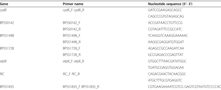
PCR Master Mix (Applied Biosystems). All experiments were conducted three times, which yielded 9 measure-ments per gene (representing 3 technical replicates of 3 biological replicates) and the relative expression ratios were calculated using REST-MCS [61]. The gene coding for cytochrome d ubiquinol oxidase subunit II, cydB, was used as the reference gene for normalization. CydB was chosen as the reference gene because it did not show any significant changes in expression in the microarray experi-ment. Primers are described in Table 3. The correlation between expression ratios obtained from the microarray and real-time qPCR was evaluated with Pearson correl-ation (Microsoft Office Excel).
Additional file
Additional file 1: List of 1259 common down-regulated genes of B. pseudomallei during intracellular growth in host macrophages relative to in vitro growth.
Competing interests
The authors declare that they have no competing interests
Authors’ contributions
SC contributed to the design of the study, performed all the experiments and data analysis and wrote the manuscript. LC contributed to the microarray interpretation and analysis and helped in performing real-time quantitative PCR. SN conceived and coordinated the study, contributed to the experimental design and wrote the manuscript. All authors read and approved the final manuscript.
Acknowledgements
This project was funded by the Ministry of Science, Technology and Innovation of Malaysia under the R&D Initiatives Grant Program awarded to S.N. We are grateful to the RNA Biology Laboratory and the Microarray Facility of the University of Aveiro for assistance in microarray data analysis, and to the Electron Microscopy Unit of Universiti Kebangsaan Malaysia and Universiti Putra Malaysia for assistance in electron microscopy.
Author details
1School of Biosciences and Biotechnology, Faculty of Science and
Technology, Universiti Kebangsaan Malaysia, Bangi 43600, Malaysia.2CESAM and Departamento de Biologia, Universidade de Aveiro, Campus de Santiago, Aveiro 3810-193, Portugal.
Received: 18 October 2011 Accepted: 23 July 2012 Published: 23 July 2012
References
1. White NJ: Melioidosis. Lancet 2003, 361:1715–1722.
2. Brett PJ, Woods DE: Pathogenesis of and immunity to melioidosis. Acta Trop 2000, 74:201–210.
3. Cheng AC, Currie BJ: Melioidosis: epidemiology, pathophysiology, and management. ClinMicrobiol Rev 2005, 18(2):383–416.
4. Currie BJ, Fisher DA, Anstey NM, Jacups SP: Melioidosis: acute and chronic disease, relapse and re-activation. Trans R Soc Trop Med Hyg 2000, 94:301–304.
5. Jenney AW, Lum G, Fisher DA, Currie BJ: Antibiotic susceptibility of Burkholderia pseudomallei from tropical northern Australia and implications for therapy of melioidosis. Int J Antimicrob Agents 2001, 17 (2):109–113.
6. Wiersinga WJ, van der Poll T, White NJ, Day NP, Peacock SJ: Melioidosis: insights into the pathogenicity of Burkholderia pseudomallei. Nat Rev Microbiol 2006, 4(4):272–282.
Table 3 List of oligonucleotides used in real-time qPCR experiments
Gene Primer name Nucleotide sequence (5’- 3’)
cydB cydB_F cydB_R GATCCGAAGAGCAGCC
CAGCCCGTGTAGAGCAG BPSS0142 BPSS0142_F ACCGATAACCTGTTCCG BPSS0142_R CGTAGATTTCCGCCATC BPSS1498 BPSS1498_F TCAAGGTCAAAGGAAAAAC BPSS1498_R AAGGCGAGGATGTGGAT BPSS1728 BPSS1728_F AGAGCCGCCAAGATCAA BPSS1728_R GCCGAGACCCGAGTTAT
atpB atpB_F atpB_R GTGGCTTTAACGATATGGC
TGATGCGAGGTGGAGAA
fliC fliC_F fliC_R CAGACGAACTACAACGGC
ATGCTTTGCGTGAGGTC
7. Harley VS, Dance DAB, Tovey G, McCrossan MV, Drasar BS: An ultrastructural study of the phagocytosis of Burkholderia pseudomallei. Microbios 1998, 94:35–45.
8. Jones AL, Beveridge TJ, Woods DE: Intracellular survival of Burkholderia pseudomallei. Infect Immun 1996, 64(3):782–790.
9. Inglis TJJ, Rigby P, Robertson TA, Dutton NS, Henderson M, Chang BJ: Interaction between Burkholderia pseudomallei and Acanthamoeba species results in coiling phagocytosis, endamebic bacterial survival, and escape. Infect Immun 2000, 68(3):1681–1686.
10. Stevens MP, Friebel A, Taylor LA, Wood MW, Brown PJ, Hardt WD, Galyov EE: A Burkholderia pseudomallei type III secreted protein, BopE, facilitates bacterial invasion of epithelial cells and exhibits guanine nucleotide exchange factor activity. J Bacteriol 2003, 185(16):4992–4996. 11. Kespichayawattana W, Rattanachetkul S, Wanun T, Utaisincharoen P,
Sirisinha S: Burkholderia pseudomallei induces cell fusion and actin-associated membrane protrusion: a possible mechanism for cell-to-cell spreading. Infect Immun 2000, 68(9):5377–5384.
12. Stevens MP, Galyov EE: Exploitation of host cells by Burkholderia pseudomallei. Int J Med Microbiol 2004, 293:549–555.
13. Stevens MP, Wood MW, Taylor LA, Monaghan P, Hawes P, Jones PW, Wallis TS, Gaylov EE: An Inv/Mxi-Spa like type III protein secretion system in Burkholderia pseudomallei modulates intracellular behaviour of the pathogen. Mol Microbiol 2002, 46(9):649–659.
14. Suparak S, Kespichayawattana W, Haque A, Easton A, Damnin S,
Lertmemongkolchai G, Bancroft GJ, Korbsrisate S: Multinucleated giant cell formation and apoptosis in infected host cells is mediated by Burkholderia pseudomallei type III secretion protein BipB. J Bacteriol 2005, 187(18):6556–6560.
15. Chen Y, Wong J, Sun GW, Liu Y, Tan GG, Gan Y: Regulation of type VI secretion system during Burkholderia pseudomallei infection. Infect Immun 2011, 79(8):3064–3073.
16. Ma AT, McAuley S, Pukatzki S, Mekalanos JJ: Translocation of a Vibrio cholerae type VI secretion effector requires bacterial endocytosis by host cells. Cell Host Microbe 2009, 5(3):234–243.
17. Mougous JD, Cuff ME, Raunser S, Shen A, Zhou M, Gifford CA, Goodman AL, Joachimiak G, Ordoñez CL, Lory S, Walz T, Joachimiak A, Mekalanos JJ: A virulence locus of Pseudomonas aeruginosa encodes a protein secretion apparatus. Science 2006, 312:1526–1530.
18. Shalom G, Shaw JG, Thomas MS: In vivo expression technology identifies a type VI secretion system locus in Burkholderia pseudomallei that is induced upon invasion of macrophages. Microbiology 2007, 153:2689–2699. 19. Pilatz S, Breitbach K, Hein N, Fehlhaber B, Schulze J, Brenneke B, Eberl L,
Steinmetz I: Identification of Burkholderia pseudomallei genes required for the intracellular life cycle and in vivo virulence. Infect Immun 2006, 74 (6):3576–3586.
20. Holden MTG, Titball RW, Peacock SJ, Cerdeño-Tárraga AM, Atkins T, Crossman LC, Pitt T, Churcher C, Mungall K, Bentley SD, Sebaihia M, Thomson NR, Bason N, Beacham IR, Brooks K, Brown KA, Brown NF, Challis GL, Cherevach I, Chillingworth T, Cronin A, Crossett B, Davis P, DeShazer D, Feltwell T, Fraser A, Hance Z, Hauser H, Holroyd S, Jagels K, Keith KE, Maddison M, Moule S, Price C, Quail MA, Rabbinowitsch E, Rutherford K, Sanders M, Simmonds M, Songsivilai S, Stevens K, Tumapa S, Vesaratchavest M, Whitehead S, Yeats C, Barrell BG, Oyston PCF, Parkhill J: Genomic plasticity of the causative agent of melioidosis, Burkholderia pseudomallei. Proc Natl Acad Sci USA 2004, 101(39):14240–14245. 21. Sun GW, Lu J, Pervaiz S, Cao WP, Gan YH: Caspase-1 dependent
macrophage death induced by Burkholderia pseudomallei. Cell Microbiol 2005, 7(10):1447–1458.
22. Harley VS, Dance DAB, Drasar BS, Tovey G: Effects of Burkholderia pseudomallei and other Burkholderia species on eukaryotic cells in tissue culture. Microbios 1998, 96:71–93.
23. Waddell SJ, Burcher PD, Stoker NG: RNA profiling in host-pathogen interactions. Curr Opin Microbiol2007, 10:297–302.
24. Du Y, Lenz J, Arvidson CG: Global gene expression and the role of sigma factors in Neisseria gonorrhoeae in interactions with epithelial cells. Infect Immun 2005, 73(8):4834–4845.
25. Grifantini R, Bartolini E, Muzzi A, Draghi M, Fringimelica E, Berger J, Ratti G, Petracca R, Galli G, Agnusdei M, Giuliani MM, Santini L, Brunelli B, Tettelin H, Rappuoli R, Randazzo F, Grandi G: Previously unrecognised vaccine candidates against group B meningococcus identified by DNA microarrays. Nat Biotechnol 2002, 20:914–921.
26. Trijbels F, Vogels GD: Allantoicase and ureidoglycolase in Pseudomonas and Penicillium species. BiochimBipphysActa 1966, 118(2):387–395. 27. Sun GW, Chen Y, Liu Y, Tan GYG, Ong C, Tan P, Gan YH: Identification of a
regulatory cascade controlling type III secretion system 3 gene expression in Burkholderia pseudomallei. Mol Microbiol 2010, 76(3):677–689.
28. Subsin B, Thomas MS, Katzenmeier G, Shaw JG, Tungpradabkul S, Kunakorn M: Role of the stationary growth phase sigma factor RpoS of
Burkholderia pseudomallei in response to physiological stress conditions. J Bacteriol 2003, 185(23):7008–7014.
29. Jangiam W, Loprasert S, Tungpradabkul S: Role of Burkholderia pseudomallei RpoS in regulation of catalase activities under hydrogen peroxide induction. Science Asia 2008, 34(1):23–29.
30. Dorman CJ: DNA supercoiling and environmental regulation of gene expression in pathogenic bacteria. Infect Immun 1991, 59(3):745–749. 31. Drlica K: Control of bacterial DNA supercoiling. Mol Microbiol 1992, 6
(4):425–433.
32. Schneider R, Travers A: The expression of the Escherichia coli fis gene is strongly dependent on the superhelical density of DNA. Mol Microbiol 2000, 38(1):167–175.
33. Rothfield LI, Shih YL, King G: Polar explorers: membrane proteins that determine division site placement. Cell 2001, 106(1):13–16. 34. Wand ME, Muller CM, Titball RW, Michell SL: Macrophage and Galleria
mellonela infection models reflect the virulence of naturally occurring isolates of B. pseudomallei, B. thailandensis and B. oklahomensis. BMC Microbiol 2011, 11:11.
35. Tuanyok A, Tom M, Dunbar J, Woods DE: Genome-wide expression analysis of Burkholderia pseudomallei infection in a hamster model of acute melioidosis. Infect Immun 2006, 74(10):5465–5476.
36. Boyce JD, Wilkie I, Harper M, Paustian ML, Kapur V, Adler B: Genomic scale analysis of Pasteurella multocida gene expression during growth within natural chicken host. Infect Immun 2002, 70(12):6871–6879.
37. Lucchini S, Liu H, Jin Q, Hinton JCD, Yu J: Transcriptional adaptation of Shigella flexneri during infection of macrophages and epithelial cells: insights into the strategies of a cytosolic bacterial pathogen. Infect Immun 2005, 73(1):88–102.
38. Chatterjee SS, Hossain H, Otten S, Kuenne C, Kuchmina K, Machata S, Domann E, Chakraborty T, Hain T: Intracellular gene expression profile of Listeria monocytogenes. Infect Immun 2006, 74(2):1323–1338.
39. Pahan K, Sheikh FG, Namboodiri AM, Singh I: Lovastatin and phenyl acetate inhibit the induction of nitric oxide synthase and cytokines in rat primary astrocytes, microglia, and macrophages. J Clin Investig 1997, 100:2671–2679.
40. Park JS, Lee EJ, Lee JC, Kim WK, Kim HS: Anti-inflammatory effects of short chain fatty acids in IFN-gamma-stimulated RAW 264.7 murine macrophage cells: involvement of NF-kappaB and ERK signaling pathways. Int Immunopharmacol 2007, 7:70–77.
41. Utaisincharoen P, Tangthawornchaikul N, Kespichayawattana W, Chaisuriya P, Sirisinha S: Burkholderia pseudomallei interferes with inducible nitric oxide synthase (iNOS) production: a possible mechanism of evading macrophage killing. Microbiol Immunol 2001, 45(4):307–313. 42. Law RJ, Hamlin JNR, Sivro A, McCorrister SJ, Cardama GA, Cardona ST: A
functional phenylacetic acid catabolic pathway is required for full pathogenicity of Burkholderia cenocepacia in the Caenorhabditis elegans host model. J Bacteriol 2008, 190(21):7209–7218.
43. Warawa J, Woods DE: Type III secretion system cluster 3 is required for maximum virulence of Burkholderia pseudomallei in a hamster infection model. FEMS Microbiol Lett 2005, 242:101–108.
44. Schell MA, Ulrich RL, Ribot WJ, Brueggemann EE, Hines HB, Chen D, Lipscomb L, Kim HS, Mrázek J, Nierman WC, Deshazer D: Type VI secretion is a major virulence determinant in Burkholderia mallei. Mol Microbiol 2007, 64(6):1466–1485.
45. Burtnick MN, Brett PJ, Harding SV, Ngugi SA, Ribot WJ, Chantratita N, Scorpio A, Milne TS, Dean RE, Fritz DL, Peacock SJ, Prior JL, Atkins TP, DeShazer D: The cluster 1 type VI secretion system is a major virulence determinant in Burkholderia pseudomallei. Infect Immun 2011, 79(4):1512–1525.
46. Stevens MP, Stevens JM, Jeng RL, Taylor LA, Wood MW, Hawes P, Monaghan P, Welch MD, Galyov EE: Identification of a bacterial factor required for actin-based motilityof Burkholderia pseudomallei. Mol Microbiol 2005, 56(1):40–53.
47. Jacob-Dubuisson F, Buisine C, Mielcarek N, Clément E, Menozzi FD, Locht C: Amino-terminal maturation of the Bordetella pertussis filamentous hemagglutinin. Mol Microbiol 1996, 19(1):65–78.
48. Abramson T, Kedem H, Relman DA: Proinflammatory and proapoptotic activities associated with Bordetella pertussis filamentous hemagglutinin. Infect Immun 2001, 69(4):2650–2658.
49. Dowling AJ, Wilkinson PA, Holden MT, Quail MA, Bentley SD, Reger J, Waterfield NR, Titball RW, Ffrench-Constant RH: Genome-wide analysis reveals loci encoding anti-macrophage factors in the human pathogen Burkholderia pseudomallei K96243. PLoS One 2011, 5(12):e15693. 50. Tolman JS, Valvano MA: Global changes in gene expression by the
opportunistic pathogen Burkholderia cenocepacia in response to internalization by murine macrophages. BMC Genomics 2012, 13:63. 51. Hayashi F, Smith KD, Hawn TR, Yi EC, Goodlett DR, Eng JK, Akira S, Underhill
DM, Aderem A: The innate immune response to bacterial flagellin is mediated by Toll-like receptor 5. Nature 2001, 410:1099–1103. 52. Feterl M, Govan BL, Ketheesan N: The effect of different Burkholderia
pseudomallei isolates of varying levels of virulence on toll-like-receptor expression. Trans R Soc Trop Med Hyg 2008, 102(Suppl 1):S82–S88. 53. Chin CY, Monack DM, Nathan S: Genome wide transcriptome profiling of
a murine acute melioidosis model reveals new insights into how Burkholderia pseudomallei overcomes host innate immunity. BMC Genomics 2010, 11:672.
54. Nanagara R, Vipulakorn K, Suwannaroj S, Schumacher HRJ: Atypical morphological characteristics and surface antigen expression of Burkholderia pseudomallei in naturally infected human synovial tissues. Mod Rheumatol 2000, 10:129–136.
55. Loprasert S, Whangsuk W, Sallabhan R, Mongkolsuk S: DpsA protects the human pathogen Burkholderia pseudomallei against organic hydroperoxide. Arch Microbiol 2004, 182(1):96–101.
56. Loprasert S, Sallabhan R, Whangsuk W, Mongkolsuk S: Compensatory increase in ahpC gene expression and its role in protecting Burkholderia pseudomallei against reactive nitrogen intermediates. Arch Microbiol 2003, 180(6):498–502.
57. Lee SH, Chong CE, Lim BS, Chai SJ, Sam KK, Mohamed R, Nathan S: Burkholderia pseudomallei animal and human isolates from Malaysia exhibit different phenotypic characteristics. Diagn Microbiol Infect Dis 2007, 58(3):263–270.
58. Abu Kwaik Y, Eisenstein BI, Engleberg NC: Phenotypic modulation by Legionella pneumophila upon infection of macrophages. Infect Immun 1993, 61(4):1320–1329.
59. Bergman NH, Anderson EC, Swenson EE, Janes BK, Fisher N, Niemeyer MM, Miyoshi AD, Hanna PC: Transcriptional profiling of Bacillus anthracis during infection of host macrophages. Infect Immun 2007, 75(7):3434–3444.
60. Dennis G Jr, Sherman BT, Hosack DA, Yang J, Gao W, Lane HC, Lempicki RA: DAVID: database for annotation, visualization, and integrated discovery. Genome Biol 2003, 4(5):P3.
61. Pfaffl MW, Horgan GW, Dempfle L: Relative expression software tool (REST©) for group-wise comparison and statistical analysis of relative expression results in real-time PCR. Nucleic Acids Res 2002, 30(9):e36. doi:10.1186/1471-2164-13-328
Cite this article as: Chieng et al.: Burkholderia pseudomallei
transcriptional adaptation in macrophages. BMC Genomics 2012 13:328.
Submit your next manuscript to BioMed Central and take full advantage of:
• Convenient online submission
• Thorough peer review
• No space constraints or color figure charges
• Immediate publication on acceptance
• Inclusion in PubMed, CAS, Scopus and Google Scholar
• Research which is freely available for redistribution
Submit your manuscript at www.biomedcentral.com/submit