Clínica Universitária de Medicina II
Prognostic value of C-Reactive Protein
during moderate to severe ARDS
João Manuel Moniz
Clínica Universitária de Medicina II
Prognostic value of C-Reactive Protein
during Moderate to Severe ARDS
João Manuel Moniz
Orientado por:
Doutora Susana Fernandes
3 RESUMO
Introdução: Apesar da melhoria do suporte de órgão oferecido a doentes críticos, a
Síndrome de Dificuldade Respiratória Aguda (ARDS) continua a estar associada a elevada mortalidade. Porém, continuam a não existir marcadores que possam prever prognóstico ou subgrupos claros de doentes que permitam modular o tipo de terapêutica efetuada. Dados recentes têm associado o fenótipo hiperinflamatório a pior prognóstico. Torna-se assim essencial desenvolver novos modelos de prognóstico em contexto de ARDS. A Proteína C-Reativa (PCR) é um marcador de inflamação medido diariamente na prática clínica.
Objetivos: Foi objetivo deste trabalho estabelecer a utilidade de medições de PCR nos
primeiros dias de ARDS e a sua relação com prognóstico. Pretendia-se ainda identificar, noutros marcadores avaliados diariamente, uma possível relação com prognóstico.
Métodos: Realizou-se um estudo retrospetivo de doentes com ARDS moderado a grave,
tratados na Unidade de Cuidados Intensivos do Hospital de Santa Maria. Foram colhidos dados demográficos, laboratoriais e de parâmetros ventilatórios. Foi efetuada uma análise descritiva, assim como de regressão logística para identificação de eventuais fatores relacionados com prognóstico.
Resultados: Foram incluídos 201 doentes na análise, dos quais 38.8% tinham ARDS
grave. O valor médio da PCR nos 3 primeiros dias não diferiu entre os 3 dias, nem se relacionou com a mortalidade, embora existisse uma tendência, estatisticamente não significativa, para os doentes que morreram terem valores mais elevados. Adicionalmente, foram criados 3 grupos diferentes de acordo com a cinética da PCR nas primeiras 48 horas (aumento, diminuição ou valor estável), sendo que nenhuma das cinéticas se relacionava com a mortalidade. De forma significativa, o rácio entre PCR/albumina no primeiro dia de ARDS relacionou-se diretamente com a mortalidade hospitalar (odds ratio de 1.03, P=0.04), mesmo quando ajustado para a gravidade e etiologia. Dos restantes parâmetros clínicos e laboratoriais avaliados nenhum se relacionou com a mortalidade.
Conclusões: Neste estudo retrospetivo de doentes com ARDS, o ratio PCR/albumina no
primeiro dia de internamento foi a única variável que se relacionou com a mortalidade. Embora seja um marcador de fácil aplicabilidade, a sua utilização deverá ser validada noutras coortes de doentes.
4 ABSTRACT
Introduction: Despite the improvement in organ support available for critical patients,
Acute Respiratory Distress Syndrome (ARDS) is still associated with high mortality rates. There are no markers that can predict ARDS prognosis or definitive subgroups that allow physicians to adapt treatment. Recent data has shown that an hyperinflammatory phenotype might be associated with a worse outcome, but this state is not easy to assess at bed-side. C-reactive protein (CRP) is an inflammatory marker that is used on daily clinical practice and might reflect this phenotype.
Objectives: The main aim of this study was to establish the value of CRP determination
on the first days of ARDS as a prognostic marker.
Methods: We did a retrospective study with moderate to severe ARDS patients treated at
the Intensive Care Unit of Hospital de Santa Maria. Demographic, laboratory and ventilatory data was gathered. Using this data, a descriptive analysis was done, as well as a logistic regression to identify possible prognosis factors.
Results: For the main analysis, 201 patients were included, of which 38,8% had severe
ARDS. The mean value of CRP in the first three days of ARDS did not differ significantly, nor was it correlated with mortality, although there was a tendency for patients who died to have higher CRP levels. Additionally, three groups were created according to the CRP kinetics during the first 48 hours (increased, decreases, maintained). None of these groups was successfully correlated with mortality. Significantly, CRP/albumin ratio on the first day of ARDS was positively correlated with hospital mortality (odds ratio of 1.03, P=0.04). This result was still significant even when adjusted for severity and etiology. None of the remaining clinical or laboratory parameters showed any correlation with mortality, namely age, gender, obesity, cause of ARDS or severity.
Conclusion: In this retrospective study, CRP/albumin ratio on the first day was the only
variable found to be successfully correlated with mortality. This ratio, as an indicator of systemic inflammation, might be applicable in the near future, separating inflammatory from non-inflammatory phenotype helping in the definition of prognostic groups.
5 ABBREVIATIONS
ARDS – Acute respiratory distress syndrome
SMI – Serviço de Medicina Intensiva
ICU – Intensive Care Unit
HSM – Hospital de Santa Maria
SOFA – Sequential Organ Failure Assessment
SAPSII – Simplified Acute Physiology Score II
ECMO – Extracorporeal Membrane Oxygenation
TC – Tomografia Computorizada
VILI – ventilator induced lung injury
𝑃𝑎𝑂2 – arterial pressure of oxygen
𝐹𝑖𝑂2– fraction of inspired oxygen
P/F - arterial pressure of oxygen/ fraction of inspired oxygen ratio
CRP – C-reactive protein
PEEP – Positive end expiratory pressure
TV – Tidal volume
PP - Prone position
DAD - Diffuse alveolar damage
6
Valor prognóstico da Proteína C-Reativa durante ARDS
moderado e grave
IntroduçãoA Síndrome da Dificuldade Respiratória Aguda (ARDS) é uma doença inflamatória do pulmão secundária a infeções respiratórias, sépsis, trauma, pancreatite, entre outros. É diagnosticada com base nos critérios de Berlim que incluem: início agudo com início até 7 dias após um insulto, infiltrados bilaterais em radiografia ou Tomografia Computorizada (TC) de tórax, P/F < 300 em doentes com PEEP > 5 cm𝐻2𝑂 e ausência de falência de ventrículo esquerdo. Afeta cerca de 10% dos doentes nos serviços de cuidados Intensivos, com mortalidades que chegam a 40% nas formas mais graves.
Não existe um tratamento específico ou terapia dirigida para ARDS pelo que a abordagem consiste essencialmente em suporte de órgão e tratamento dirigido à causa original. Inclui ventilação mecânica protetora e em casos mais graves, decúbito ventral, bloqueio neuromuscular, restrição de fluidos e oxigenação extracorporal por membrana (ECMO).
Em termos de prognóstico, não existem marcadores que consigam prever a mortalidade ou modular o tratamento destes doentes, sendo essencial a pesquisa de novas variáveis. Alguns grupos de marcadores já foram estudados, mas a sua utilização não é prática na actividade clínica diária:
• Marcadores endoteliais: VEGF, VWF e angiopoietina 2 correlacionam-se com a extensão do dano endotelial, causas indiretas de ARDS e maior mortalidade.
• Marcadores epiteliais: a lesão alveolar aumenta as concentrações séricas de SP-D, relacionado com causas diretas de ARDS, sem relação com a mortalidade.
• Marcadores inflamatórios: LTB4, IL-6, IL-8 estão associados a mau prognóstico. Alguns marcadores do dia-a-dia podem ser bons preditores de mortalidade, nomeadamente a albumina e a LDH. Valores diminuídos de albumina, especialmente se < 20g/L, traduzem pior prognóstico, tal como valores elevados de LDH foram associados a uma maior mortalidade em 28 dias.
7
O grupo Calfee sugeriu recentemente a divisão de grupos fenotípicos de ARDS, baseando-se em variáveis clínicas e laboratoriais. Foram, assim, identificados 2 grupos:
• Tipo 1, hipoinflamatório: mais frequentemente causado por trauma, beneficiava de PEEPs menores e com menor mortalidade.
• Tipo 2, hiperinflamatório: maior concentração de marcadores de inflamação (IL6, IL8, PAI-1), maior frequência cardíaca, frequência respiratória, causas indiretas de ARDS (eg: sépsis), uso de vasopressores, menor pressão arterial e bicarbonato. Este fenótipo associou-se a maior mortalidade, menos dias livres de ventilação e dias sem falência de órgão.
A proteína C reativa (PCR) é frequentemente usada na prática clínica como marcador de estado inflamatório, sendo pertinente o seu estudo como fator prognóstico. É uma proteína de fase aguda produzida em resposta a citocinas, principalmente Il-6. Apesar de estudada anteriormente, o seu valor continua incerto no ARDS, havendo estudos que demonstram que PCR elevada nas primeiras 48h se associa a bom prognóstico, enquanto que outros estudos referem a sua associação com um fenótipo hiperinflamatório e, por isso, a pior prognóstico. Perante o exposto, a nossa hipótese é que os níveis de PCR nas primeiras 72h podem ser usados para prever prognóstico.
Métodos
Foi realizado um estudo retrospetivo no Serviço de Medicina Intensiva (SMI) do Hospital de Santa Maria (HSM). Foram identificados os doentes com ARDS moderado a grave entre dezembro de 2012 e dezembro de 2017. O diagnóstico de ARDS foi confirmado pela nota de alta dos doentes ou verificado quando P/F < 200 com PEEP > 5cm𝐻2𝑂, com um insulto nos 7 dias anteriores e sem falência ventricular esquerda. Não foram revistos exames de imagem destes doentes.
Foram colhidos dados demográficos assim como variáveis clínicas, laboratoriais e ventilatórias. Foram excluídos doentes sem critérios de ARDS moderado ou grave, doentes com morte antes das 24h e, na análise final, doentes com menos de 3 determinações de PCR. Também foram excluídos doentes com alguns dados em falta.
8
O dia 1 de ARDS foi considerado quando os doentes cumpriram critérios de ARDS moderado. O endpoint primário foi a mortalidade no SMI e os endpoints secundários foram a mortalidade hospitalar e dias livres de ventilação mecânica.
O STATA foi usado para análise descritiva e, para a análise comparativa, foi feito o t-test ou 𝑥2 ou testes não paramétricos, conforme a variável. O odds ratio e intervalo de confiança foram calculados e foi considerado significativo um p-value 0,05.
Resultados
Foram incluídos 201 doentes com diagnóstico de ARDS, independentemente da etiologia. Dos 245 avaliados, foram excluídos 44. Da amostra, 134 eram homens, com idade media de 60, IMC de 28,1; SOFA na admissão de 9 e SAPSII de 46. A causa mais comum foi sépsis, em 59% dos doentes e a segunda trauma, em 13% das admissões. O tempo médio de internamento até diagnóstico de ARDS foi 1,49 dias. Dos casos avaliados, 78 eram moderados e 123 graves. O número de dias sobre ventilação mecânica foi 13.
Dos doentes incluídos, a média de PCR no primeiro dia foi de 22.0 ± 13.0 mg/dL, no dia 2 de 24 ±13.4 mg/dL, e no dia 3 de 22.5 ±14.8mg/dL. Não houve diferença significativa nas médias durante estes 3 dias (p = 0,05). A análise da variação de PCR, durante as primeiras 72h, demonstrou que 25 doentes tinham redução de, pelo menos, 40%, 99 não tinham variação significativa e 42 tinham aumento de 40%.
Foi feita uma descrição detalha das características demográficas dos doentes, com distribuição por morte ao terceiro dia e categorias de PCR. Destaca-se que a maioria dos doentes sem alterações significativas de PCR eram doentes de trauma, com tempo até diagnóstico de ARDS significativamente superior.
Da análise dos parâmetros laboratoriais, destaca-se que os doentes que tiveram aumento da PCR nos primeiros dias partiram de valores inicialmente mais baixos e com albumina mais alta, sugerindo menor inflamação. Os níveis de PCR, após uma semana, não diferiram significativamente. A PCR isoladamente não variou significativamente de acordo com a mortalidade dos doentes. A variação da PCR também não apresentou essa correlação. O rácio PCR/Albumina no primeiro dia, relacionou-se positivamente com a mortalidade. Das variáveis clínicas colhidas, nenhuma foi positivamente associada com a mortalidade dos doentes.
9 Discussão
Este estudo incluiu 201 doentes com diagnóstico de ARDS, com a mortalidade global de 43,8%. A PCR, por si só, ou a sua variação nas primeiras 48 horas não se revelaram bons indicadores de mortalidade. No entanto, quando analisado o rácio PCR/Albumina no dia 1, existiu uma associação significativa com a mortalidade. Este rácio já foi previamente estudado e positivamente correlacionado com a mortalidade em doentes críticos, sendo um melhor preditor de estado inflamatório do que a PCR ou albumina isoladamente. Salienta-se ainda que o valor de PCR não se associou a nenhuma etiologia, embora doentes com trauma estivessem concentrados no grupo em que não se verifica cinética da mesma nas primeiras 48 horas. Apesar da associação noutros estudos entre sépsis e mortalidade em doentes com ARDS, nesta coorte essa associação não foi estabelecida. Das variáveis demográficas analisadas (idade, sexo, IMC, SOFA, SAPSII), nenhuma delas demonstrou relação com a mortalidade, apesar de serem fatores previamente descritos noutros estudos. Isto pode dever-se a um efeito relativamente pequeno sobre a mortalidade ou por deficiente amostra, com número de doentes insuficiente para demonstrar o efeito.
Das medidas terapêuticas consideradas cruciais nestes doentes, o volume corrente (VC) e bloqueio neuromuscular não se associaram à mortalidade. Isto pode ser devido ao desenho de estudo, que não foi esquematizado para demonstrar este efeito, ou ainda dever-se ao facto de que os tratamentos foram aplicados apenas em doentes que deles beneficiavam. Também o uso de vasopressores foi previamente associado a maior mortalidade, o que não se verificou neste estudo.
Das limitações do estudo, é de referir ser um estudo retrospetivo, realizado no mesmo centro hospitalar, não podendo ser excluído um viés de informação. Para o diagnóstico de ARDS, não foram considerados os critérios imagiológicos, podendo ter sido incluídos doentes com outras patologias respiratórias. Por outro lado, mesmo quando são aplicados de forma estrita os critérios de ARDS, existe um subgrupo significativo de doentes em que não é efetuado diagnóstico histológico de lesão alveolar difusa posteriormente. Assim se pode explicar a heterogeneidade de resultados em doentes com esta síndrome. Por fim, dado tratar-se de uma doença de evolução muito rápida, é extremamente difícil definir com rigor absoluto o dia 1 de ARDS, o que pode enviesar conclusões sobre cinética ou mesmo sobre o valor de PCR no primeiro dia.
10
Concluindo, ARDS continua a ser uma síndrome complexa e os conhecimentos sobre esta patologia estão longe de ser completos. No futuro, seria relevante poder fazer um novo estudo, prospetivo, com maior população, com melhor protocolo de colheita de dados para minimizar erros e maximizar o poder estatístico do estudo. A PCR, apesar de ser prontamente usada, continua a ter um papel pouco claro no ARDS, não sendo suficiente para prever o prognóstico dos doentes. Por outro lado, o ratio PCR/albumina, como marcador mais específico de inflamação, poderá ter um papel relevante num futuro próximo em identificar doentes com fenótipo hiperinflamatório e assim definir grupos prognósticos.
11 INDEX
1. INTRODUCTION ... 12
2. METHODS ... 15
2.1. Subjects and data collection ... 15
2.2. Terms and measures ... 15
2.3. Statistical testing ... 16
3. RESULTS... 16
3.1. General description ... 16
3.2. Correlation with outcome ... 20
4. DISCUSSION ... 23
12 1. INTRODUCTION
Acute respiratory distress syndrome (ARDS) is an inflammatory disease of the lung that often occurs after seven days of an inflammatory stimulus such as pneumonia, influenza infection, sepsis, trauma, pancreatitis, among others. (1-4)It is characterized by an increase in pulmonary vascular permeability that causes a non-cardiogenic pulmonary edema. (1,2,5) Since 2012, its diagnosis is based on the Berlin criteria which include: acute onset, bilateral infiltrates on chest x-ray or CT-scan, hypoxemia with a P/F ratio < 300 in patients submitted to a PEEP of at least 5cm𝐻2𝑂, and no major evidence of left ventricular failure, which could explain by itself the pulmonary edema. (1,2,3)
Its physiopathology is characterized by three main phases: an initial exudative phase, with protein-rich pulmonary edema and loss of normal surfactant causing alveolar collapse; proliferative phase, with manifest disorganization of the parenchyma, necrosis and proliferation of both type I and II alveolar cell as well as fibroblasts which are needed to start the regenerative process. Lastly, the fibrotic phase, with an increase in total lung collagen, that might result in the scarring of the lung. (5,6)
There are no targeted therapies or specific treatments for ARDS and therefore treatment is mainly aimed at the causative event plus supportive care. (7,8)In general it includes protective mechanical ventilation, and for more severe cases prone position, neuromuscular blockade, fluid restriction, and extracorporeal membrane oxygenation (ECMO).(9-14)
ARDS is present in approximately 10% of ICU patients, with mortality rates reaching 40% in its most severe form. (2,15) Furthermore, even if patients survive, they can suffer from debilitating sequels such as pulmonary fibrosis, obstructive disease, diffusion restriction and neurocognitive disorders (insomnia, anxiety, cognitive decline). (16)
There is no optimal marker to predict prognosis or to modulate ARDS treatment which makes the study of new possible variables so essential. Some biomarkers for ARDS have already been associated with outcome. We can separate them in groups according to the type of marker, but they mainly include:
● Endothelial markers: higher values of Vascular Endothelial Growth Factor (VEGF), Von Willebrand Factor (VWF) and angiopoietin 2 correlate with a higher mortality. Hence, it could be inferred that a more extensive endothelial
13
lesion is responsible for a worse outcome. According to the Calfee group studies, these markers are associated with indirect causes of ARDS (eg: nonpulmonary sepsis, pancreatitis…).
● Alveolar epithelial markers: alveolar lesion increases the serum level of Surfactant Protein D (SP-D), indicating direct lung injury. SP-D has not associated been with a higher mortality rate.
● Inflammatory markers: such as LTB4, IL-6, IL-8 were linked with a worse outcome. (17-19)
It is also possible that every day markers have a role in predicting the prognosis of patients, specifically albumin and Lactate Dehydrogenase (LDH). Albumin is a negative acute phase protein and low levels of albumin in the first week after ARDS correlated with a lower chance of survival, especially when < 20g/L. Moreover, higher LDH levels were associated with higher 28 days mortality rates. (20) Procollagen III, a precursor of collagen, is another very promising biomarker in ARDS. Despite not being routinely used, it can be used as an indicator that the scarring process can begin fairly early in the pathophysiology of ARDS, predicting a worse outcome. (21)
Recently, Calfee et al suggested that different phenotype groups identified based on clinical and laboratory variables respond differently to support therapy, namely level of PEEP or fluid restriction.(22) They identified a hyperinflammatory phenotype (type 2) associated with higher levels of inflammatory biomarkers (IL6, IL8, PAI-1), higher heart rate and minute ventilation, higher use of vasopressors, lower systolic blood pressure, lower bicarbonate and higher prevalence of sepsis as the causative insult. Type 1 phenotype, the hypoinflammatory type, on the other hand, was more often caused by trauma, benefited from lower PEEP strategies and was associated with lower mortality rate. (23,24) When it comes to prognosis, the inflammatory phenotype is predisposed to a worse outcome with higher mortality rate and fewer ventilator and organ failure-free days. Of relevance, there was no single marker that could be used to promptly distinguish between these two phenotypes. Nonetheless, when used together, some clinical and laboratorial variables pointed for a specific phenotype. (22)
C-Reactive protein (CRP), being easily available in the daily practice and associated with an inflammatory state, would be a pertinent candidate to be addressed as a prognostic factor. (25,26) It is an acute phase protein produced chiefly by the liver in response to
14
cytokines, particularly Il-6 and IL-1. Previous studies have shown that the inflammatory cytokines responsible for CRP induction, namely Il-1, Il-6 and TNF-a are increased in ARDS. (27) The significance of increased CRP levels during ARDS remains uncertain, and in the limited studies available it has been associated with both better and worse outcomes. On the one hand, there have been studies that demonstrate that high RCP levels in the first 48h translate into a better outcome. (26) On the other hand, as an acute phase protein it has been associated with more inflammatory phenotypes which in turn, have been associated with a worse outcome. (19,22)
Consequently, there is still doubt about the utility of daily measurements of CRP during the first days of ARDS and if its levels can correlate with prognosis. Thus, we hypothesized that CRP levels during the first 72 hours of ARDS could be used to predict prognosis.
15 2. METHODS
2.1. Subjects and data collection
A non-interventive retrospective cohort study was made at the SMI of HSM. We identified patients discharged from the ICU with a moderate to severe ARDS diagnosis between December 2012 and December 2017.
We recorded data from all patients with the clinical diagnosis of ARDS based on discharge notes and recorded them in the SMI’s clinical database. It was then confirmed that the diagnosis was in the written record. In case it was not assumed in the clinical file, we confirmed if patients had a P/F < 200 with PEEP > 5cmH2O, without left ventricular dysfunction and if a clinical insult was present in the previous 7 days. For the purposes of this study, we did not review the imagological findings, assuming the evaluation made by the assistant physicians was correct.
We collected the following specific parameters from the patients: date of admission; days of mechanical ventilation; SOFA at admission; SAPS II; age; height; weight; sex; reason of admission; first diagnosis; day of diagnosis of ARDS; blood arterial gas (pH, bicarbonate, pCO2, lactates) from the day of ARDS diagnosis; P/F ratio; CRP on day 1, day 2, day 3 and day 8; procalcitonin; platelets; albumin; bilirubin; ventilatory settings (tidal volume, PEEP, respiratory rate); use and dosing of vasopressors; neuromuscular blockade; support with extracorporeal membrane oxygenation (ECMO).
We used as exclusion criteria all patients that had a length of stay shorter than 24 hours, patients who did not meet the criteria for moderate or severe ARDS and for the final analysis patients with less than 3 determinations of CRP. We also excluded patients that had crucial missing data (eg.: blood arterial gas data). We did not collect any data that could identify patients, maintaining our database anonymous.
2.2. Terms and measures
We used standard classification for moderate ARDS (P/F < 200) and severe ARDS (P/F < 100). We considered day 1 as the day when the patient first met at least moderate ARDS diagnosis criteria after ICU admission. The primary endpoint was ICU mortality and secondary endpoints were hospital mortality and mechanical ventilation free-days.
16 2.3. Statistical testing
We performed a statistical descriptive analysis using STATA (software of statistics and Data Science) and the comparative analysis was done using t-test or 𝑥2, or using non-parametric tests according to the variable and its distribution. In order to identify factors that could be relevant for the mortality and secondary endpoints we used a uni and multivariable logistic regression using a forward method.
The results were expressed with mean values ± standard deviation, median and interquartile ranges or percentage as applicable. The odd ratio and confidence interval at 95% were calculated, and a p value of less than 0,05 was considered significant.
3. RESULTS
3.1. General description
We included in this study 201 patients with the clinical diagnosis of ARDS admitted to the ICU of HSM, regardless of the etiology. From the 245 screened patients, 44 were excluded because of missing data (n = 4); deceased in less than 24h (n = 16); no moderate or severe ARDS criteria (n = 24). (Figure 1)
Figure 1 – Number of patients screened and included in the primary analysis. A total of 44 patients were
17
From the study’s population, 134 were male (66%), with a mean age of 60 (from 18 to 92); median BMI of 28,1 (24.4 – 31.6); median SOFA on admission of 9 (6-12); median SAPSII of 46 (34-63).
The most common cause for ARDS was sepsis (n = 119; 59%) and trauma, which was responsible for 13% of admissions. The mean time in the ICU until ARDS was 1.49 days ± 2.8, with most of the patients with an ARDS diagnosis established at the first day of ICU stay. Overall, 78 were moderate (38.8%) and 123 were severe (61.2%) with a median P/F of 88 (IQ range: 65-118). The mean number of days under mechanical ventilation was 13.1 ± 12 days, with a median of 9 days (4-19).
The patients included in this study had a mean CRP at day 1 of 22.0 ± 13.0 mg/dL, at day 2 of 24 ±13.4 mg/dL, and at day 3 of 22.5 ±14.8mg/dL. There was no significant difference in the means during these three days (P=0.05). Nevertheless, as there were patients with different CRP kinetics during the first 3 days, we chose to analyzed patients according to its variation in the first 72 hours. Decreased CRP by at least 40% was observed in 25 patients; 99 patients had no significant variation; and 42 increased CRP at least 40%.
Accordingly, a more detailed demographic description of the study population is shown in Table 1, with distributions sorted by death by day 3 and CRP categories. Of note, patients with no change on CRP were mainly trauma patients and spent significantly longer in the ICU until ARDS criteria were met.
18 Table 1. Patient characteristics divided by CRP kinetics and death at day 3
Total Death at day 3 CRP categories P Decrease in 48h No variation Increased in 48 h N 201 35 25 99 42 Age, years 60 (18-92) 66 (54-74) 60 (45-68) 57 (45-70) 59 (48-68) 0.57 Male Gender 66% 69% 68% 72% 47% 0.79 BMI 28.1 (24.4-31.6) 27.5 (25.4-31.1) 28.7 (24.1-33.6) 28.3 (23.5-31.6) 26.6 (24.4-31.6) 0.29 SAPSII 46 (34-63) 72 (56-96) 42 (32-51) 44 (33-53) 45 (34-56) 0.61 First SOFA 9 (6-12) 14 (10-16) 8 (5-11) 8 (6-11)) 9 (7-11) 0.51 Sepsis, % 59 57 76 56 57 0.07 Lung infection, n (%) 80 (67) 7 (35%) 14 (73.6%) 38 (68%) 21 (88%) 0.19 Trauma 13% 2.8% 0% 19% 2.9% 0.02
Time to ARDS (days) 1.49 ± 2.8 0.94 ±1.6 1.48 ± 4.8 1.66 ±2.4 1.5 ±2.6 < 0.001
PaO2/FiO2 88 (65-118) 77 (61-105) 84 (70-114) 91 (65-125) 85 (68-112) 0.27 Vt/IBW 6.5 (5.7-7.4) 7.1 (6.5-7.9) 5.9 (5.4-6.9) 6.5 (5.7-7.3) 6.1 (5.4-7.1) 0.28 Neuromuscular blockage, n (%) 109 (55.9) 13 (39.4) 21 (84) 49 (51) 26 (63) < 0.001 ECMO 16% 0 % 32% 16% 21% 0.09 Vasopressor usage 146 (72.6) 35 (100) 19 (76) 61 (61) 31 (74) 0.22 Noradrenaline dose 0.5 (0.2-1.4) 1.7 (0.7-2.3) 0.3 (0.2-0.7) 0.4 (0.2-1.0) 0.4 (0.2-0.8) 0.52 BMI – body mass index; Medians and interquartile range are plotted for age, BMI, SAPSII, fist SOFA, and PaO2/FiO2. Mean ± standard deviation is plotted for time in the ICU until development of ARDS. Categories were based on variation > 40%. P values refers to test using ANOVA comparing the differences between CRP kinetics categories.
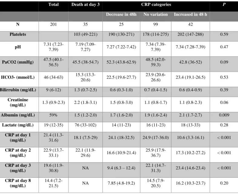
We then analyzed the different laboratory parameters, according to the category of CRP. Of note, patients who had increased CRP in the first days of ARDS had lower levels
19
initially, as well as higher albumin levels suggesting that ARDS was diagnosis in a less inflammatory environment. Furthermore, CRP levels after one week, were not statistically different between groups regardless of the previous kinetics.
Table 2 - Laboratory variables during the first 48h
Total Death at day 3 CRP categories P
Decrease in 48h No variation Increased in 48 h
N 201 35 25 99 42 Platelets 103 (49-221) 190 (130-271) 178 (114-275) 202 (147-288) 0.59 pH 7.31 (7.23-7.39) 7.19 (7.09-7.27) 7.27 (7.22-7.42) 7.34 (7.39-7.39) 7.34 (7.28-7.39) 0.47 PaCO2 (mmHg) 47.5 (40.1-56.5) 45.5 (38-54.7) 52.3 (43.8-62.9) 48.5 (42.0-59.3) 42.8 (36-52) 0.09 HCO3- (mmol/L) 46 (34-63) 15.3 (13.3-20.6) 22.5 (19.6-27.7) 23.9 (20.6-26.6) 23.4 (19.1-26.5) 0.53 Bilirrubin (mg/dL) 9 (6-12) 1.3 (0.7-2.5) 0.6 (0.3-1.0) 0.7 (0.4-1.5) 0.6 (0.4-0.9) 0.39 Creatinine (mg/dL) 1.3 (0.9-2.3) 2.2 (1.8-3.1) 1.5 (0.8-3.0) 1.1 (0.8-1.7) 1.1 (0.8-2.3) 0.06 Albumin (mg/dL) 59% 1.5 (1.2-2.0) 1.7 (1.6-2.0) 1.9 (1.6-2.4) 2.1 (1.7-2.7) 0.009 Lactate (mg/dL) 19 (12-35) 76 (33-102) 14 (11-23) 16 (11-23) 18 (13-33) 0.28 CRP at day 1 (mg/dL) 21.4 (11.3-31.6) 18.1 (7.5-29) 24.1 (18-32.5) 24.9 (17-36.0) 10.6 (3.3-16.1) < 0.001 CRP at day 2 (mg/dL) 22.9 (13.7-33.1) 22.1 (11.9-29.6) 16.6 (10.9-21.4) 25.9 (17.9-36.7) 17.3 (10.2-27.2) < 0.001 CRP at day 3 (mg/dL) 19.6 (11.9-30.8) NA 9.4 (6.3 – 12.4) 22.1 (14.7-31.3) 23.4 (14.6-23.4) < 0.001 CRP at day 8 (mg/dL) 14.4 (7.2-21.5) NA 7.85 (4.8-19.2) 14.5 (7.9-20.5) 16.2 (10.3-23.7) 0.20 CRP – C-reative protein. Categories were based on variation > 40%. P values refers to test using ANOVA comparing the differences between CRP kinetics categories
20 3.2. Correlation with outcome
We then analyzed if the CRP levels differed significantly according to the outcome, and found no difference between levels of CRP, although there was a tendency for higher CRP levels in the patients who died (Figure 2). CRP/albumin ratio, which has been previously tested as a more sensitive marker of an inflammatory state (28) was also compared between survivors and non-survivors and found to be significantly different between groups.
Figure 2. CRP levels and CRP/albumin levels after ARDS diagnosis. Medians levels are plotted.
We further analyzed if CRP kinetics in the first 72 hours was linked to prognosis and found no association between CRP kinetics groups or CRP kinetics as a continuous variable and ICU mortality, hospital mortality or time under mechanical ventilation. (Table 3). 1 2 3 8 1 2 3 8 0.1 1 10 100 1000 C -R e a c ti v e p ro te in (m g /d L )
Day after ARDS diagnosis Non-survivors Survivors 1 3 1 3 0 20 40 60 C -r e a c ti v e p ro te in / a lb u m in r a ti o P = 0.03
21 Table 3 – Correlation of CRP categories with outcome.
Total CRP categories P
Decrease in 48h No variation Increased in 48 h
N 201 25 99 42
ICU mortality 43.8 48% 41.4 45 0.81
Hospital mortality 45.2 56 41.8 46.3 0.44
Days of mechanical ventilation 9 (4-19) 13 (8-22) 13 (7-20) 10 (4-26) 0.06 P values refers to test using ANOVA comparing the differences between CRP kinetics categories
Finally, we addressed if any of the collected variables (clinical or laboratory) were linked to prognosis (Table 4). In this retrospective cohort we could not associate outcome to any of the known prognostic factors namely PaO2/FiO2 categories or severity scores. The CRP/albumin ratio at day 1 was positively linked to hospital mortality, even after adjustment for SAPSII (P=0.041) or sepsis as cause of ARDS (P=0.032).
22 Table 4 - Predictors of hospital mortality in ARDS patients
N used Odds ratio 95% Confidence Interval P
SAPSII 199 1.00 0.99 – 1.02 0.78 SOFA at admission 199 0.99 0.94-1.06 0.96 Sepsis cause 199 0.72 0.41-1.28 0.27 Obesity 191 0.91 0.50-1.64 0.75 PF ratio 199 1.01 0.99-1.01 0.12 Bilirubin 198 1.06 0.93-1.21 0.41 Albumin 184 0.75 0.46-1.21 0.24 Creatinine 199 0.97 0.78-1.21 0.79 pH 198 0.61 0.05-6.42 0.69 CRP at day 1 199 1.01 0.99-1.04 0.17 CRP at day 3 164 1.01 0.99-1.04 0.20
CRP/albumin ratio at day 1 184 1.03 1.00-1.07 0.04
CRP/albumin ratio at day 3 154 1.03 0.99-1.07 0.08 2 patients with hospital outcome unknown. Univariate logistic regression was used.
23 4. DISCUSSION
This study included a total of 201 critical care patients that received the clinical diagnosis of moderate to severe ARDS, featuring an overall mortality rate of 43.8%. From the data collected, we aimed to correlate demographic, clinical and laboratory variables with prognosis, having a keen interest in CRP. For this last analysis we only considered 166 patients, excluding the ones who did not have 3 determinations of CRP.
CRP did not show to be a good predictor for mortality in these patients when used alone and in the first three days of ARDS. Additionally, CRP kinetics in the first 48 hours was also not correlated with prognosis, meaning that it seems irrelevant in the early stages if the patient is in the upper or lower curve of the inflammatory state. Nevertheless, when considering the CRP/albumin ratio on day 1, it did have a statistically significant power to predict mortality. This has been previously shown to be a more sensitive indicator of an inflammatory milieu since it combines a positive and a negative acute phase protein. Importantly, this ratio has been previously shown to be correlated with mortality, in a heterogeneous population of critical patients. (28)
Sepsis was the main primary injury for ARDS which is consistent with other published studies. (1-4) Since CRP is frequently used to follow patients with infectious diseases and to access response to treatment, it is relevant that in our analysis, CRP and its variation was not different between etiologies. This reinforces the value of this marker to identify inflammation but not infection. Of note, the group in which CRP decrease had an almost significant over-representation of sepsis patients suggesting that ARDS had only developed in the phase of inflammation improvement. We cannot make any other consideration, since we did not assess the timepoint of sepsis course at which the patient developed ARDS. On the contrary, there was an over-representation of trauma patients in the no variation group of CRP, which might mean that ARDS developed at the peak of inflammation after trauma. Relevantly, neither sepsis or trauma were associated with prognosis, despite what has been previously shown.(22)
We also analyzed whether demographics and clinical variables had any impact on the outcome of patients. From the variables tested, age and sex, SOFA, SAPSII and BMI, none showed any correlation with mortality. However, previous studies have associated older age, higher SOFA and SAPSII scores with a worse outcome. (2, 4, 19) It is possible, that in this cohort we did not have enough statistical power to demonstrate this effect. On
24
the other hand, higher BMI which is associated with younger age, lower APACHE III and SAPS II scores, higher PaO2/FIO2 ratio, and lower levels of pro-inflammatory cytokines has been associated with milder disease and better outcome. (29) Again, we could not find any correlation of BMI with prognosis in our cohort. Importantly, it is possible that obese people are often misdiagnosed with ARDS, because of a greater incidence of atelectasis which might explain a better outcome in cohorts applying a less strict ARDS criteria. (30)
All the patients were offered the same treatment, as guided by what is the standard care at the ICU. Tidal Volume (TV) was administered according to a protective ventilation paradigm and was not associated with prognosis, as expected. According to LUNG SAFE, clinical recognition of ARDS is associated with higher PEEP, greater use of neuromuscular blockade and prone positioning, increasing with the severity of the syndrome. (31) Despite having been associated with a better outcome in previous RCTs, when analyzing neuromuscular blockade, there was no reduction in mortality when used as an adjuvant. This might be explained by an adequate usage of neuromuscular blockade, being impossible to estimate its effect with this study design. Also, vasopressor use has been systematically associated with a worse outcome being often used in patients with an hyperinflammatory phenotype and indirect lung injury. This correlation could not be ascertained in our study.
In our cohort, P/F ratio was not correlated with mortality, nor was the division between moderate and severe ARDS, as has been previously shown in the LUNG SAFE study. Taking this into consideration, mortality rates in that very large cross-sectional study did overlap (40.3% with 95%CI, 37.4%-43.3%, for those with moderate, and 46.1% with 95% CI, 41.9%-50.4% for severe cases), meaning it is possible that we might find similar mortality rate between groups. Our study did not have the power to detect such a small difference. (31)
Our study has some limitations: firstly, it was done retrospectively, in the same hospital center, with the data recorded at the ICU, and therefore, we cannot exclude an information bias.
When considering ARDS diagnosis, the Berlin Criteria still include many patients that do not have diffuse alveolar damage. On the contrary, it can also include patients with other pulmonary injuries that are not ARDS cases. Additionally, there is some degree of
25
subjectivity in the evaluation of X-ray findings, as well as in the way the ventilator is set which might influence P/F ratio. This might place in the same group patients with very different diseases, which make prognosis even harder to envisage. Furthermore, it is very hard to define retrospectively day 1 of ARDS (ie.: when the patients met all the criteria). We considered ARDS to be present based on clinical criteria alone, and imagological findings were not confirmed which might again have led to some misclassification of patients.
Finally, heterogeneous sampling of patients with many different causes of ARDS makes drawing conclusions that would be true for a specific set of patients impossible. This specific limitation could be overcome by grouping patients with a specific set of features that would make each group more homogenous and possible to infer from.
In conclusion, it is clear that ARDS is an intricate clinical syndrome, and the medical and scientific community’s knowledge is far from complete. In the future, we hope to do a prospective cohort study, increasing the number of patients with a better-defined protocol of data gathering to minimize collection bias. Although several studies have demonstrated potential prognosis factors, it has been difficult to reproduce them, and the true effect of these variables remains to be determined. CRP, although readily available in the clinical settings, is not enough to predict outcome in ARDS patients. However, CRP/albumin ratio, as an indicator of systemic inflammation, might be applicable in the near future, shining some new light on how we tackle ARDS.
26 5. REFERENCES
1. Pneumotikos, I. and Papaioannou V. E., The new Berlin definition: what is,
finally, the ARDS?, PNEUMON, Number 4, Vol 25, October – December 2012.
2. Confalonieri M., Salton F., Fabiano F., Acute Respiratory Distress Syndrome, European Respiratory Review, volume 26, 2017.
3. Bellani, G., Laffey J. G., Pham T. et al, Epidemiology, Patterns of Care, and
Mortality for Patients With Acute Respiratory Distress Syndrome in Intensive Care Units in 50 Countries, JAMA, Volume 315, Number 8, 2016.
4. De Prost N., Pham., Carteaux G., et al, Etiologies, diagnostic work-up and
outcomes of acute respiratory distress syndrome with no common risk factor: a prospective multicenter study, Annals of Intensive Care, 7:69, 2017.
5. Bellingan G. L., The pulmonary physician in critical care: the pathogenesis of
ALI/ARDS, Thorax, number 57, 2002, 540-546.
6. Blondonnet R., Constantin J., Sapin V., Jabaudon M., A pathophysiologic
approach to biomarkers in acute respiratory distress syndrome, Disease Markers,
2016.
7. Gattinoni L. and Quintel M., How ARDS should be treated, Gattinoni and Quintel Critical Care, 2016.
8. Gattinoni L., Marini J. J., Pesenti A., Quintel M., Mancebo J., Brochard L., The
“baby lung” became an adult, Intensive Care Med, 42:663–673, 2016.
9. Gattinoni L. et al, Ventilator-related causes of lung injury: the mechanical power, Intensive Care Med, 42:1567–1575, 2016
10. Gill S. E., Yamashita C. M. and Veldhuizen R. A. W., Lung remodeling
associated with recovery from acute lung injury, Cell Tissue Res, DOI
10.1007/s00441-016-2521-8, 2016.
11. Nieman G., Satalin J., Andrew P., et al, Personalizing mechanical ventilation according to physiologic parameters to stabilize alveoli and minimize ventilator induced lung injury (VILI), Intensive Care Medicine Experimental, 5:8, 2015.
12. Meamu R. and Martin G., Fluid Management in acute respiratory distress
27
13. Aokage T., Palmér K., Ichiba S., Takeda S., Extracorporeal membrane
oxygenation for acute respiratory distress syndrome, Journal of Intensive Care,
2015.
14. Combes A., Hajage D., Capellier G., et al, Extracorporeal membrane
Oxygenation for severe Acute Respiratory Distress Syndrome, The new England
Journal of Medicine, volume 378, 1965-1975, 2018.
15. Rocco P. and Pelosi P., Pulmonary and extrapulmonary acute respiratory distress
syndrome: myth or reality? Current Opinion in Critical Care, volume 14, 50-55,
2008.
16. Li T., Gomersall C., Joynt G., Chan D., et al, Long term outcome of acute
respiratory distress syndrome caused severe acute respiratory syndrome (SARS): an observational study, Critical Care and resuscitation, Volume 8, number 4,
302-308, December 2006.
17. Grigoryev D., Cheranova D., Chaudhary S., Identification of new biomarkers for
Acute Respiratory Distress Syndrome by expression-based genome-wide associaciation study, BMC Pulmonary Medicine, 2015.
18. Todd M., Brendan C., McFann, K., Moss M., Pulmonary Vascular Dysfunction Is Associated with Poor, Outcomes in Patients with Acute Lung Injury, American
Journal of Respiratory and Critical Care Medicine, Volume 182, 2010
19. Chen W. and Ware L. B., Prognosis factors in the acute respiratory distress
syndrome, Chen and Ware Clinical and Translational Medicine, 4:23, 2015
20. Hoeboer S., Straaten H., Groeneveld A., Albumin rather than C-reactive protein
may be valuable in predicting and monitoring the severity and course of acute respiratory distress syndrome in critically ill patients with or at risk for the syndorme after new onset of fever, BMC Pulmonary Medicine, 15:22, 2015.
21. Chestnutt A., Matthay M., Tibayan F., Clark J., Early detection of type III
procollagen peptide in acute lung injury, American Journal of Respiratory and
Critical Care Medicine, Volume 156, 840-845, 1997.
22. Calfee C., Delucchi K., Parsons., et al, Latent Class Analysis of ARDS
Subphenotypes: Analysis of data from two randomized controlled trials, Lancet
Respiratory Medicine, volume 2, 2-8, 2014.
23. Calfee C. et al, Acute respiratory distress syndrome subphenotypes and
differential response to simvastatin: secondary analysis of a randomised controlled trial, Lancet Respiratory Medicine, 2016.
28
24. Calfee., Janz D., Bernard G., et al, Distinct molecular phenotypes of direct vs
Indirect ARDS in single center and multicenter studies, Chest, volume 147,
1539-1548, 2015.
25. Mahours A., Hassanien A., Atta M., Predictive value of C-reactive Protein in
critically ill patients who develop acute lung injury, Egyptian Journal of Chest
Diseases and Tuberculosis, volume 64, 225-236, 2015.
26. Bajwa E., Khan U., Januzzi J., et al, Plasma C-reactive protein levels are
associated with improved outcome in ARDS, Chest volume 136, 2009.
27. Bauer T., Montón C., Torres A. et al, Comparison of systemic cytokine levels in
patients with acute respiratory distress syndrome, severe pneumonia, and controls, Thorax, volume 55, 46-52, 2000.
28. Park J. et al, The C-Reactive Protein/albumin Ratio as a Predictor of Mortality in
Critically Ill Patients, Journal of Clinical Medicine, volume 333, 2018.
29. Yue N., Luo J., Yu H., Wang Y., et al, Can body mass index predict clinical
outcomes for patients with acute lung injury/acute respiratory distress syndrome?
A meta-analysis, Critical Care, 21:36, 2017.
30. Jon A., Verzilli D., Jaber S., ARDS in obese patients: specificities and
management, Critical Care, volume 23, 2019.
31. Laffey J. et al, Potentially modifiable factors contributing to outcome from acute
respiratory distress syndrome: the LUNG SAFE study, Intensive Care Med;
volume 42, 1865–187, 2016.
32. Blazer F., Menk M., Zielger J., et al, Predictors of survival in critically ill patients
with acute respiratory distress syndrome (ARDS): an observational study, BMC
Anesthesiology, 16:108, 2016.
33. Monchi M., Bellenfant F., Cariou A., et al, Early predictive factors of survival in
the acute respiratory distress syndrome, American Journal of Respiratory and