Article
*e-mail: [email protected]
Novel Lipid Constituents Identified in Seeds of
Nigella sativa
(Linn)
B. K. Mehta,* Manjul Verma and Meenal Gupta
School of Studies in Chemistry and Biochemistry, Vikram University, Ujjain 456010, M.P., India
Lipídeos inéditos foram isolados de matéria insaponificável extraída de sementes de Nigella sativa Linn, usando-se n-hexano. O dienoato e os dois monoésteres inéditos foram identificados por técnicas espectrométricas, incluindo IV, RMN de 1H e 13C, espectrometria de massas, e análise
química. O dienoato (1) foi identificado como metilnonadeca-15, 17-dienoato e os dois monoésteres
foram identificados como pentil hexadec-12-enoato (2) e pentil pentadec-11-enoato (3). Ácido
linoleico, ácido oléico, b-sitosterol e stigmasterol foram identificados como parte das estruturas dos lipídeos. Todos os compostos mostraram atividade moderada contra Staphylococcusaureus e baixa atividade contra shigellaspp e Klebsiellapneumoniae.
Novel lipids were isolated from the unsaponifiable matter extracted from seeds of Nigella sativa Linn by using n-hexane. The new dienoate and two monoesters were the new lipids identified by spectral (IR, 1H- and 13C-NMR spectra, mass spectrum, elemental analysis) and chemical analysis.
The dienoate (1) was identified as methylnonadeca-15,17-dienoate and two monoesters were
identified as pentyl hexadec-12-enoate (2) and pentyl pentadec-11-enoate (3). Linoleic acid, oleic
acid, b-sitosterol and stigmasterol were identified as part of the lipid structures. All compounds exhibited moderate activity against Staphylococcus aureus and poor activity against shigella spp, and Klebsiella pneumoniae.
Keywords:Nigella sativa, seeds, Ranunculaceae, aliphatic esters
Introduction
Nigella sativa Linn., a plant belonging to the family
Ranunculaceae, grows as a small herb and is cultivated throughout India and other tropical regions of the world.1,2 It is commonly known as black cumin seed and
kalaungi in Hindi in India. The seed contains alkaloids nigellidine,3 nigellimineand nigellicine,4,5 tannin, steroids
a-spinasterol,6 campesterol, cholesterol,
stigmasta-7-en-3b-ol, stigmasterol and b-sitosterol, flavonoids of trigillin quercetin-3-glucoside, saponin and nigellone.7,.8 Recent
pharmacological investigations of the seed extract revealed that a wide spectrum of biological activities including anti-inflammatory, antidiabetic, analgesic, antibacterial, antifungal, anti-helmintic,9 bronchodilatory,10 hypertensive
and immunoprotecting activities.11 The seeds are believed
to have carminative, diuretic, lactogoge, stimulatory and diaphoretic properties and are used in the treatment of bronchial asthma and eczema.12-15 The hexane and
benzene extracts of its seeds were exhibited 67% and 40%
post–coital anti–implantation activity at the dose level of 500 mg kg-1per day, respectively.16
The present study reports the isolation and structural elucidation of three new lipids (Figure 1) isolated from seeds of Nigella sativa Linn. Linoleic acid, oleic acid,
b-sitosterol and stigmasterol were identified as part of the structures of the new lipids.
Results and Discussion
The novel natural compounds were identified mainly by their 1D (1H and 13C NMR), 2D (1H-1H COSY), DEPT
and mass spectrometry analysis, including comparison with literature data.
The mass spectrum and elemental analysis of methyl nonadeca-15,17-dienoate (1) indicated the molecular ion
peak at m/z 308 suggesting its molecular formula C20H36O2.
IR spectrum showed absorption bands for ester group (1730 cm-1), for unsaturation (1651, 1627 cm-1) and long chain
aliphatic nature (1020, 730-720 cm-1).17,181H NMR spectrum
carbomethoxyl (-COOCH3).19 The methylene protons adjacent
to carbomethoxyl (-CH2-COO-) moiety were resonated as triplet at d 2.36. A multiplet at d 1.56 was due to the presence of methylene protons b- to double bond and ester group. A multiplet at d 5.35(4H, J 5.4Hz) was due to the unsaturated
protons which are in cis configuration, confirmed by absorption band at 730 cm–1 in IR spectrum.17 Rests of the methylenes
were resonated at d 1.25 as an intense singlet.
The 13C NMR spectrum showed the presence of terminal
methyl carbon at 19.9 ppm. The carbon of ester group was resonated at 174.0 ppm. The olefinic carbons at C-15 and C-18 were resonated at 118.0 ppm while C-16 and C-17 were resonated at 122.0 ppm which revealed presence of conjugated double bond. The methylene carbons, a- and b- to ester group were resonated at 34.5 and 32.0 ppm
respectively and rest of the methylene carbons were resonated at 29.8 ppm. The coupling exhibited in the COSY spectrum between hydrogen at 1.60 (d) to the olefinic protons at d 5.35 confirmed the presence of terminal methyl adjacent to the double bond. The d 2.36 and cross peak d 1.56, showed connectivity between methylene and b-CH2- group in the molecule. The base peak at m/z 81
was due to b- cleavage to the double bond and abundant peak at m/z 235 was due to a-cleavage to ester group. The
other abundant peaks at m/z 179, 136, 109, 95, 67, 65, 55
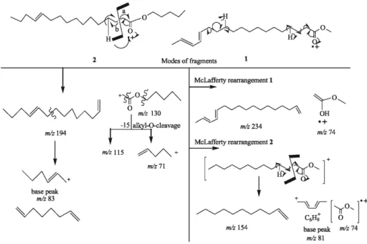
and 41 were in agreement with the proposed structure. The fragmentation pattern was given in fluxogram (Figure 2).
The alkaline hydrolysis of dienoate (1), yielded a
mixture of alcohol and carboxylic acid identified as methanol and dienoic acid. The unsaturated acid was brominated to yield 15, 16, 17, 18-tetrabromo derivative.20,21
The formation of tetrabromo derivative was revealed by the appearance of a pentet in mass spectrum, thus confirming two double bonds in the molecule.22
Thus on the basis of above evidences the dienoate (1)
was characterized as methylnonadeca-15, 17-dienoate, it is a novel compound and being reported first time by us.
The mass spectral analysis of monoester (2) gave the
molecular formula as C21H40O2.
IR Spectrum showed absorption bands for ester group (1740 cm-1), unsaturation (1627 cm-1) and long chain
aliphatic nature (730-720 cm-1).1H NMR Spectrum showed
a peak at d 0.90 as triplet for six protons was due to the terminal methyl groups.19 Methylene protons of -CH
2
-O-CO- and -CH2-CO-O-moieties were resonated as two
Figure 2. Fluxogram showing the fragmentation patterns proposed for dienoate 1 and monoester 2.
triplets at d 4.14 and 2.36 respectively. A triplet at d 5.35 was due to unsaturated protons. A multiplet at d 1.56 was due to the presence of methylene protons b- to the ester group and double bond, while rests of the methylenes were resonated at d 1.25 as an intense singlet.
The 13C NMR spectrum of monoester (2) showed the
presence of terminal methyl carbon at 19.9 ppm. The carbon of ester group was resonated at 174.0 ppm. The olefinic carbons at C-12 resonated at 118.0 ppm. The methylene carbons, a- and b- to ester group were resonated at 34.5 and 32.0 ppm respectively and rests of the methylene carbons were resonated at 29.8 ppm.
In mass spectrum the base peak at m/z 83 was obtained
due to b-cleavage to unsaturation and the abundant peak at m/z 115 was due to cleavage to ester group, suggested
the position of double bond and ester group. The other abundant peaks obtained at m/z 253, 227, 195, 166, 145,
55 and 41 were inconsistent with the proposed structure. The fluxogram (Figure 2) showed the fragmentation pattern of monoester (2).
On alkaline hydrolysis of (2), a mixture of alcohol and
carboxylic acid was obtained and identified as pentanol and acid. The unsaturated acid was brominated and identified as 12,13-dibromo hexadecanoic acid.20,21 The formation of
dibromo derivative was confirmed by the appearance of a triplet in mass spectrum.22
Thus on the basis of above evidences the monoester 2
was characterized as a pentyl hexa deca-12-enoate. It is a novel compound and reported first time by us. The molecular formula of naturally isolated monoester (3) was
C20H38O2.IR absorption bands (1740, 1627, 720 cm–1 ) and
peaks in NMR spectrum and fragmentation modes of mass spectra indicated that chemical structure of (3) was similar
to (2), i.e., long chain aliphatic ester. The position of ester
group was determined by mass fragmentation pattern (m/z,
241, 195, 166, 115, 83 (base peak), 55 and 41.
Alkaline hydrolysis of monoester (3), yielded a mixture
of alcohol and carboxylic acid, identified as pentanol and pentadeca-11-en-oic acid respectively.
Thus on the basis of above evidences the compound (3)
was characterized as a pentyl pentadeca-11-enoate. The known natural compounds linoleic acid, oleic acid, b-sitosterol and stigmasterol were identified as part of the lipid structures by analysis of IR, 1H NMR and 13C NMR
(DEPT) and mass spectral analysis and comparison with 1H
NMR and 13C NMR data of literature (see experimental).
Screening of antimicrobial activity
Agar diffusion technique was used for the screening of antimicrobial activity, using paper disc method as reported
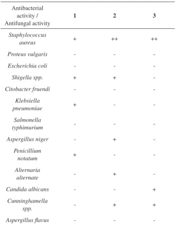
in the literature. 23,24 The results are shown in Table1. The
results have shown that all compounds exhibited moderate activity against Staphylococcus aureus and poor activity
against shigella spp. and K. pneumoniae.
Experimental
General experimental procedures
Melting points (mp) are uncorrected. 1H NMR was
recorded on 300 MHz Varian XL spectrometer and 400 MHz Brucker WM spectrometer, 13C NMR spectra were recorded
on Varian XL 75 MHz spectrometer. 1H-1H COSY NMR
was performed on the same spectrometer, using standard Varian pulse sequences. IR spectra were recorded in KBr discs on Perkin-Elmer-377 spectrometer, EIMS on Jeol-JMS D 300 mass spectrometer. Chromatography was performed using alumina grade III for column and silica gel G for TLC. The purity of the compounds were checked by
1H and 13C NMR spectral analysis and TLC plate, revealed
with vanillin (0.5 g) in H2SO4+EtOH(4:1).
Table 1. Antimicrobial assays for the 1-3
Antibacterial activity / Antifungal activity
1 2 3
Staphylococcus
aureus + ++ ++
Proteus vulgaris - -
-Escherichia coli - - -
Shigella spp. + + -
Citobacter fruendi - - -
Klebsiella
pneumoniae + -
-Salmonella
typhimurium - - -
Aspergillus niger - +
-Penicillium
notatum + -
-Alternaria
alternate - + -
Candida albicans - - +
Cunninghamella
spp. - + +
Aspergillus flavus - -
Plant material
The seeds of N. sativa Linn were collected from the
nearby area of Ujjain city, identified from IEMPS, Vikram University, Ujjain.
Extraction and isolation of the constituents
The seeds (6 kg) of were shade dried, cleaned, coarse powdered and extracted with hexane in soxhlet extractor for 72 h. The extract was concentrated by rotary evaporator to afford (3500 mL) oil. The oil was saponified by alcoholic potash method.25 Usual work up yielded (116 g)
unsaponifiable matter which was separated by repeated column chromatography on alumina grade III. The column was eluted by gradient elution in increasing order of polarity. The fractions were collected in bulk and monitored by TLC. The residue (6.8 g) of hexane fraction was subjected to rechromatographed on alumina on basis of increasing order of polarity of eluents. Fractions 5-6 (hexane:benzene, v/v, 8:2 and 7:3) were purified and identified as linoleic acid and oleic acid respectively. Presence of these acids were analyzed by IR, 1H NMR and mass spectrometry, and
compared with literature data.26-29 Fractions 7-12 afforded a
mixture of dienoate1 and monoester 2 and other impurities.
It was further rechromatographed, eluent hexane:benzene (4:1 and 3:1, v/v) was eluted (1) and (2) in pure form,
respectively. Fraction 13 yielded monoester 3 which was
in crystal form. Rest of the fractions (hexane:benzene, v/v, 1:1) yielded b-sitosterol and stigmasterol. The sterols were identified by comparison with literature data.30-32
Methyl nonadeca-15, 17-dienoate (1)
M+ 308, C
20H36O2 (40 mg, methanol), mp 90-92 oC
(Found: C, 77.9; H, 11.68; Calc.: C, 77.7; H, 11.65%), isolated from hexane: benzene (4:1, v/v) fraction. On TLC examination it showed single spot using hexane: ether: acetic acid (9:1:1, v/v) as solvent system. IR (KBr) λmax/cm-1:
2958, 2919, 2854, 1730, 1651, 1627, 1460, 1170, 1020, 730-720 cm-1. 1H NMR (200 MHz, CDCl
3 , TMS, d) 1.60
(3H, d, -CH3, J 7.5 Hz), 3.64 (3H, s, -CH3), 2.36 (2H, t,
-CH2-CO-O-, J 6.0 Hz), 1.56 (4H, m, 2x-CH2, b to double
bond and ester group), 5.35 (4H, m, 2x-CH=CH-), 1.25 (18H, s, 9x-CH2). EIMS m/z (rel. int.,%) 308 [M+] (1.1),
279 (5.1), 263 (1.2), 250 (1.3), 236 (3.5), 207 (7.2), 180 (7.7), 164 (17.9), 147 (18.8), 136 (26.1), 123 (26.5), 121 (31.1), 109 (28.6), 95 (62.6), 81 (100), 67 (92.0), 55 (95.8), 41 (78.2), 29 (38.1).13C NMR (75 MHz, CDCl
3, TMS, d)
174.0,118.0, 122.0 ,34.5, 32.0, 29.8,19.9.
Dienoate 1 (2.0 mg) was refluxed with ethanolic
KOH (1.3 mL, 5%) for 1 h. At the end of the reaction, the
mixture was diluted with water (3.0 mL) and extracted with chloroform. The chloroform layer was dried over anhydrous magnesium sulphate and concentrated. To separate both the compounds it was put in deep freezer. After 4 days some semi solid mass was separate out. After usual work-up, the solid was identified as an acid (IR: 3420, 1720 cm-1)and
liquid gave positive test for alcohol.
(1.0 mg) of an acid in diethyl ether (10%) was treated with drop wise addition of 2-3 mL of bromine in cold condition (-10 oC) from a finely drawn pipette, with constant
stirring the reaction mixture.20,21 Addition of bromine was
continued until a persistent reddish-yellow color was obtained. After standing 25 minutes, excess of bromine was destroyed by adding b-amylene and then it was kept in cold condition till over night. After usual work up, the solid was crystallized by adding cold petro naptha in ethereal solution and identified as 15, 16, 17, 18-tetrabromo nonadecanoic acid.
Pentyl hexadec-12-enoate (2)
M+ 324, C
21H40O2 (35 mg, methanol), mp 101-102 oC
(Found: C, 77.7; H, 12.3; Calc.: C, 77.5; H, 12.1 %), isolated from hexane: benzene (3:1, v/v) fraction. On TLC examination it showed a single homogenous spot using hexane: ether: acetic acid (9.5:0.5:0.5, v/v). IR (KBr) λmax/cm-1: 2920, 2860, 1740, 1627, 1470, 1390, and
730-720 cm-1. 1H NMR (200 MHz, CDCl
3 , TMS, d) 0.90
(6H, t, 2x-CH3, J 7.5 Hz), 4.14 (2H, t, -CH2-O-CO-, J 6.0
Hz), 2.36 (2H, t, -CH2-CO-O-, J 6.0 Hz), 1.56 (8H, m,
4x-CH2, b to double bond and ester group), 5.35 (2H, t, -CH=CH-, J 7.0 Hz), 1.25 (20H, s, 10x-CH2). EIMS m/z
(rel.int., %) 324 [M+] (1.1), 279 (2.2), 253 (3.1), 239 (1.5),
227 (44.8), 195 (52.4), 177 (3.0), 166 (30.0), 145 (38.6), 115 (91.2), 95 (33.1), 83 (100), 71(26.7), 67 (29.5), 55 (77.7), 41 (38.1). 13C NMR (75 MHz, CDCl
3, TMS, d)
174.0, 118.0, 34.5, 32.0, 29.8, 19.9.
Monoester 2(2.0 mg) was refluxed with ethanolic
KOH (1.3 mL, 5%) for 1 h. At the end of the reaction, the mixture was diluted with water (3.0 mL) and extracted with chloroform. The chloroform layer was dried over anhydrous magnesium sulphate and concentrated. To separate both the compounds it was put in deep freezer. After 4 days some semi solid mass was separate out. After usual work-up, they were identified as an acid (IR: 3420, 1720 cm-1) andliquid
gave positive test for alcohol.
Pentyl pentadec-11-enoate (3)
M+ 310, C
20H38O2 (40 mg, methanol) mp 82-83 oC
clear spot. IR (KBr) λmax/cm-1: 2920, 2860, 1740, 1627,
1470, 1390, and 730-720 cm-1. 1H NMR (200 MHz, CDCl 3
, TMS, d) 0.90 (6H, t, 2x-CH3, J 7.5 Hz), 4.14 (2H, t, -CH2
-O-CO-, J 6.0 Hz), 2.36 (2H, t, -CH2-CO-O-, J 6.0 Hz), 1.56
(8H, m, 4x-CH2, b to double bond and ester group), 5.35 (2H, t, -CH=CH-, J 7.0 Hz), 1.25 (20H, s, 10x-CH2). EIMS m/z (rel.int., %) 310 [M+] (1.1), 279 (5.2), 242 (2.1), 241
(47.3), 212 (6.3), 195 (51.0), 166 (27.8), 145 (30.0), 124 (17.5), 115 (99.7), 95 (38.2), 83 (100), 69 (31.1), 67 (31.2), 55 (81.5), 41 (39.8).13C NMR (75 MHz, CDCl
3, TMS, d)
174.0, 118.0, 34.8, 32.5, 29.8, 19.9.
Monoester 3 (2.0 mg) was refluxed with ethanolic
KOH (1.3 mL, 5%) for 1 h. At the end of the reaction, the mixture was diluted with water (3.0 mL) and extracted with chloroform. The chloroform layer was dried over anhydrous magnesium sulphate and concentrated. To separate both the compounds it was put in deep freezer. After 4 days some semi solid mass was separate out. After usual work-up, it was identified as an acid (IR: 3420, 1720 cm-1)andliquid
gave positive test for alcohol.
Screening of antimicrobial activity
Agar diffusion technique was used for the screening of antibacterial and antifungal activities using paper disk method.23,24 The results are being shown in Table 1.
Acknowledgments
Authors are grateful to RSIC, CDRI, Lucknow and RSIC; IIT Bombay, Mumbai for spectral analysis and UGC, New Delhi for financial assistance.
References
1. Satavati, G. V.; Gupta, A. K.; Medicinal Plants of India,
Dehradun, 1987, vol.1.
2. Anon; Wealth of India, CSIR: New Delhi, 1991, vol. 3,
pp. 63-65.
3. Atta-Ur-Rahaman; Malik, S.; Hasan, S. S.; Chaudhary, M. I.; Nie, N.; Clardy, J.; Tetrahedron Lett. 1995, 1993.
4. Atta-Ur- Rahaman; Malik, S.; Zaman, K.; J. Nat. Prod. 1992,
55, 676.
5. Atta-Ur-Rahaman; Malik, S.; Cun-heng-H.; Clardy, J.;
Tetrahedron Lett.1985, 2759.
6. Salma, R. B.; Planta Med. 1973, 24, 375.
7. Ansar, A. B.; Hassan, S.; Kanne, L.; Atta-Ur-Rahaman; Wohler, T.; Phytochemistry1988, 27, 3977.
8. Chakravarti, N.; Annu. Allergy1993, 70, 237.
9. Al-Naggar, T. B.; Gomez-Serranillos, M. P.; Carreto, M. E.; Villar, A. M.; J. Ethnopharmacol.2003, 88, 63.
10. Sharma, P. V.; Dravyaguana-Vijnana, Chaukhambha Bharti Academi:Varanasi, 1998, vol. 2, pp. 596, 597.
11. Hailat, N.; Bataineh Z.; Lafi, S.; Rowdily, E.; Aquel, M.; Muhamad, Al-Katib; Hanash, S.; Int. J. Pharmacog. 1995, 33,
16.
12. Chopra, R. N.; Nayer, S. L.; Chopra, I. C.; Glossary of Indian
Medicinal Plants, CSIR: NewDelhi, 1956, pp. 176,177.
13. El-Alfy, T. S.; El-Fatatry, H. M.; Toama, M. A.; Pharmazie
1975, 30, 109.
14. Ali, B. H.; Blunden, G.; Phytother. Res.2003, 17, 299.
15. Boulos, L.; Reference Publication1983,103.
16. Mehta, B. K.; Singh, N.; Keshri, G.; Chaudhary, S. R.; Biol.
Memories1999, 25, 38.
17. Dyer, J. R. Application of Absorption Spectroscopy of Organic
Compound, Prentice Hall of India Ltd.: New Delhi, 1984,
p.35.
18. Bellamy, L. J.; The Infrared Spectra of Complex Molecules, Chapman and Hall: London, 1975, p. 39.
19. Silverstein, R. M.; Webster, F. X.; Spectrometric Identification of
Organic Compounds 6th ed., John Wiley and Sons: New York,
2003, pp.160-253.
20. Birosel, D. M.; Univ. Philippines Nat. Appl. Sci. Bull. 1932,
2,103.
21. Kass, J. P.; Burr, G. O.; J. Am. Chem. Soc.1939, 61, 1062.
22. Mohan, J.; Organic Spectroscopy Principles and Applications, Narosa Publishing House: New Delhi, 2000, p.366.
23. Maruzzella, I. C.; Henry, P. A.; J. Am. Pharm. Assoc.1958, 47, 294.
24. Vincent, J. G.; Vincent, H. W.; Proc. Soc. Expt. Biol. Med. 1995,
55, 712.
25. Mohan, J.; Organic Analytical Chemistry, Narosa Publishing House: New Delhi, 2003, p. 533.
26. Huber, W. F.; J. Am. Chem. Soc.1951, 73, 2732.
27. Swern, D.; Coleman, J. E.; J. Am. Oil Chem. Soc.1955, 32,
539.
28. Bernhard, K.; Helv. Chim. Acta1948, 31, 977.
29. Crombie, L.; J. Chem. Soc.1962, 2449.
30. Francis, G. W.; Veland, K.; J. Chromatogr.1981, 219, 379.
31. Pouchert, C. J.; Behnke, J.; The Aldrich Library of 13C and 1H
NMR Spectra, 1st ed., 1993, vol.1, 2.
32. Maitra, S. K.; Chatterjee, B. N.; Chakravarti, D.; Maiti, B. C.;
J. Indian Chem. Soc. 2006, 83, 513.