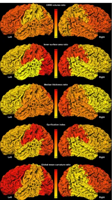
Development of Cortical Morphology Evaluated with Longitudinal MR Brain Images of Preterm Infants.
Texto
Imagem


Documentos relacionados
This study evaluated the morphological and chemical composition of the following bone substitutes: cancellous and cortical organic bovine bone with macro and microparticle size
The evaluated Parameters included: total pore count, total pore volume, mean pore volume, total porosity (% of pore volume in relation to total sealer volume) and mean pore
KEY WORDS: malformations of cortical development, focal cortical dysplasias, neuronal migration disorders, cortical organization disorders.. Malformações do
The brain regions where abnormalities are observed in studies of diffusion tensor, volumetry, spectroscopy and cortical thickness are the same involved in neurobiological theories of
In a study which evaluated the development of 55 infants with corrected chronological age between 4 and 5 months, born preterm, found that infants with lower gestational age
Number of positive, negative and contaminated samples in culture of cortical bone and bone marrow without treatment (unprocessed) and in cortical bone subjected to
In this context, the objective of the present study is to measure the exogenous components of the cortical auditory evoked potential (CAEP) in term and preterm newborns and compare
Longitudinal local volume ratios (percent, y-axis) during follow-up examination (months, x-axis) of different cortical and subcortical regions that revealed no significant


