Antibacterial Evaluation of Synthetic Thiazole
Compounds
In Vitro
and
In Vivo
in a
Methicillin-Resistant
Staphylococcus aureus
(MRSA) Skin Infection Mouse Model
Haroon Mohammad1, Mark Cushman2, Mohamed N. Seleem1*
1Department of Comparative Pathobiology, Purdue University College of Veterinary Medicine, West Lafayette, Indiana, United States of America,2Department of Medicinal Chemistry and Molecular Pharmacology, Purdue University College of Pharmacy, and the Purdue Center for Cancer Research, West Lafayette, Indiana, United States of America
Abstract
The emergence of community-associated methicillin-resistantStaphylococcus aureus
(MRSA), including strains resistant to current antibiotics, has contributed to an increase in the number of skin infections reported in humans in recent years. New therapeutic options are needed to counter this public health challenge. The aim of the present study was to examine the potential of thiazole compounds synthesized by our research group to be used topically to treat MRSA skin and wound infections. The broth microdilution method con-firmed that the lead thiazole compound and four analogues are capable of inhibiting MRSA growth at concentrations as low as 1.3μg/mL. Additionally, three compounds exhibited a synergistic relationship when combined with the topical antibiotic mupirocin against MRSA
in vitrovia the checkerboard assay. Thus the thiazole compounds have potential to be used alone or in combination with mupirocin against MRSA. When tested against human kerati-nocytes, four derivatives of the lead compound demonstrated an improved toxicity profile (were found to be non-toxic up to a concentration of 20μg/mL). Utilizing a murine skin infec-tion model, we confirmed that the lead compound and three analogues exhibited potent antimicrobial activityin vivo, with similar capability as the antibiotic mupirocin, as they reduced the burden of MRSA present in skin wounds by more than 90%. Taken altogether, the present study provides important evidence that these thiazole compounds warrant fur-ther investigation for development as novel topical antimicrobials to treat MRSA skin infections.
Introduction
Ten percent of all hospital admissions in the United States each year are due to patients suffer-ing from skin and soft tissue infections (SSTIs) [1]. SSTIs can range from simple abscesses,
OPEN ACCESS
Citation:Mohammad H, Cushman M, Seleem MN (2015) Antibacterial Evaluation of Synthetic Thiazole CompoundsIn VitroandIn Vivoin a Methicillin-ResistantStaphylococcus aureus(MRSA) Skin Infection Mouse Model. PLoS ONE 10(11): e0142321. doi:10.1371/journal.pone.0142321
Editor:Herminia de Lencastre, Rockefeller University, UNITED STATES
Received:May 22, 2015
Accepted:October 19, 2015
Published:November 4, 2015
Copyright:© 2015 Mohammad et al. This is an open access article distributed under the terms of the
Creative Commons Attribution License, which permits unrestricted use, distribution, and reproduction in any medium, provided the original author and source are credited.
Data Availability Statement:All relevant data are within the paper.
Funding:This work was supported by the Showalter Research Trust (Grant 205994). The funder had no role in study design, data collection and analysis, decision to publish, or preparation of the manuscript.
cellulitis, and traumatic wound infections to complicated infections (infected burns, diabetic foot ulcers, and major abscesses) [1]. SSTIs are often caused by the bacterial pathogen methicil-lin-resistantStaphylococcus aureus(MRSA) [2,3]. Indeed, 58% of all SSTIs treated in the United States alone were caused by MRSA, according to an epidemiological study of one national health care system [4]. This agrees with a 2004 study conducted in emergency room departments in 11 cities where MRSA was responsible for 59% of patients presenting with a SSTI [5]. The large number ofS.aureus-based SSTIs has placed a significant economic burden on the healthcare system. A recent report examining the increase inS.aureus-SSTI hospitaliza-tions in the United States documented a dramatic rise in the annual cost of treating infected patients from $3.36 billion to $4.50 billion (from the years 2001 through 2009) [6]. A recent increase in skin abscesses has been observed and has been associated with a rise in strains of community-associated MRSA (CA-MRSA) [7]. Of these strains, MRSA USA300 has been linked most frequently to skin infections in the United States [5,8].
According to guidelines provided by the Infectious Diseases Society of America, treatment of moderate to severe skin infections caused by MRSA involves incision and drainage of the affected region combined with administration of empirical antibiotics (such as clindamycin, vancomycin, linezolid, and mupirocin) [9,10]. However, strains of MRSA exhibiting resistance to several of these antibiotics including vancomycin [11,12], clindamycin [13,14], and topical ointments like mupirocin [14–16], indicate that such therapies may be rendered ineffective in the future. Therefore, development of novel antimicrobials capable of treating MRSA-induced SSTIs is an important step necessary to circumvent the burden of this public health issue.
Previous research by our group has identified a lead disubstituted phenylthiazole compound (compound1,Fig 1) that exhibited potent antimicrobial activityin vitroagainst a diverse array of clinically-significant strains of MRSA [17]. Derivatives of the lead1were synthesized to elu-cidate the structure-activity relationships of this compound. These derivatives revealed that the aminoguanidine moiety at thiazole-C5 is critical for antibacterial activity [18]. Furthermore, a nonpolar, hydrophobic group is favored at thiazole-C2. Analogues to the lipophilic alkyl tail of the lead1were subsequently constructed in order to enhance the antimicrobial activity of these thiazole compounds, to improve their toxicity profile, and to refine their physicochemical properties [18]. These particular phenylthiazole compounds possess several excellent charac-teristicsin vitroincluding rapid bactericidal activity against MRSA, a low frequency of bacterial resistance developing, and they have been shown to possess the ability to be used in combina-tion with currently approved antibiotics, such as vancomycin, against MRSA [19]. Studies per-formed to date indicate these compounds have potential to be used as topical antimicrobials for treatment of MRSA skin infections. The objectives of the current study were to assess the antibacterial activity of the lead thiazole compound and four analogues (Fig 1)in vitroagainst antibiotic-resistantS.aureusstrains isolated from/responsible for skin infections, to assess the ability of these compounds to be paired with mupirocin as a treatment option against MRSA, to confirm the compounds have limited toxicity to human keratinocytes, and to verify the thia-zole compounds can retain their antimicrobial activityin vivoin an established murine MRSA skin infection model. Confirmation of the ability of these compounds to successfully treat mice infected with a MRSA skin infection will lay the foundation for further assessment of these compounds as novel antimicrobials for treatment of MRSA skin infections.
Materials and Methods
Synthesis of thiazole compounds 1
–
5
Synthetic schemes, spectral data, and purity (>95%, determined by HPLC) of thiazole
Bacterial strains and reagents used in this study
Clinical isolates ofS.aureuswere obtained through the Network of Antimicrobial Resistance inStaphylococcus aureus(NARSA) program (Table 1). Clindamycin hydrochloride monohy-drate (TCI America, Portland, OR, USA,>98.0% purity) and mupirocin (pure USP)
(Appli-Chem, St. Louis, MO, USA) powders were purchased commercially and dissolved in dimethyl sulfoxide (DMSO) (for clindamycin) or ethanol (for mupirocin) to prepare a stock solution (10μg/mL). Lipoderm was purchased from the Professional Compounding Centers of America
(Houston, TX, USA). Cation-adjusted Mueller-Hinton broth (CAMHB) (Sigma-Aldrich, St. Louis, MO, USA), Tryptic soy broth (TSB) (Becton, Dickinson and Company, Franklin Lakes, NJ, USA), mannitol salt agar (Hardy Diagnostics, Santa Maria, CA, USA), phosphate-buffered saline (PBS) (Sigma-Aldrich, St. Louis, MO, USA), Dulbeco’s modified Eagle’s medium (DMEM) (Sigma-Aldrich, St. Louis, MO, USA), fetal bovine serum (FBS) (USA Scien-tific, Inc., Orlando, FL, USA), petroleum jelly (Equate [Walmart, Inc.], Bentonville, AR, USA), and 96-well plates (CellTreat Scientific Products, Shirley, MA, USA) were all purchased from commercial vendors.
Determination of minimum inhibitory concentration (MIC) against
drug-resistant
S
.
aureus
strains
The MIC of thiazole compounds1–5, clindamycin, and mupirocin was determined against five different MRSA strains and one highly mupirocin-resistantS.aureusstrain isolated from skin wounds, using the broth microdilution method, following the guidelines outlined by the Clini-cal and Laboratory Standards Institute [21]. A bacterial suspension equivalent to a McFarland
Fig 1. Chemical structures of thiazole compounds 1–5 presented in this study.
doi:10.1371/journal.pone.0142321.g001
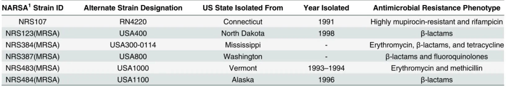
Table 1. Drug-resistant clinical isolates ofStaphylococcus aureusused in this study.
NARSA1Strain ID Alternate Strain Designation US State Isolated From Year Isolated Antimicrobial Resistance Phenotype
NRS107 RN4220 Connecticut 1991 Highly mupirocin-resistant and rifampicin
NRS123(MRSA) USA400 North Dakota 1998 β-lactams
NRS384(MRSA) USA300-0114 Mississippi - Erythromycin,β-lactams, and tetracycline
NRS387(MRSA) USA800 Washington - β-lactams andfluoroquinolones
NRS483(MRSA) USA1000 Vermont 1993–1994 Erythromycin and methicillin
NRS484(MRSA) USA1100 Alaska 1996 β-lactams
1NARSA, Network on Antimicrobial Resistance inStaphylococcus aureus.
standard of 0.5 was prepared and subsequently diluted 1:300 in CAMHB. This bacterial sus-pension (~1 × 10⁵colony forming unit (CFU/mL)) was then added to each well of a 96-well microtiter plate. Compounds1–5, clindamycin, or mupirocin were added (in triplicate) to the first row of the plate and then serially diluted down the ordinate. Plates were incubated at 37°C for 18–20 hours and then the MIC was ascertained. The MIC was classified as the lowest con-centration of each test agent where bacterial growth could not be visualized.
Assessment of synergistic relationship between thiazole compounds
and mupirocin against MRSA
The checkerboard assay was utilized to asses if the most potent thiazole compounds (1–3) have potential to be combined with mupirocin for treatment of MRSA infections [22]. Briefly, a bac-terial suspension (1 × 10⁵CFU/mL) in CAMHB was added to each well of a 96-well microtiter plate. Compounds1–3and mupirocin were diluted in CAMHB in order to reach the desired starting concentration (2 × or 4 × MIC). Mupirocin was serially diluted along the horizontal axis of the plate while compound1,2, or3was diluted along the vertical axis. Plates were incu-bated for at least 18 hours at 37°C and the MIC of each compound was recorded. The fractional inhibitory concentration index (ƩFIC) was computed for each combination using the following equation:
X
FIC ¼ MICthiazole compound in combination with mupirocin
MICthiazole compound alone
þ MICmupirocin in combination with thiazole compound
MICmupirocin alone
A FIC index less than or equal to 0.50 was classified as synergism, as described previously [19]. FIC values above 0.50 but less than 4.00 were classified as indifference, while FIC values greater than 4.00 were indicative of antagonism.
In vitro
cytotoxicity analysis of thiazole compounds against HaCaT cells
Compounds1–5were assayed (at concentrations of 5μg/mL, 10μg/mL, 20μg/mL, and
40μg/mL) against a human keratinocyte (HaCaT) cell line (Catalogue Number: T0020001,
AddexBio, San Diego, CA, USA) to determine the potential toxic effect to mammalian skin cellsin vitroas described before [18]. Briefly, cells were cultured in DMEM supplemented with 10% FBS at 37°C with CO2(5%). Control cells received DMSO alone at a concentration equal
to that in drug-treated cell samples. The cells were incubated with the compounds (in tripli-cate) in a 96-well plate at 37°C with CO2(5%) for two hours prior to addition of the assay
reagent MTS 3-(4,5-dimethylthiazol-2-yl)-5-(3-carboxymethoxyphenyl)-2-(4-sulfophenyl)-2H-tetrazolium) (Promega, Madison, WI, USA). Absorbance readings (at OD490) were taken
using a kinetic microplate reader (Molecular Devices, Sunnyvale, CA, USA). The quantity of viable cells after treatment with each compound was expressed as a percentage of the viability of DMSO-treated control cells (average of triplicate wells ± standard deviation). The toxicity data was analyzed via a one-way ANOVA, with post hoc Dunnet’s multiple comparisons test (P<0.05), utilizing GraphPad Prism 6.0 (GraphPad Software, La Jolla, CA).
In vivo
assessment of antimicrobial activity of thiazole compounds 1
–
5
and mupirocin in a MRSA skin infection mouse model
number: 1207000676) and carried out in strict accordance with the recommendations in the Guide for the Care and Use of Laboratory Animals of the National Institutes of Health. To ini-tiate the formation of a skin wound, eight groups (n = 5) of eight-week old female Balb/c mice (obtained from Harlan Laboratories, Indianapolis, IN, USA) were disinfected with ethanol (70%) and shaved on the middle of the back (approximately a one-inch by one-inch square region around the injection site) one day prior to infection, similar to what has been described elsewhere [23,24]. To prepare the bacterial inoculum, an aliquot of overnight culture of MRSA USA300 was transferred to fresh TSB and shaken at 37°C until an OD600value of ~1.0 was
achieved. The cells were centrifuged, washed once with PBS, re-centrifuged, and then re-sus-pended in PBS. Mice then received an intradermal injection (20μL) containing ~2.76 × 10⁸
CFU/mL MRSA USA300. An open wound formed at the site of injection, 48 hours post-infec-tion. Topical treatment was initiated subsequently with each group of mice receiving the fol-lowing: compound1–5(2%, using petroleum jelly as the vehicle), mupirocin (2%, using petroleum jelly as the vehicle), compound1(2%, using Lipoderm as an alternative vehicle), and a control group receiving the control vehicle (20 mg, petroleum jelly) alone. Each group of mice receiving a particular treatment regimen was housed separately in a ventilated cage with appropriate bedding, food, and water. Mice were checked twice daily during infection and treatment to ensure no adverse reactions were observed. In the event a mouse was observed to become severely ill, the subject was euthanized per the IRB protocol. Mice were treated twice daily for three days. Mice were humanely euthanized via CO2asphyxiation 24 hours after the
last dose was administered. The region around the skin wound was lightly swabbed with etha-nol (70%) and excised. The tissue was subsequently homogenized in TSB (1 mL). The homoge-nized tissue was then serially diluted in PBS before plating onto mannitol salt agar plates. The plates were incubated for 20–22 hours at 37°C before viable CFU were counted and MRSA reduction in the skin wound post-treatment was determined for each group. Data were ana-lyzed using a one-way ANOVA, with post hoc Holm-Sidak’s multiple comparisons test (P<0.05), utilizing GraphPad Prism 6.0.
Results and Discussion
Antimicrobial activity of thiazole compounds 1
–
5 against MRSA strains
isolated from skin wounds
Previous work has established thiazole compounds1–5possess potent antimicrobial activity against MRSA (particularly isolates derived from healthcare-associated MRSA cases). To con-firm these compounds maintain their antibacterial activity against CA-MRSA strains and MRSA isolates derived from patients presenting with infected wounds (Table 1), the broth microdilution assay was utilized to determine the lowest concentration each compound was able to inhibit the growth of these strains (denoted as the minimum inhibitory concentration or MIC).
When tested against these important clinical isolates of drug-resistantS.aureus, the thiazole compounds exhibited strong antimicrobial activity similar to (and in several cases better than) mupirocin. As presented inTable 2, the lead thiazole1exhibits the most potent activity with a MIC value of 1.3μg/mL against all six drug-resistant staphylococcal strains tested. The
biphe-nyl and butyne analogues (2and3,respectively) possess MIC values ranging from 2.8 to 5.6μg/mL. All five thiazole compounds possess antimicrobial activity against MRSA strains
exhibiting resistance to an array of antibiotics includingβ-lactams, fluoroquinolones
(MIC values range from 1.3 to 13.3μg/mL), a strain exhibiting a high-level of resistance to
mupirocin (MIC of 1024.0μg/mL). Furthermore, compounds1and2(MIC of 1.3 and 2.8μg/
mL, respectively) are more active than mupirocin (MIC of 4.0μg/mL) against three additional
MRSA strains (USA800, USA1000, and USA1100). Clindamycin, when tested against four of the five MRSA strains, was found to have a MIC of 0.1μg/mL. This MIC value is similar to
what has been reported elsewhere for clindamycin [25]. Collectively, the results confirm that the thiazole compounds do possess potent antimicrobial activity against important CA-MRSA strains and MRSA isolates responsible for infected wounds in patients.
Combination therapy using thiazole compounds with mupirocin against
MRSA
The susceptibility analysis performed with the thiazole compounds indicated they have poten-tial to be used alone for the treatment of MRSA skin/wound infections. While the use of a sin-gle agent to treat such infections is often used in the clinical setting, combination therapy using two or more antibiotics is favorable for multiple reasons. Among these reasons include that combination therapy has the potential to slow down the emergence of resistant bacterial strains to antibiotics, to reduce potential negative side effects to patients (by using lower concentra-tions/doses of each drug), and to alleviate the morbidity related to bacterial infections [26,27]. Given that multiple topical treatments for skin infections involve a combination of more than one antibiotic, such as Neosporin (consisting of bacitracin, neomycin, and polymyxin B sul-fate) and Polysporin ointment (consisting of bacitracin, polymyxin B sulfate, and gramicidin) [28], the identification of compounds to pair with known antibiotics has good potential to expand the available treatment options. Mupirocin has been a key ally in the treatment of MRSA skin infections; however, isolates exhibiting moderate to high-level of resistance to mupirocin (MIC512μg/mL) have emerged, particularly in environments where this
antibi-otic has been extensively utilized [15,29,30]. Identifying agents that can be partnered with mupirocin has the potential to extend the usage of this particular antimicrobial in the clinical setting.
In an earlier study, Alouet al, demonstrated that mupirocin forms a synergistic relationship with amoxicillin-clavulanate against MRSA isolates testedin vitrovia the checkerboard assay [31]. Amoxicllin is aβ-lactam antibiotic that interferes with bacterial cell wall synthesis by inhibiting crosslinking of peptidoglycan subunits in the bacterial cell wall [32]. Preliminary studies conducted with our thiazole compounds indicate they also interfere with cell wall syn-thesis in bacteria; thus we were curious to assess if the thiazole compounds could be used in combination with mupirocin against MRSA, similar to what was found with
amoxicillin-Table 2. Minimum inhibitory concentration (MIC inμg/mL) of thiazole compounds 1–5, clindamycin, and mupirocin (tested in triplicate) against
five methicillin-resistantStaphylococcus aureus(MRSA) and one mupirocin-resistantS.aureus(NRS107) strain isolated from skin wounds.
S.aureusStrain Number
Compound Number/Name NRS107 USA300 USA400 USA800 USA1000 USA1100
1 1.3 1.3 1.3 1.3 1.3 1.3
2 2.8 2.8 2.8 2.8 2.8 2.8
3 2.8 5.6 5.6 5.6 2.8 5.6
4 13.3 13.3 13.3 13.3 13.3 13.3
5 6.4 6.4 12.8 6.4 12.8 6.4
Clindamycin 0.1 1.8 0.1 0.1 0.1 0.1
Mupirocin 1024.0 1.0 1.0 4.0 4.0 4.0
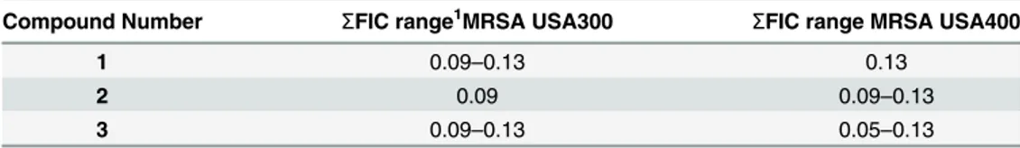
clavulanate. Using the checkerboard assay, it was discovered that the most potent thiazole com-pounds (1–3) exhibited a strong degree of synergy (FIC index0.50) with mupirocin against two of the most prevalent MRSA strains responsible for skin infections (Table 3). Against MRSA USA300, all three compounds exhibited a fractional inhibitory concentration (FIC) index ranging from 0.09 to 0.13 when combined with mupirocin. A similar trend was observed when this combination was tested against MRSA USA400, with FIC values ranging from 0.05 to 0.13. The data provide evidence that supports the prospect that these particular thiazole compounds can be successfully paired with mupirocin to treat MRSA infections (and poten-tially prolong the utility of mupirocin in the clinical setting).
Toxicity analysis of thiazole compounds to human keratinocytes
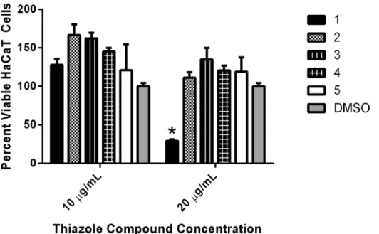
Selective toxicity is important to ensure compounds with promising antimicrobial activity don’t possess negative side effects to mammalian tissues. Certain regimens (in particular anti-septics) used for treatment of skin infections and wounds have been found to exhibit toxicity to human keratinocytes and impair wound healing, thus limiting their use as therapeutic options [33–36]. Prior to validating the antimicrobial activity of the thiazole compounds in a MRSA skin infection model, it was critical to confirm the thiazole compounds were not toxic to human keratinocytes. Using the MTS assay with a human keratinocyte (HaCaT) cell line, it was confirmed that thiazole compounds1–5were not toxic at a concentration of 10μg/mL
(Fig 2). Interestingly, the four analogues constructed from compound1demonstrated an improved toxicity profile, as they were found to be non-toxic to HaCaT cells up to a concentra-tion of 20μg/mL. Taken altogether, the data indicate the most potent thiazole compoundsin
vitro(1–3) are not toxic to human keratinocytes at concentrations up to seven-fold higher than the compounds’MIC values determined against MRSA.
Assessment of topical application of thiazole compounds
in vivo
via a
murine MRSA skin infection model
As thiazole compounds1–5exhibited excellent activity against MRSAin vitroand displayed no toxicity to human keratinocytes at the compounds’MIC, we moved to confirm that these compounds could maintain their antimicrobial activityin vivo, using an established MRSA murine skin infection model. After the formation of an open wound (infected with MRSA) in the dorsal region of infected mice, each group of mice was treated with a suspension of com-pounds1–5(2%), mupirocin (2% suspension), or petroleum jelly (20 mg, used as a vehicle for topical delivery of the compounds/antibiotic) twice daily for three days. The reduction in bac-terial burden present in the wounds of infected mice was determined after cessation of treat-ment. Reduction of bacterial burden in infected wounds is critical to promote proper wound
Table 3. Combination testing of thiazole compounds 1–3 with mupirocin against clinically-prevalent
strains of community-acquired methicillin-resistantStaphylococcus aureus(CA-MRSA).
Compound Number ƩFIC range1MRSA USA300 ƩFIC range MRSA USA400
1 0.09–0.13 0.13
2 0.09 0.09–0.13
3 0.09–0.13 0.05–0.13
1
ƩFIC, fractional inhibitory concentration index. Results for the FIC index (ƩFIC) are as follows:0.50, synergistic;>0.50 to4.00, indifference;>4.00, antagonistic.ƩFIC range provided is from two independent experiments.
repair and to prevent a severe inflammatory response from being triggered that may negatively impact healing of wounded tissues [37].
As presented inFig 3, four thiazole compounds mimic mupirocin’s ability to drastically reduce the burden of MRSA present in skin wounds. Compounds3–5produce a 1.47 to 1.62 log10reduction in MRSA CFU; this corresponds to a greater than 96% reduction in the
bacte-rial burden, as compared to mice receiving only the vehicle alone (petroleum jelly) for treat-ment. The lead1exceeds the effect of mupirocin, producing a 2.27 log10reduction in MRSA
CFU in the skin wound (relative to the 2.07 log10reduction observed with mupirocin). The
emergence of increasing resistance to mupirocin, a drug of choice, amongst MRSA strains makes it extremely important to find alternative options for treatment (particularly for skin infections), such as these thiazole compounds. Interestingly, one of the most potent com-pounds against MRSA USA300in vitro(the biphenyl analogue2, MIC of 2.8μg/mL) is the
least effective compoundin vivo(produces a 0.47 log10reduction in MRSA CFU, that was
found to not be statistically significant); this provides a stark reminder that the behavior of compoundsin vitroneeds to be validated within vivostudies to confirm their viability as novel treatment options.
Antimicrobial compounds that can be administered topically (such as thiazole compounds 1,3,4, and5) for treatment of localized skin lesions have certain advantages over their systemic counterparts. These advantages include the ability to avoid adverse systemic side effects, the ability to localize/concentrate the drug at the target site of infection (providing increased con-centration of the drug), lower treatment costs, and a reduced likelihood of inducing bacterial resistance to the treatment agent [36,38]. Overall, the results garnered from the present study
Fig 2. Toxicity analysis of thiazole compounds against human keratinocytes (HaCaT).Percent viable mammalian cells (measured as average absorbance ratio (test agent relative to DMSO)) for cytotoxicity analysis of thiazole compounds1,2,3,4, and5(tested in triplicate) at 10 and 20μg/mL against HaCaT cells using the MTS 3-(4,5-dimethylthiazol-2-yl)-5-(3-carboxymethoxyphenyl)-2-(4-sulfophenyl)-2H-tetrazolium) assay. Dimethyl sulfoxide (DMSO) was used as a negative control to determine a baseline measurement for the cytotoxic impact of each compound. The absorbance values represent an average of a minimum of three samples analyzed for each compound. Error bars represent standard deviation values for the absorbance values. A one-way ANOVA, with post hoc Dunnet’s multiple comparisons test, determined statistical difference between the values obtained for compound1and DMSO (denoted by the asterisk) (P<0.05).
indicate the thiazole compounds (in particular the lead1) do warrant further investigation as a topical treatment option for MRSA-infected skin wounds.
Impact of changing vehicles in reduction of MRSA burden present
in vivo
in infected skin wounds
After confirming four thiazole compounds (1,3–5) have potential for use as novel topical anti-microbials against MRSA, we examined if changing the vehicle used for delivery may further enhance the reduction in bacterial burden present in infected wounds. To assess this, a 2% sus-pension of the most potent compound (1), using Lipoderm as an alternative vehicle, was tested using the murine MRSA skin infection model described above. Lipoderm has been used com-mercially as a transdermal delivery vehicle to enhance permeation of active pharmaceutical compounds through the skin [39]. It was hypothesized that switching vehicles (from petroleum jelly) to Lipoderm would enhance penetration of the thiazole compounds into the skin wound, thus permitting a greater reduction in the bacterial burden present. AsFig 3demonstrates, changing vehicles from petroleum jelly to Lipoderm does enhance the reduction in the bacterial load in the skin wound of mice that is achieved by compound1. A 0.4-log10improvement in
the reduction of MRSA CFU for compound1is observed when Lipoderm is used. This
Fig 3. Average log10-reduction in MRSA USA300 burden in infected murine skin wounds.Evaluating
the effectiveness of treatment of MRSA skin lesions in mice with mupirocin (2%), thiazole compounds1–5 (2%), and compound1(2%, using Lipoderm as the vehicle) twice daily for three days. The average log10-reduction in bacterial burden (relative to the negative control group (petroleum jelly)) was calculated and presented in the figure. Error bars represent standard deviation values. A one-way ANOVA, with post hoc Holm-Sidak’s multiple comparisons test revealed statistical difference (denoted by asterisk) between compounds1,3,4,5,1(using Lipoderm as the vehicle), and mupirocin relative to the negative control (P<0.05).
corresponds to a>99.6% reduction in MRSA present in the skin wound after treatment. Thus
switching vehicles from petroleum jelly to Lipoderm appears to permit enhanced penetration of the thiazole compounds into skin wounds, leading to an increased reduction in MRSA burden.
Conclusion
In this study, we demonstrate that five novel synthetic phenylthiazole compounds exhibit potent antimicrobial activityin vitroagainst clinically-relevant strains of MRSA responsible for skin and wound infections. Additionally, compounds1–3exhibit a strong synergistic relation-ship when combined with mupirocin against two highly prevalent strains of CA-MRSA. Fur-thermore, three compounds are not toxic to human keratinocytes at a concentration seven times higher than their MIC against MRSA. The antimicrobial activity of compounds1,3,4, and5is confirmedin vivoin a murine MRSA skin infection model (>96% reduction in
bacte-rial load observed, post-treatment). Substitution of the vehicle from petroleum jelly to Lipo-derm permits a nearly 0.4-log10additional reduction in bacterial load achieved by compound
1, indicating this vehicle may be more suitable for enhanced penetration of the compound into infected tissues. Collectively, the results provide valuable information to further develop these thiazole compounds as topical antimicrobial agents for treatment of skin infections and wounds infected by MRSA. Future work with these thiazole compounds includes constructing additional analogues of the lead compound1in an effort to improve its potency against MRSA and enhance its toxicity profile with human keratinocytes. Additionally, addressing the limited physicochemical properties of these compounds (through structural modifications of lead compound1) is an important next step in order to expand the therapeutic potential of these compounds so they can be administered orally/intravenously for treatment of invasive MRSA infections (both complicated skin infections and systemic infections).
Acknowledgments
The authors would like to thank the Network of Antimicrobial Resistance inStaphylococcus aureus(NARSA) program supported under NIAID/NIH Contract # HHSN272200700055C for providing the MRSA strains used in this study.
Author Contributions
Conceived and designed the experiments: HM MC MS. Performed the experiments: HM. Ana-lyzed the data: HM MC MS. Contributed reagents/materials/analysis tools: MC MS. Wrote the paper: HM MC MS.
References
1. Giordano P, Weber K, Gesin G, Kubert J. Skin and skin structure infections: treatment with newer gen-eration fluoroquinolones. Therapeutics and clinical risk management. 2007; 3(2):309–17. PMID: 18360639; PubMed Central PMCID: PMC1936312.
2. Otter JA, French GL. Molecular epidemiology of community-associated meticillin-resistant Staphylo-coccus aureus in Europe. The Lancet infectious diseases. 2010; 10(4):227–39. doi: 10.1016/S1473-3099(10)70053-0PMID:20334846.
3. Nagao M, Iinuma Y, Suzuki M, Matsushima A, Takakura S, Ito Y, et al. First outbreak of methicillin-resistant Staphylococcus aureus USA300 harboring the Panton-Valentine leukocidin genes among Japanese health care workers and hospitalized patients. Am J Infect Control. 2010; 38(9):E37–E9. doi: 10.1016/j.ajic.2010.04.214PMID:WOS:000283582500003.
2005–2010. JAMA: the journal of the American Medical Association. 2012; 308(1):50–9. doi:10.1001/ jama.2012.7139PMID:22760291.
5. Moran GJ, Krishnadasan A, Gorwitz RJ, Fosheim GE, McDougal LK, Carey RB, et al. Methicillin-resis-tant S-aureus infections among patients in the emergency department. New England Journal of Medi-cine. 2006; 355(7):666–74. doi:10.1056/Nejmoa055356PMID:ISI:000239784500005.
6. Suaya JA, Mera RM, Cassidy A, O'Hara P, Amrine-Madsen H, Burstin S, et al. Incidence and cost of hospitalizations associated with Staphylococcus aureus skin and soft tissue infections in the United States from 2001 through 2009. Bmc Infect Dis. 2014; 14. doi:10.1186/1471-2334-14-296PMID: WOS:000337310900001.
7. Qualls ML, Mooney MM, Camargo CA Jr., Zucconi T, Hooper DC, Pallin DJ. Emergency department visit rates for abscess versus other skin infections during the emergence of community-associated methicillin-resistant Staphylococcus aureus, 1997–2007. Clin Infect Dis. 2012; 55(1):103–5. doi:10. 1093/cid/cis342PMID:22460965.
8. McDougal LK, Steward CD, Killgore GE, Chaitram JM, McAllister SK, Tenover FC. Pulsed-field gel electrophoresis typing of oxacillin-resistant Staphylococcus aureus isolates from the United States: establishing a national database. Journal of clinical microbiology. 2003; 41(11):5113–20. PMID: 14605147; PubMed Central PMCID: PMC262524.
9. Liu C, Bayer A, Cosgrove SE, Daum RS, Fridkin SK, Gorwitz RJ. Clinical Practice Guidelines by the Infectious Diseases Society of America for the Treatment of Methicillin-Resistant Staphylococcus aureus Infections in Adults and Children (vol 52, pg e18, 2011). Clin Infect Dis. 2011; 53(3):319–. doi: 10.1093/Cid/Cir353PMID:WOS:000293025000026.
10. Stevens DL, Bisno AL, Chambers HF, Dellinger EP, Goldstein EJ, Gorbach SL, et al. Executive sum-mary: practice guidelines for the diagnosis and management of skin and soft tissue infections: 2014 update by the infectious diseases society of america. Clin Infect Dis. 2014; 59(2):147–59. doi:10.1093/ cid/ciu444PMID:24973422.
11. Graber CJ, Wong MK, Carleton HA, Perdreau-Remington F, Haller BL, Chambers HF. Intermediate vancomycin susceptibility in a community-associated MRSA clone. Emerging infectious diseases. 2007; 13(3):491–3. doi:10.3201/eid1303.060960PMID:17552110; PubMed Central PMCID: PMC2725904.
12. Rybak MJ, Leonard SN, Rossi KL, Cheung CM, Sader HS, Jones RN. Characterization of vancomycin-heteroresistant Staphylococcus aureus from the metropolitan area of Detroit, Michigan, over a 22-year period (1986 to 2007). Journal of clinical microbiology. 2008; 46(9):2950–4. doi: 10.1128/JCM.00582-08PMID:18632899; PubMed Central PMCID: PMC2546725.
13. Kaplan SL, Hulten KG, Gonzalez BE, Hammerman WA, Lamberth L, Versalovic J, et al. Three-year sur-veillance of community-acquired Staphylococcus aureus infections in children. Clin Infect Dis. 2005; 40 (12):1785–91. doi:10.1086/430312PMID:WOS:000229204300012.
14. Han LL, McDougal LK, Gorwitz RJ, Mayer KH, Patel JB, Sennott JM, et al. High frequencies of clinda-mycin and tetracycline resistance in methicillin-resistant Staphylococcus aureus pulsed-field type USA300 isolates collected at a Boston ambulatory health center. Journal of Clinical Microbiology. 2007; 45(4):1350–2. doi:10.1128/Jcm.02274-06PMID:ISI:000245779300049.
15. Simor AE, Stuart TL, Louie L, Watt C, Ofner-Agostini M, Gravel D, et al. Mupirocin-resistant, methicillin-resistant Staphylococcus aureus strains in Canadian hospitals. Antimicrobial agents and chemother-apy. 2007; 51(11):3880–6. doi:10.1128/AAC.00846-07PMID:17724154; PubMed Central PMCID: PMC2151460.
16. Mulvey MR, MacDougall L, Cholin B, Horsman G, Fidyk M, Woods S, et al. Community-associated methicillin-resistant Staphylococcus aureus, Canada. Emerging infectious diseases. 2005; 11(6):844– 50. doi:10.3201/eid1106.041146PMID:15963278; PubMed Central PMCID: PMC3367573.
17. Mohammad H, Mayhoub AS, Ghafoor A, Soofi M, Alajlouni RA, Cushman M, et al. Discovery and char-acterization of potent thiazoles versus methicillin- and vancomycin-resistant Staphylococcus aureus. Journal of medicinal chemistry. 2014; 57(4):1609–15. doi:10.1021/jm401905mPMID:24387054. 18. Mohammad H, Reddy P.V.N., Monteleone D., Mayhoub A.S., Cushman M., Seleem M.N. Synthesis
and antibacterial evaluation of a novel series of synthetic phenylthiazole compounds against methicil-lin-resistant Staphylococcus aureus (MRSA). European Journal of Medicinal Chemistry. 2015; 94:306– 16. doi:10.1016/j.ejmech.2015.03.015PMID:25771109
19. Mohammad H, Mayhoub AS, Cushman M, Seleem MN. Anti-biofilm activity and synergism of novel thi-azole compounds with glycopeptide antibiotics against multidrug-resistant Staphylococci. The Journal of antibiotics. 2014. doi:10.1038/ja.2014.142PMID:25315757.
21. Institute CaLS. Methods for Dilution Antimicrobial Susceptibility Tests for Bacteria That Grow Aerobi-cally—Ninth Edition: Approved Standard M07-A9. Wayne, PA2012.
22. Orhan G, Bayram A, Zer Y, Balci I. Synergy tests by E test and checkerboard methods of antimicrobial combinations against Brucella melitensis. Journal of clinical microbiology. 2005; 43(1):140–3. Epub 2005/01/07. doi:10.1128/JCM.43.1.140–143.2005PMID:15634962; PubMed Central PMCID: PMC540140.
23. Mohamed MF, Seleem MN. Efficacy of short novel antimicrobial and anti-inflammatory peptides in a mouse model of methicillin-resistant Staphylococcus aureus (MRSA) skin infection. Drug design, devel-opment and therapy. 2014; 8:1979–83. doi:10.2147/DDDT.S72129PMID:25378910.
24. Cho JS, Pietras EM, Garcia NC, Ramos RI, Farzam DM, Monroe HR, et al. IL-17 is essential for host defense against cutaneous Staphylococcus aureus infection in mice. J Clin Invest. 2010; 120(5):1762– 73. doi:10.1172/Jci40891PMID:WOS:000277248000040.
25. LaPlante KL, Leonard SN, Andes DR, Craig WA, Rybak MJ. Activities of clindamycin, daptomycin, doxycycline, linezolid, trimethoprim-sulfamethoxazole, and vancomycin against community-associated methicillin-resistant Staphylococcus aureus with inducible clindamycin resistance in murine thigh infec-tion and in vitro pharmacodynamic models. Antimicrobial agents and chemotherapy. 2008; 52 (6):2156–62. doi:10.1128/AAC.01046-07PMID:18411321; PubMed Central PMCID: PMC2415789. 26. Dumitrescu O, Badiou C, Bes M, Reverdy ME, Vandenesch F, Etienne J, et al. Effect of antibiotics,
alone and in combination, on Panton-Valentine leukocidin production by a Staphylococcus aureus ref-erence strain. Clinical microbiology and infection: the official publication of the European Society of Clinical Microbiology and Infectious Diseases. 2008; 14(4):384–8. doi:10.1111/j.1469-0691.2007. 01947.xPMID:18261123.
27. Stevens DL, Ma Y, Salmi DB, McIndoo E, Wallace RJ, Bryant AE. Impact of antibiotics on expression of virulence-associated exotoxin genes in methicillin-sensitive and methicillin-resistant Staphylococcus aureus. J Infect Dis. 2007; 195(2):202–11. doi:10.1086/510396PMID:17191165.
28. Suzuki M, Yamada K, Nagao M, Aoki E, Matsumoto M, Hirayama T, et al. Antimicrobial ointments and methicillin-resistant Staphylococcus aureus USA300. Emerging infectious diseases. 2011; 17 (10):1917–20. doi:10.3201/eid1710.101365PMID:22000371; PubMed Central PMCID: PMC3310646.
29. Netto dos Santos KR, de Souza Fonseca L, Gontijo Filho PP. Emergence of high-level mupirocin resis-tance in methicillin-resistant Staphylococcus aureus isolated from Brazilian university hospitals. Infec-tion control and hospital epidemiology: the official journal of the Society of Hospital Epidemiologists of America. 1996; 17(12):813–6. PMID:8985770.
30. Miller MA, Dascal A, Portnoy J, Mendelson J. Development of mupirocin resistance among methicillin-resistant Staphylococcus aureus after widespread use of nasal mupirocin ointment. Infect Cont Hosp Ep. 1996; 17(12):811–3. PMID:WOS:A1996WA10500017.
31. Alou L, Cafini F, Sevillano D, Unzueta I, Prieto J. In vitro activity of mupirocin and amoxicillin-clavula-nate alone and in combination against staphylococci including those resistant to methicillin. Int J Antimi-crob Ag. 2004; 23(5):513–6. doi:10.1016/j.ijantimicag.2003.09.030PMID:WOS:000221720400017. 32. Weber DJ, Tolkoffrubin NE, Rubin RH. Amoxicillin and Potassium Clavulanate—an Antibiotic
Combina-tion—Mechanism of Action, Pharmacokinetics, Antimicrobial Spectrum, Clinical Efficacy and Adverse-Effects. Pharmacotherapy. 1984; 4(3):122–36. PMID:WOS:A1984SV36900002.
33. Boyce ST, Warden GD, Holder IA. Cytotoxicity testing of topical antimicrobial agents on human kerati-nocytes and fibroblasts for cultured skin grafts. The Journal of burn care & rehabilitation. 1995; 16(2 Pt 1):97–103. PMID:7775517. doi:10.1016/S1052-3057(10)80161-2
34. Damour O, Hua SZ, Lasne F, Villain M, Rousselle P, Collombel C. Cytotoxicity evaluation of antiseptics and antibiotics on cultured human fibroblasts and keratinocytes. Burns. 1992; 18(6):479–85. PMID: 1489497.
35. Lineaweaver W, Howard R, Soucy D, McMorris S, Freeman J, Crain C, et al. Topical antimicrobial toxic-ity. Archives of surgery. 1985; 120(3):267–70. PMID:3970664.
36. Lio PA, Kaye ET. Topical antibacterial agents. The Medical clinics of North America. 2011; 95(4):703– 21, vii. doi:10.1016/j.mcna.2011.03.008PMID:21679788.
37. Guthrie HC, Martin KR, Taylor C, Spear AM, Whiting R, Macildowie S, et al. A pre-clinical evaluation of silver, iodine and Manuka honey based dressings in a model of traumatic extremity wounds contami-nated with Staphylococcus aureus. Injury. 2014; 45(8):1171–8. doi:10.1016/j.injury.2014.05.007 PMID:24908627.