İntraserebral Kanama / Intracerebral Hemorrhage
Delayed Traumatic Intracerebral Hemorrhage: For How Many
Hours Should Patients with Mild Head Trauma be Observed?
Gecikmiş Travmatik İntraserebral Kanama:
Hafif Kafa Travmalı Hastalar Kaç Saat İzlenmeli?
DOI: 10.4328/JCAM.3841 Received: 17.08.2015 Accepted: 16.09.2015 Printed: 01.10.2015 J Clin Anal Med 2015;6(suppl 5): 707-9 Corresponding Author: Sevdegul Karadas, Department of Emergency Medicine, Yuzuncu Yil University, Faculty of Medicine, Van, Turkey.
T.: +90 4322150470/6765 F.: +90 4322167519 E-Mail: sevdegulkaradas@gmail.com
Özet
Gecikmiş travmatik intraserebral hematom (GTİH) kafa travmasının nadir görü-len bir komplikasyondur. GTİH’un oluşum mekanizması tam olarak bilinmemekte-dir. Biz burada hafif kafa travmalı bir olguda gelişen GTİH olgusu rapor ettik. 25 yaşındaki erkek hasta futbol maçı yaparken düşmüştü. Başvuru anında baş ağrısı ve baş dönmesi vardı. Acil serviste yaklaşık 12 saat boyunca gözlem altında tutul-du. Kafa travmasından 26 saat, bulantı, kusma ve baş ağrısı şikayetleri ile tekrar hastanemize başvurdu. Kontrol beyin bilgisayarlı tomografi çekildi ve frontal böl-gede travmatik intraserebral hematom tespit edildi. Sonuç olarak GTİH fatal ola-bilir. Yakın gözlem ve tekrarlayan BBT komplikasyonları ve mortaliteyi azaltaola-bilir.
Anahtar Kelimeler
İntraserebral Kanama; Kafa Travması; Gözlem
Abstract
Delayed traumatic intracerebral hematoma is a rare complication of head injury. The etiopatogenesis of DTIH is not precisely known. We herein report a case of delayed traumatic intracerebral hematoma, with mild HT. This 25-year-old male fell down while playing in a footbal match. He had headache and vertigo. He was kept under observation for about 12 hours at the emergency department. At the 26th hour ater the HT incident, he presented to our hospital again with the com-plaints of nausea, vomiting and headache. A control brain computed tomography was performed and a traumatic intracerebral hematoma was determined in the frontal region. As a result, DTIH may be fatal. Close observation and repeat CBT scanning may reduce complications and mortality.
Keywords
Intracerebral Hemorrhage; Head Trauma; Observation
Sevdegul Karadas1, Hayriye Gonullu1, Ismail Gulsen2, Huseyin Baltacıoglu1 1Department of Emergency Medicine, 2Department of Neurosurgery, Yuzuncu Yil University, School of Medicine, Van, Turkey
| Journal of Clinical and Analytical Medicine İntraserebral Kanama / Intracerebral Hemorrhage
2
Introduction
Head trauma (HT) is one of the most important public health problems. The incidence of minor HT has been reported to range from 130 to 200 cases per 100,000 people per year, although the estimates have been as high as 500 cases per 100,000 people per year [1]. Traumatic intracerebellar hema-toma accounts for 0.6-0.82 % of all head traumas [2]. Patients with HT are evaluated in diferent groups according to their Glasgow Coma Scale (GCS) score. GCS of 13-15 indicates mild HT, 9-12 indicates moderate HT and 3-8, severe HT [3]. Delayed traumatic intracerebral hemorrhage (DTIH) is rare and is shown by brain computed tomography (BCT), where the he-matoma develops several hours ater the trauma [2]. Its condi-tion and etiopatogenesis are still controversial and there is no standardization in terms of incidence, classiication and treat-ment [2,4]. The irst case of DTIH was described in 1891 by Bollinger. The author said that areas of sotening developed in the cerebrum and medulla ater head trauma. In later years, another author emphasized that vascular spasm acted a role in the etiology of DTIH [4].
In patients with DTIH, early diagnosis and treatment are very important to decrease mortality and morbidity. Therefore, a control BCT should be performed 4-8 hours ater trauma in pa-tients with severe head trauma, coagulation disorders, cranial fracture and patients over the age of ity [4]. Observation for at least 6-24 hours in patients with minor HT can be suggested. Furthermore, a control BCT should be obtained before discharg-ing these patients [5]. Here, we report a case of delayed trau-matic intracerebral hematoma, with mild HT.
Case Report
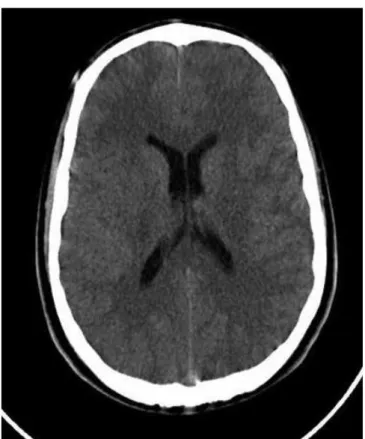
A 25-year-old male fell down while playing in a footbal match. He had headache and vertigo. Upon admission to the hospital, his arterial blood pressure was 130/80 mm Hg, his respiratory rate was 20/min, temperature was 36°C and his O2 saturation level was 98%. His heart rate was 82/min during auscultation, and the other system examinations revealed normal results. He had no history of chronic illness , and no drug or alcohol use. His Glasgow Coma Scale (GCS) score was 15 (E-4, V-5, M-6). His pupils were isochoric and the light relex was bilaterally positive during his physical examination. No focal inding, no evidence of meningeal irritation or pathologic relex was noted. On ad-mission, an initial BCT scan was performed revealing normal indings (Figure 1). He was kept under observation for about 12 hours at the emergency department and discharged from the hospital with normal neurological examination signs, normal signs of second BCT (Figure 2) and without complaints. He had no history of abnormally high systemic blood pressure occurring during the observation period. Clinically, no attacks of vertigo or nausea and focal neurological sign occurred. However, at the 26th hour ater the HT incident, he presented to our hospital again with the complaints of nausea, vomiting and headache. GCS was 15. A control BCT was performed and a traumatic intracerebral hematoma was determined in the frontal region (Figure 3). He was transferred to the neurosurgery clinic and he was discharged from the hospital without any sequela on the seventh day of his hospital stay.
Discussion
DTIH is a rare complication of head injury [2]. The etiopato-genesis of DTIH is not precisely known. However, the possible pathogenic mechanisms may include impairment of autoregu-lation, necrosis, structural abnormalities of the vessel walls af-ter injury, traumatic aneurysm, metabolic changes at the cellu-lar level, vasospasm, venous congestion as a result of increased intracranial pressure and coagulopathy at the region of trauma [4].
Figure 1. An initial BCT scan normal indings
Figure 2. Normal neurological examination signs, normal signs of second BCT ( About 12 hours at the emergency department)
| Journal of Clinical and Analytical Medicine 708
| Journal of Clinical and Analytical Medicine İntraserebral Kanama / Intracerebral Hemorrhage
3
BCT and MRI (Magnetic resonance imaging) is useful in diag-nosing intracerebral hematomas. BCT has been particularly helpful at the emergency department in increasing the diag-nosed patients population [4,6]. Fukamachi et al. reported that DTIH developed within 48 hours in 50 % of patients in whom CBT was normal or when there was a minimal hyperdense le-sion within the irst 6 hours [6]. Nagata et al. said that DTIH oc-cured in 54.5 % of patients who had normally appearing areas on the initial BCT [2].
Kaplan et al. stated that DTIH is commonly demonstrated with-in the irst post-traumatic 10 days, particularly with-in the irst 3 days [7]. Nagata et al. said that the time of occurrence of DTIH ranged from 4 hours to 4 days, with a mean of 31.5 hours [2]. In the literature, it has been stated that DTIH usually occurs within 72 hours ater HT [4] . In a pediatric patient group with 397 patients, Hamilton et al. reported the rate of DTIH as % 4.3 [1]. However, Nagata et al. [2] reported an average age of 25.5 and reported no gender predominance. In our study, the was 28 years of age and ICH was established 26 hours ater the HT event.
Nagata et al. [2] stated that DTIH was usually found in the pos-terior fossa and that its prognosis was known as unfavorable. However, in another study, it reported that the most common locations for DTIH were the frontal and the temporal lobes [4]. Kaplan et al. [7] said that DTIH was located in the occipito-parietal region by a countercoup mechanism. In our case, the intracerebral hemorrhage was located in the frontal region and he was discharged on the seventh day of hospitalization. Fortu-nately, our patient completely recovered.
In a study, While the clinical appearance is initially stable in these patients, a suddenly decreased GCS, increased focal neu-rological sign or focal seizures can ocur [4]. In Kutlay’s study, neurological examination was normal in 50% of cases with DTIH [4]. In our study, the patient’s clinical situation was stable and his neurological examination was normal. We performed CBT initially and 12 hours ater admission. He had normal CBT indings. However, he presented again to our emergency
de-partment 26 hours ater the HT event and an intracerebral he-matoma was determined.
The use of emergency CBT is still controversial in patients with mild HT [8]. In previous studies, it has been reported that the rates of detection of abnormal signs on the CBT varied in pa-tients with mild HT [8]. Furthermore, mortality and neurosurgi-cal intervention are rare in mild HT. Nevertheless, Kreitzer et al. reported that patients with mild HT demonstrated a low rate of adverse outcomes when observed for 6-24 hours [5].
Our case had a favorable outcome without surgery, but DTIH may be fatal. Close observation and repeat CBT scanning ren-der more favorable results. Therefore, patients with mild HT should undergo close observation and they should be given ad-vice regarding HT ater discharge. Furthermore, the observa-tion period may be extended and an outpatient control should be proposed within three days for patients with mild HT. Conlict of interest; The authors further declare that we have no inancial arrangement as product in the case report, sources of funding, institutional ailiations, and any possible inancial or personal conlicts of interest.
Competing interests
The authors declare that they have no competing interests.
References
1. Hamilton M, Mrazick M, Johnson D. Incidence of Delayed Intracranial Hem-orrhage in Children Ater Uncomplicated Minor Head Injuries. Pediatrics 2010; 1(126) : e33
2. Nagata K, Ishikawa T, Ishikawa T, Shigeno T, Kawahara N, Asano T, et al. Delayed traumatic intracerebellar hematoma: correlation between the location of the hematoma and the pre-existing cerebellar contusion: case report. Neurol Med Chir 1991;31(12):792-6.
3. Cete Y, Pekdemir M, Oktay C, Eray O, Bozan H, Ersoy FF. The role of computed tomography for minor head injury. Ulus Travma Dergisi 2001;7(3):189-94. 4. Kutlay M, Kıbıcı K, Demircan N. Early diagnosis, Follow up and Treatment of Delayed Traumatic intracerebral haematomas. Turkish Journal of Trauma- Emer-gency Surgery 1998;4(1):33-8.
5. Kreitzer N, Lyons MS, Hart K, Lindsell CJ, Chung S, Yick A, et al. Repeat neuro-imaging of mild traumatic brain-injured patients with acute traumatic intracranial hemorrhage: clinical outcomes and radiographic features. Acad Emerg Med 2014 ;21(10):1083-91.
6. Fukamachi A, Nagaseki Y, Kohno K, Wakao T. The incidence and developmental process of delayed traumatic intracerebral haematomas. Acta Neurochir (Wien) 1985;74(1-2):35-9.
7. Kaplan M, Ozveren MF, Topsakal C, Erol FS, Akdemir I. Asymptomatic interval in delayed traumatic intracerebral hemorrhage: report of two cases. Clin Neurol Neurosurg 2003 ;105(3):153-5.
8. Aygün D, Güven H, İncesu L, Şahin H, Doğanay Z, Altıntop L. The incidence of pathologic indings of the cranial computed tomography in patients with minor head trauma and its correlation with age and clinical features. Ulus Travma Derg 2003;9(2):129-33.
How to cite this article:
Karadas S, Gonullu H, Gulsen I, Baltacıoglu H. Delayed Traumatic Intracerebral Hemorrhage: For How Many Hours Should Patients with Mild Head Trauma be Observed? J Clin Anal Med 2015;6(suppl 5): 707-9.
Figure 3. A traumatic intracerebral hematoma was determined in the frontal re-gion
Journal of Clinical and Analytical Medicine | 709