CerebriformintradermalnevusasaCauseofcutis verticis gyrata
639
Rev Assoc Med Bras 2010; 56(6): 639-41
Cutis verticis gyrata (CVG) is a rare disease that is characte-rized by excess scalp skin, producing thick folds that form creases and ridges similar in appearance to the whorls of the cerebral cortex. It was first described in the literature by Robert in 1843 and the name cutis verticis gyrata was proposed by Unna in 1907 and remains the accepted name to date. 1,2 It has an estimated
prevalence of one case in every 100,000 men and 0,026 cases for every 100,000 women. 3 It can be an isolated manifestation
that is present from birth or it can be part of other syndromes.
Cutis verticis gyrata is classified as primary (subdivided into essential and non essential) or secondary. The essential primary form is not associated with neurological or ophthalmological disorders, and only the folds on the scalp mimicking the cerebral whorls appear. It appears in puberty and affects more men than women. The non essential primary form affects 0.5% of patients with mental retardation. Cerebral palsy, epilepsy, cataracts and blindness may also be present. 2,4,5,6
The secondary forms of CVG generally occur as a result of inflammatory or neoplastic processes that cause changes to the structure of the scalp. 2,6 One of the rarest forms of secondary
CVG is the nevoid form, in which it is melanocytic intradermal nevi that cause the cutaneous hypertrophy. This condition is known as cerebriform intradermal nevus (CIN) and was first linked with CVG in 1937 by Hammond and Ransom. 7 Patients
affected by CIN have normal intelligence and are free from other local or systemic diseases.
The objective of this paper is to describe a rare case of cere-briform intradermal nevus, discussing its histopathological and clinical features.
C
aseA white female patient, 43 years old, presented at the Instituto de Dermatologia Professor Azulay with large dimension lesions on the scalp. She stated that at birth she had had a small normal colored macula on her scalp and that it had gradually developed,
reaching its largest size during puberty. On physical examina-tion, multiple patches of alopecia were observed in addition to a normal-colored convoluted mass covering the right parietal, right temporal and right occipital regions and small areas of foul-smelling seborrheic dermatitis (Figures 1A and 1B). Neurological and ophthalmological test results were normal. Family history included no relevant details.
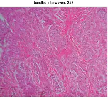
X-rays and computerized tomography of the head did not reveal damage to the calvarium. Blood tests and liver, kidney and thyroid function were normal. A cutaneous biopsy revealed mela-nocytic intradermal nevus with progressive maturation inwards towards the depth of the tissues. Nests and bundles reached the deep dermis and parts of the subcutaneous zone, thereby indicating the intradermal cerebriform subtype (Figures 2 and 3).
Figures 1 A and1 B
Convoluted, normal-colored mass with areas of alopecia
D
isCussionCutis verticis gyrata is a rare disease that is characterized by excess scalp skin, producing folds that are reminiscent of the
Cerebriform
intraDermal
nevus
as
a
Cause
of
cutis
verticis
gyrata
aguinalDo bonalumi filho1*, luiz guilherme Darrigo Jr2, João Carlos regazzi avelleira3, bernarD Kawa KaC4, DaviD rubem azulay5
Study conducted at Instituto de Dermatologia Professor Rubem David Azulay, Santa Casa de Misericórdia do Rio de Janeiro, RJ, Brazil
1- Professor da Pós-graduação de Dermatologia do Hospital Naval Marcilio Dias e Professor Correspondente do curso de Pós-graduação de Dermatologia do Instituto de Dermatologia Professor Rubem David Azulay da Santa Casa de Misericórdia do Rio de Janeiro, Rio de Janeiro, RJ
2- Doutorando em Medicina pela Faculdade de Medicina de Ribeirão Preto, Universidade de São Paulo, São Paulo, SP
3- Professor associado de Dermatologia do Instituto de Dermatologia Professor Rubem David Azulay da Santa Casa de Misericórdia do Rio de Janeiro, Rio de Janeiro, RJ 4- Professor do Curso de Pós-graduação de Dermatologia do Instituto de Dermatologia Professor Rubem David Azulay da Santa Casa de Misericórdia do Rio de Janeiro,
Rio de Janeiro, RJ
5-Chefe do Instituto de Dermatologia Professor Rubem David Azulay da Santa Casa de Misericórdia do Rio de Janeiro; Professor Titular do Curso de Pós-Graduação da Pontifícia Universidade Católica – RJ e Professor de Dermatologia da Fundação Técnico Educacional Souza Marques - FTESM e da Universidade Federal do Rio de Janeiro – UFRJ, Rio de Janeiro, RJ
filho ab etal.
640
Rev Assoc Med Bras 2010; 56(6): 639-41whorls of the cerebral cortex.2 The different forms of CVG are
classified as follows: 2,8,9
1. Primary
A. Essential: extremely rare form in which only the cutaneous manifestations are observed.
B. Non essential: associated with mental deficiency, schizo-phrenia, epilepsy, cerebral palsy, cranial abnormalities (espe-cially microcephaly) and cataracts.
2. Secondary
A. Inflammatory acute or chronic: dermatosis (eczema, psori-asis, impetigo, erysipelas, keloid folliculitis and pemphigus).
B. Proliferative: cerebriform intradermal nevus, lipoma, neurofibroma dermatofibroma.
C. Miscellaneous: acromegaly, pachydermoperiostosis, acro-megaly, syphilis, myxedema, cretinism, leukemia, Ehlers-Danlos syndrome, acanthosis nigricans, tuberous sclerosis, amyloidosis and diabetes mellitus.
D. Trauma.
Therefore, CIN is a rare form of secondary CVG, characterized by well-delimited lesions, that primarily affects the parietal region of the calvarium and prefers females. 7 Lesions are normally
present at birth as normal-colored or over-pigmented macules that increase in size and stand further proud as the years pass. The lesions gradually begin to take on their brain-like appearance which leads to alopecia in addition to the possibility of itching, bleeding, secondary infections and foul smell, due to bacterial proliferation in the folds. 8 The social and esthetic repercussions
of CIN make it extremely important.
With the patient described here, the lesions located in the parietal, temporal and occipital areas are in line with the majority
of reports in the literature. 10 Furthermore, although rare, the
brain-like lesions can affect other areas and cases have been described where the neck and scrotum were involved as has a case where a malignant peripheral melanoma was associated with a giant melanocytic intradermal nevus. 11,12,13 With relation
to size, CIN can vary from 2 x 3 cm to 22.5 x 25 cm 7,14
Diagnosis of CIN is based on clinical findings and confirmed by histopathology. 7 Characteristics of a positive histopathological
result are: nevocytes arranged intradermally with a minimal junctional component, possibly affecting superficial parts of the hypodermis; adjacent structures enveloped by nevocytes; loss of the typical arrangement of nests in stretches; a prevalence of type C nevus cells with large areas of neuroid differentiation; irregular arrangement and distribution of intracellular melanin pigment and perivascular nevocyte aggregation. 15
O differential diagnosis is to rule out primary CVG, the other forms of secondary CVG and other pathologies such as: cerebri-form nevus sebaceous and aplasia cutis congenita. 7
Treatment consists of surgical excision with plastic recon-struction where necessary. Small lesions can be removed by simple excision and suture. Where lesions are larger, flap rotation or serial surgeries should be used, with or without an expander or other techniques, minimizing esthetic deformities. 10
The possibility that these lesions may undergo considerable growth during puberty means that early excision and suture should be considered in order to avoid extensive surgery later in life. Another argument in favor of removal of CIN is the possibility of melanomas, although they are rare. 10
r
eferenCes1. Unna PG. Cutis verticis gyrata. Monatsschr Prakt. 1907;45:227-33.
Figure 2 - Nests and bundles of nevus cells, showing accentuated focal intracellular melanin pigmentation, with dermal collagen
bundles interwoven. 25X
Figure 3 - Presence of nests of irregularly pigmented nevocytes, arranged in the reticular dermis . The sections are tangential,
CerebriformintradermalnevusasaCauseofcutis verticis gyrata
641
Rev Assoc Med Bras 2010; 56(6): 639-41
2. Schenato LK, Gil T, Carvalho LA, Ricachnevsky N, Sanseverino A, Halpern R. Cutis verticis gyrata primária essencial. J. Pediatr (Rio J). 2002;78(1):75-80. 3. Larsen F, Birchall N. Cutis verticis gyrata: three cases with different aeti-ologies that demonstrate the classification system. Australas J Dermatol. 2007;48(2):91-4.
4. Tan O, Ergen D. Primary essential cutis verticis gyrata in an adult female patient: a case report. J Dermatol. 2006;33(7):492-5.
5. Radwanski HN, Almeida MW, Pitanguy I. Primary essential cutis verticis gyrata- a case report. J Plast Reconstr Aesthet Surg. 2009;62(11):e 430-3. 6. Azulay RD, Azulay DR, Azulay LA. Dermatologia. 5ª ed. Rio de Janeiro:
Guanabara Koogan; 2008.
7. von Geest AJ, Berretty PJ, Klinkhamer PJ, Neumann HA. Cerebriform intra-dermal naevus (a rare form of secondary cutis verticis gyrata). J Eur Acad Dermatol Venereol. 2002;16(5):529-31.
8. Orkin M, Frichot BC 3rd, Zelickson AS. Related Articles, Links. Cerebri-form intradermal nevus. A cause of cutis verticis gyrata. Arch Dermatol. 1974;110(4):575-82.
9. Prado C, Hernando V, Esperon G, Cardama, Rial C, Cimadevila P. Intradermal cerebriform nevus. Actas Dermosifiliogr. 1980;71(3-4):143-50.
10. Hamm JC, Argenta LC. Related Articles, Links. Giant cerebriform intradermal nevus. Ann Plast Surg. 1987;19(1):84-8.
11. Jeanfils S, Tennstedt D, Lachapelle JM. Related Articles, Links. Cerebriform intradermal nevus. A clinical pattern resembling cutis verticis gyrata. Derma-tology. 1993;186(4):294-7.
12. Gross PR, Carter DM. Malignant melanoma arising in a giant cerebriform nevus. Arch Dermatol. 1967;96(5):536-9.
13. Goldstone S, Samitz MH, Carter DM. Giant cerebriform nevus of the scalp with malignant melanoma and metastases. Arch Dermatol. 1967;95(1):137. 14. Tabata H, Yamakage A, Yamazaki S. Related Articles, Links Cerebriform
intra-dermal nevus. Int J Dermatol. 1995;34(9):634.
15. Magnin PH, Schroh RG, Cajas R, Pacheco E, Aguirre L. Particularidades del nevo melanocitico cerebriforme. Rev Argent Dermatol. 1987;68(5):341-7.
*Correspondence: