Abstract
Objectives: To determine the frequency of hypomagnesaemia in pediatric patients after spinal fusion, to verify whether postoperative magnesium levels were lower than preoperative levels and, if so, to identify possible causes and assess the clinical repercussions for patients.
Methods: This was a retrospective descriptive study of pediatric patients admitted to a pediatric intensive care unit (ICU) after spine fusion surgery, between March 1 and August 31, 2011. Preoperative magnesium, phosphorus and total and ionized calcium concentrations were compared with the results of tests conducted during the irst 24 hours after admission to the ICU.
Results: A total of 45 patients were enrolled on the study. Median age was 13.1 years. Preoperative mean serum magnesium was 1.8±0.2 mg/dL and postoperative serum magnesium was 1.4±0.2 mg/dL, which was a signiicant reduction between the two periods (p < 0.001). The frequency of hypomagnesaemia rose from 1 patient (2%) in the preoperative period to 31 patients (68%) during the postoperative period. There were also signiicant reductions in concentrations of phosphorus (p < 0.001) and total calcium (p < 0.001). There was a signiicant correlation between magnesium reductions and the volume of luids administered during the surgery (p = 0.03), transfused blood volume (p < 0.001) and number of vertebrae fused (p < 0.05). Seven of the 31 patients with hypomagnesemia exhibited symptoms (22%).
Conclusion: There was an elevated frequency of hypomagnesemia in patients who underwent spinal fusion. Serum magnesium should be assayed when patients are admitted to the pediatric ICU, so appropriate supplementation can be initiated immediately, minimizing the risk of complications.
J Pediatr (Rio J). 2012;88(3):227-32: Hypomagnesemia, pediatric ICU, spine fusion. Copyright © by Sociedade Brasileira de Pediatria
227
Introduction
Although hypomagnesemia is one of the most common electrolyte disorders among hospitalized patients, especially among the critically ill1,2 and during the postoperative period,1 it is also considered the most underdiagnosed of these disorders.3 It affects around 11% of hospitalized patients; 20% of patients admitted to Intensive Care Units (ICU); and 61% of patients admitted to surgical ICUs.3 It is associated with prolonged hospitalization, with increased length of stay in an ICU and with increased mortality, both among clinical and surgical patients.1
There are few reports in the literature of orthopedic patients with hypomagnesemia,1,3 but it has been observed to affect an elevated proportion of patients receiving postoperative care after surgical procedures with major bone involvement, such as spinal fusion.1,3,4 In such cases, the etiology of magnesium deiciency is considered to be multifactorial: it may be blood-transfusion related (the citrate chelates magnesium, forming magnesium citrate)1,3 it may involve dilution, resulting from administration of large volumes of hypotonic luids during surgery;1,3,4
O
riginala
rticleHypomagnesemia after spinal fusion
Larissa Rossato Chrun,1Paulo Ramos David João2
1. Resident physician, Unidade de Terapia Intensiva Pediátrica, Hospital Pequeno Príncipe, Curitiba, PR, Brazil. 2. Head, Unidades de Terapia Intensiva Pediátrica e Cirúrgica, Hospital Pequeno Príncipe, Curitiba, PR, Brazil.
No conflicts of interest declared concerning the publication of this article.
Suggested citation: Chrun LR, João PR. Hypomagnesemia after spinal fusion. J Pediatr (Rio J). 2012;88(3):227-32. Manuscript submitted Oct 16 2011, accepted for publication Jan 18 2012.
and it can be secondary to a transient parathyroid dysfunction, caused by disruption of the periosteum and bone tissue.4 Irrespective of etiology, patients with hypomagnesemia can present with the following principal signs and symptoms: atrial and ventricle arrhythmia,2 myocardial ischemia,3,5 heart failure2 and neurological manifestations, such as convulsive crises2,4,6,7 and reduced level of consciousness.6
Place et al.3 demonstrated a signiicant decrease in serum magnesium levels during the postoperative period after spinal fusion, when compared with the results of tests performed immediately before surgery, and reported hypomagnesemia incidence of 71%. In contrast, a study conducted by Chang et al.1 reported a lower incidence of hypomagnesemia (13%), but the reduction in serum magnesium levels from the preoperative to the postoperative period was also signiicant in their study. Both studies investigated adults.
Verive et al.4 conducted one of the few studies to investigate pediatric patients, assessing 463 children admitted to ICUs with clinical and surgical pathologies. On admission, hypomagnesemia was detected in 11% of patients, but the greatest frequency (72%) and the lowest magnesium levels were found in patients who had undergone surgery involving bone tissues.
The objectives of this study were to determine the frequency of hypomagnesemia among patients admitted to a pediatric ICU for postoperative care after spinal fusion, to investigate whether they suffered signiicant falls in serum magnesium levels between the preoperative and postoperative periods and, if so, to identify possible causes and assess the clinical repercussions for the patients.
Methods
We reviewed the medical records of patients admitted for immediate postoperative care after spinal fusion to a surgical ICU at the Hospital Infantil Pequeno Príncipe (Curitiba, PR, Brazil) from 1st March to 31st August of 2011.
During this period, 52 patients underwent spinal fusion surgery and were admitted to the surgical ICU after the procedure. Seven of these were excluded because of data missing from medical records. None of the patients were being administered oral or intravenous magnesium supplementation and none had renal dysfunction, which would have been grounds for exclusion. The inal number of patients enrolled on the study was therefore 45.
This was a retrospective descriptive study. The project was approved by the Research Ethics Committee at the Hospital Pequeno Príncipe.
We investigated whether there was a signiicant decrease in serum magnesium levels from the preoperative to postoperative period; whether there were associations
between magnesium reductions and the volume of luids administered during the procedure (mL/kg), the volume of blood transfused (mL/kg) or the number of vertebrae fused during the operation; and whether there was a signiicant difference between patients with and without symptoms of hypomagnesemia in terms of the reduction in magnesium levels. In view of the strict relationship between magnesium, phosphorous and calcium metabolism,4,8 phosphorous and calcium assays performed before and after the operation were analyzed for signiicant changes in electrolyte levels.
The following ranges were deined as normal: 1.6 to 2.6 mg/dL for magnesium, 2.5 to 4.5 mg/dL for phosphorous, 8.8 to 10.7 mg/dL for total calcium and 1.16 to 1.32 mmol/L for ionized calcium.
Initially, a descriptive analysis was conducted using frequencies, means and standard deviations. Student’s t test for repeated measures was used to compare preoperative and postoperative electrolyte assay results (magnesium, phosphorous, total and ionized calcium).
Pearson’s linear correlation test was used to correlate reductions in serum magnesium with blood transfusion volumes and luids volumes administered intraoperatively. Spearman’s correlation was used to test for correlations between reductions in magnesium levels and the number of vertebrae fused during surgery. Finally, the Mann-Whitney test was used to compare patients with and without symptoms of hypomagnesemia on the basis of reductions in serum magnesium levels.
Graphs were plotted using Statistica and tables were constructed in Excel 2007. The signiicance level was set at 5%.
Results
We enrolled a total of 45 patients on the study who had been admitted to the surgical ICU at the Hospital Pequeno Príncipe during the immediate postoperative period after spinal fusion operations. Thirty-three of them (n = 33, 73.3%) were female, 12 (26.6%) were male and their ages ranged from 9 to 17 years (mean: 13.1±2.2).
All of the patients had scoliosis, which was the reason they were referred for spinal fusion surgery, but 20 of them also had comorbidities: nine had cerebral palsy (one of these nine also had congenital heart disease); three had myelomeningocele; two had neuroibromatosis; three had spinal muscular atrophy type 2; one had a cognitive deicit with no deinite etiology; one had an unidentiied genetic syndrome; and one had a history of cancer that had been treated (rhabdomyosarcoma).
Table 1 - Means, standard deviations and probability (p) according to Student’s t test: comparison of preoperative and postoperative serum magnesium, phosphorous, total and ionized calcium assay results
* p < 0.05.
Preoperative Postoperative
Variable Mean Standard deviation Mean Standard deviation p
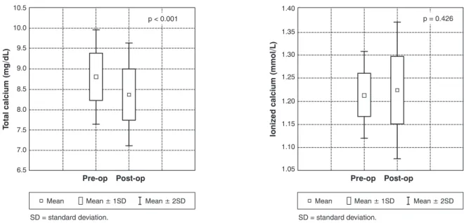
Total calcium 8.8 0.5 8.3 0.6 < 0.001*
Phosphorous 4.6 0.6 3.9 0.8 < 0.001*
Ionized calcium 1.21 0.05 1.22 0.07 0.426
Magnesium 1.8 0.2 1.4 0.2 < 0.001*
Table 2 - Correlation coefficients (r) and coefficients of determination (R2) and p values according to the Pearson and Spearman tests: associations between reductions in serum magnesium and luids volume, blood transfusion volume and number of vertebrae fused
Variable r R2 p
Fluids volume 0.357 12.7% 0.030*
Blood transfusion volume 0.540 29.2% < 0.001*
Number of vertebrae 0.491 24.1% < 0.05*
* p < 0.05. with normal total calcium; and none of the patients had
hypophosphatemia.
In contrast, postoperative electrolyte assays conducted during the irst 24 hours after admission to the ICU showed that 31 patients (68% of the sample), had hypomagnesemia. Hypophosphatemia was detected in just two patients (4%); total hypocalcemia was found in 13 patients (28%), although all of these had normal ionized calcium; and ionized hypocalcemia was present in nine patients (20%). Magnesium, phosphorous and total calcium all suffered signiicant reductions between the preoperative and postoperative periods, but this was not true of ionized calcium levels (Table 1).
Just three (9%) of the 31 patients with postoperative hypomagnesemia also had other electrolyte disorders while in the ICU: one had hypokalemia, hypophosphatemia and hypocalcemia; one had hypophosphatemia; and one had hyponatremia.
The mean volume of luids administered intraoperatively was 101.4±51 mL/kg. Thirty-six patients (n = 36, 80%) needed blood transfusions during the procedure and were given a mean volume of 12.5±7.9 mL/kg. The number of vertebrae fused during surgery ranged from four to 22, with a mean of 12.3±4.2. There were signiicant correlations between reductions in serum magnesium and the volume of luids given during surgery, transfused blood volume and number of vertebrae fused, but the coeficients of determination for all three variables were below 30% (Table 2).
Symptoms attributable to hypomagnesemia were observed in seven (22%) of the 31 patients with the imbalance. Disorders of cardiac rhythm and function were the most common indings: three patients had bradycardia, one of whom also had extrasystoles; one had tachyarrhythmia (supraventricular tachycardia); one had a left ventricle systolic dysfunction, with acute pulmonary edema and required reintubation. Other symptoms
observed were a reduced level of consciousness in one patient and convulsive crises in another. There was no signiicant difference between patients with symptomatic and asymptomatic hypomagnesemia in terms of the magnitude of the reduction in serum magnesium (p = 0.16).
All of the patients with postoperative hypomagnesemia were given appropriate intravenous magnesium supplementation, according to the routine protocol at our service (0.4 mEq/kg of magnesium, in a 50% magnesium sulphate presentation, given with maintenance saline over 24 hours). All of the symptomatic patients exhibited improvement in hypomagnesemia in the ICU, while being given intravenous magnesium in addition to treatment for their speciic case where needed.
Figure 1 - Comparison of preoperative and postoperative serum total calcium and serum ionized calcium assay results
SD = standard deviation. SD = standard deviation.
p < 0.001 p = 0.426
Figure 2 - Comparison of preoperative and postoperative serum magnesium and serum phosphorous assay results
SD = standard deviation. SD = standard deviation.
p < 0.001 p < 0.001
Discussion
In this study we observed signiicant decreases in serum magnesium, total calcium and phosphorous levels during the postoperative period after spinal fusion surgery, when compared with preoperative baselines. However, in the majority of patients only magnesium reduced suficiently to drop below the lower limit of normality and there was no signiicant fall in ionized calcium assay results (Figures 1 and 2).
An association between hypomagnesemia and other electrolyte disorders is described in the literature and has been observed by several authors,2,4,6,8,9 but was not signiicant in this sample, since just three patients had other disorders. This could be because spinal fusion surgery is elective; patients are not critically ill and it is therefore less likely that other electrolyte disorders will occur.
Serum magnesium levels can be affected by other factors, such as: prolonged hospitalization, prior nutritional status and medications (loop diuretics, aminoglycosides, amphotericin B, cyclosporine),3,4 which can cause hypomagnesemia prior to surgery.6,8 However, once again, since this is an elective procedure, these factors are less common. Indeed, in the sample described here, just one patient had preoperative hypomagnesemia and this case had no other comorbidities. Even the nine patients with cerebral palsy, which is often associated with malnutrition, had normal preoperative magnesium levels.
The cause for the reduction in serum magnesium levels observed postoperatively has not yet been fully elucidated.1,3 The association described here between the reduction in magnesium levels and the volumes of blood transfused and liquids administered intraoperatively has been described in the literature1-4 and has been shown in previous studies, such as one published by Chang et al.;1 but contrasts with results published by Verive et al.4 and Tatari et al.,10 who did not detect this relationship in practice.
The administration of large quantities of hypotonic luids, which leads to hemodilution, and massive transfusions, which involve magnesium citrate formation, both contribute to the hypomagnesemia observed after spinal fusion surgery, but not suficiently to explain the entire reduction,1 since the same rate of hypomagnesemia has not been observed after other types of surgery during which similar volumes of luids and blood were administered.4
Another cause of postoperative hypomagnesemia that has been described with relation to spinal fusion surgery is an association with the syndrome of inappropriate antidiuretic hormone hypersecretion.4,11,12 However, hypomagnesemia has also been observed in connection with other bone surgery procedures that cannot be linked with this factor, as is the case of surgery to correct craniostenosis and craniofacial anomalies.4 None of the patients enrolled on the study had signs of the syndrome.
Thus, the best explanation for the decrease in serum magnesium observed in this study involves changes to normal calcium, phosphorous and magnesium metabolism, caused by disruption of bone tissues and periosteum, which is where the greater part of free magnesium is found in the body.4 Spinal fusion surgery involves disruption of a considerable quantity of bone tissue, which results in rapid release of magnesium, calcium and phosphorous into the extracellular space, and consequent transient dysregulation of parathyroid function.4
Transient increases in serum ionized calcium levels could be enough to reduce parathyroid hormone secretion and increase calcitonin levels,8 which would in turn reduce tubular resorption of magnesium and increase excretion of calcium, phosphorous and magnesium.8 Hypomagnesemia would therefore primarily be the result of renal excretion, caused by this change to parathyroid metabolism, triggered by the insult to bone tissue.4 Urinary magnesium assays were not conducted, which we consider the principal limitation of this study.
On this basis, it is to be expected that there would be an association between the reduction in magnesium after surgery and the number of vertebrae fused during surgery, since the greater the bone tissue involvement, the greater the changes to parathyroid metabolism would be expected to be, and, consequently, the greater the reduction in serum magnesium. Both the volume of luids administered and the volume of blood transfused intraoperatively correlated signiicantly with the reduction in magnesium levels, but neither had a determination coeficient (R²) greater than 30%. It can therefore be stated that a correlation exists, but it is not particularly strong. These results contrast with those found by Tatari et al.,10 who conducted a study in which no association was found with blood transfusions or the number of vertebrae fused.
The inal effect, as observed here, is a reduction in serum levels of magnesium, phosphorous and total calcium during the postoperative period, although only magnesium fell far enough to have clinical repercussions. Ionized calcium did not decrease signiicantly, which can be explained by parathyroid metabolism,6,8,9 which is capable of maintaining the ionized fraction of calcium (which is the biologically active fraction)5 within normal limits. The reduction in total calcium did not therefore have clinical repercussions despite falling below normal levels in 13 patients during the postoperative period, since the ionized fraction remained normal in these cases.
It is worth remembering that, although there is an ionized fraction of magnesium and this is the biologically active fraction, the ionized fraction is directly proportional to the total magnesium level,5 which means there is no need to assay it, since decreases can be estimated from the reduction in total magnesium.5,13 This is why total magnesium was used in this study.
Just seven (22%) of the 31 patients with hypomagnesemia exhibited symptoms; which is a considerably higher percentage than that observed by Place et al.,3 who reported 5%. The fact that the majority of patients with hypomagnesemia remain asymptomatic means that there is still debate on the need for intravenous magnesium supplementation in patients who do not exhibit clinical repercussions of the disorder.1-3
Correspondence: Larissa Rossato Chrun Rua Emilio Cornelsen, 398/503 CEP 80540-220 - Curitiba, PR - Brazil
Tel.: +55 (41) 9996.6327, +55 (41) 3044.4760 E-mail: lary_rc@yahoo.com.br
References
1. Chang CH, Nam SB, Lee JS, Han DW, Lee HK, Shin CS. Changes in ionized and total magnesium concentration during spinal surgery. Korean J Anesthesiol. 2007;52:S37-41.
2. Soliman HM, Mercan D, Lobo SS, Mélot C, Vincent JL. Development of ionized hypomagnesemia is associated with higher mortality rates. Crit Care Med. 2003;31:1082-7.
3. Pace HM, Enzenauer RJ, Muff BJ, Ziporin PJ, Brown CW. Hypomagnesemia in postoperative spine fusion patients. Spine (Phila Pa 1976). 1996;21:2268-72.
4. Verive MJ, Irazuzta J, Steinhart CM, Orlowski JP, Jaimoviclh DG. Evaluating the frequency rate of hypomagnesemia in critically ill pediatric patients by using multiple regression analysis and a computer-based neural network. Crit Care Med. 2000;28:3534-9.
5. Koch SM, Warters RD, Mehlhorn U. The simultaneous measurement of ionized and total calcium and ionized and total magnesium in intensive care unit patients. J Crit Care. 2002;17:203-5. 6. Marino PL. Magnesium. In: Marino PL, Sutin KM, eds. The ICU Book.
Philadelphia: Lippincott Williams & Wilkins; 2007. p. 625-38. 7. Deshmukh CT, Rane SA, Gurav MN. Hypomagnesaemia in
paediatric population in an intensive care unit. J Postgrad Med. 2000;46:179-80.
8. Bossolan RM, Ernesto LC, Hirschheimer MR. Distúrbios hidroeletrolíticos do cálcio, do fósforo e do magnésio. In: Carvalho WB, Hirschheimer MR, Matsumoto T, eds. Terapia Intensiva Pediátrica. 3ª edição. São Paulo: Atheneu; 2010. p. 743-63. 9. Baran DT, Aronin N. Disorders of mineral metabolism. In: Irwin RS,
Rippe JM, eds. Irwin and Rippe’s Intensive Care Medicine. 6th ed. Philadelphia: Lippincott Williams & Wilkins; 2008. p. 1287-93. 10. Tatari H, Işlekel H, Altekin E, Göçen S, Ozcan C, Ergör A. Serum
magnesium, copper, and zinc alterations following spinal fusion. Biol Trace Elem Res. 2001;80:33-42.
11. Lieh-Lai MW, Stanitski DF, Sarnaik AP, Uy HG, Rossi NF, Simpson PM, et al. Syndrome of inappropriate antidiuretic hormone secretion in
children following spinal fusion. Crit Care Med. 1999;27:622-7. 12. Bell GR, Gurd AR, Orlowski JP, Andrish JT. The syndrome of
inappropriate antidiuretic-hormone secretion following spinal fusion. J Bone Joint Surg Am. 1986;68:720-4.
13. Motoyama E, Yamada A, Cicarelli DD, Benseñor FE, Vieira JE. Relação entre magnésio sérico total e mortalidade em pacientes com síndrome da resposta inlamatória sistêmica em unidade de terapia intensiva pós-operatória. Rev Bras Ter Intensiva. 2005;17:262-4.
disorders, such as increased PR interval and QRS complex, atrioventricular block and asystole.3 These conditions are however rare as long as supplementation dosages are appropriate.5 In this sample, not only did we fail to detect any collateral effects resulting from supplementation, but all patients exhibited improvement of symptoms, as was the case in the study conducted by Place et al.3
There was no signiicant difference between symptomatic and asymptomatic patients in terms of reduction in magnesium levels. Therefore, the magnitude of the reduction from the preoperative to the postoperative period did not predict which patients would suffer clinical repercussions from hypomagnesemia.
There is an elevated incidence of hypomagnesemia among patients subjected to spinal fusion surgery, with signiicant reductions in serum magnesium against baseline preoperative levels. The causes of this reduction can be considered multifactorial, but have a direct relationship with disruption of bone tissue.
Although the majority of patients do not exhibit symptoms attributable to the disorder, there is a risk of potentially lethal complications, which is why these patients should have their magnesium levels tested soon after the procedure.4
In view of the good results observed with appropriate treatment and considering that supplementation is administered in an ICU environment, with due monitoring of heart rhythm and periodic laboratory tests, the beneits undoubtedly outweigh the risks. When hypomagnesemia is diagnosed at the time of admission to the ICU, appropriate supplementation should be initiated promptly, reducing the risk of complications.5