ISSN 1806-3713 © 2017 Sociedade Brasileira de Pneumologia e Tisiologia
http://dx.doi.org/10.1590/S1806-37562016000000295
Multiple cavitated nodules
Edson Marchiori1, Bruno Hochhegger2,3, Gláucia Zanetti1
1. Universidade Federal do Rio de Janeiro, Rio de Janeiro (RJ) Brasil. 2. Santa Casa de Misericórdia de Porto Alegre, Porto Alegre (RS) Brasil.
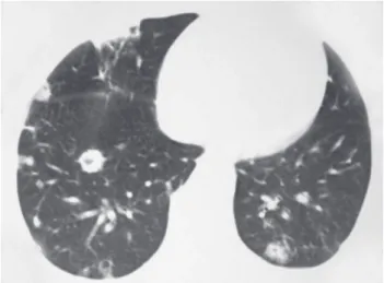
3. Universidade Federal de Ciências da Saúde de Porto Alegre, Porto Alegre (RS) Brasil. A 24-year-old male patient, who was an injection cocaine user, presented with about a 10-day history of cough and fever. A CT scan (Figure 1) showed multiple nodules, some of which were cavitated, with a predominant peripheral distribution.
The differential diagnosis of multiple cavitated nodules includes neoplastic diseases (metastases, lymphomas, etc.) and infectious diseases (septic embolism, granulomatous diseases, etc.), as well as other less frequent etiologies (nodular sarcoidosis, rheumatoid nodules, Wegener’s granulomatosis, nodular amyloidosis, etc.).
The most common causes are cavitated metastases and septic embolism. The frequency of cavitation in metastatic nodules is much lower than is that observed in primary tumors. Squamous cell carcinomas are the tumors that most commonly cause cavitated metastases, accounting on average for 70% of such cases. Tumors of the head, neck, reproductive system, and large intestine are the most common primary sites, although any primitive tumor can in principle cause cavitated metastases. In metastases, cavitations originate both from tumor necrosis and from the formation of a check-valve mechanism, because of
neoplastic iniltration into the distal airways. The cavitation
walls are more frequently thick and irregular, but they can also be thin, similar to cysts.
Septic embolism is caused by embolization of microor-ganism-infected fragments to the lungs. The disease is most commonly secondary to right-sided endocarditis or to septic thrombophlebitis, but it can occur secondary to the use of infected intravascular catheters, to suppurative processes of the skin, head or neck, or to contamination
related to intravenous drug use. CT indings consist of
multiple, predominantly peripheral, bilateral, well- or
ill-deined nodules exhibiting varying degrees of cavitation.
Findings of associated peripheral triangles frequently correspond to infarction due to vascular occlusion. Septic embolism can be accompanied by unilateral or bilateral pleural effusion.
Clinical signs are very important for differential diagnosis. The presence of a known primary tumor should raise the suspicion of lung metastases. Patients with metastases are frequently asymptomatic from a pulmonary standpoint. Septic embolism is clinically characterized by fever, dyspnea, cough, and pleuritic pain. Blood culture can be positive. Laboratory tests can be key to diagnosing rheumatoid nodules or Wegener’s granulomatosis. The patient in the case reported here had clinical signs of an infectious process, and blood culture was positive for Streptococcus viridans. The inal diagnosis was septic embolism secondary to intravenous drug use.
Figure 1. CT scan with lung window settings at the level of the lung bases, showing multiple nodules of various sizes, many of which were cavitated, with a predominant peripheral distribution.
RECOMMENDED READING
Fraser RS, Müller NL, Colman NC, Pare PD, editors. Diagnosis of Diseases of the Chest. 4th ed. Philadelphia: WB Saunders Company; 1999.
J Bras Pneumol. 2017;43(2):85-85
85