PetCO
2
, VCO
2
and CorPP Values in the Successful
Prediction of the Return of Spontaneous
Circulation: An Experimental Study on
Unassisted Induced Cardiopulmonary Arrest
Ana Carolina Longui Macedo
1, MSc; Luiz Claudio Martins
1, MD, PhD; Ilma Aparecida Paschoal
1, MD, PhD; Carlos
Cesar Ivo Sant’Ana Ovalle
2, PhD; Sebastião Araújo
1, MD, PhD; Marcos Mello Moreira
1, PhD
Abstract
Introduction: During cardiac arrest, end-tidal CO2 (PetCO2), VCO2 and coronary perfusion pressure fall abruptly and tend to return to normal levels after an effective return of spontaneous circulation. Therefore, the monitoring of PetCO2 and VCO2 by capnography is a useful tool during clinical management of cardiac arrest patients.
Objective: To assess if PetCO2, VCO2 and coronary perfusion pressure are useful for the prediction of return of spontaneous circulation in an animal model of cardiac arrest/cardiopulmonary resuscitation treated with vasopressor agents.
Methods: 42 swine were mechanically ventilated (FiO2=0.21). Ventricular fibrillation was induced and, after 10 min, unassisted cardiac arrest was initiated, followed by compressions. After 2 min of basic cardiopulmonary resuscitation, each group received: Adrenaline, Saline-Placebo, Terlipressin or Terlipressin + Adrenaline. Two minutes later (4th min of cardiopulmonary resuscitation), the
animals were defibrillated and the ones that survived were observed for an additional 30 min period. The variables of interest were
recorded at the baseline period, 10 min of ventricular fibrillation, 2nd
min of cardiopulmonary resuscitation, 4th min of cardiopulmonary
resuscitation, and 30 min after return of spontaneous circulation. Results: PetCO2 and VCO2 values, both recorded at 2 min and 4 min of cardiopulmonary resuscitation, have no correlation with the return of spontaneous circulation rates in any group. On the other hand, higher values of coronary perfusion pressure at the 4th min of cardiopulmonary resuscitation have been associated with increased return of spontaneous circulation rates in the adrenaline and adrenaline + terlipressin groups.
Conclusion: Although higher values of coronary perfusion pressure at the 4th min of cardiopulmonary resuscitation have been associated with increased return of spontaneous circulation rates in the animals that received adrenaline or adrenaline + terlipressin, PetCO2 and VCO2 have not been shown to be useful for predicting return of spontaneous circulation rates in this porcine model.
Keywords: Heart Arrest, Induced. Cardiopulmonary Resuscitation. Capnography. Epinephrine.
DOI: 10.5935/1678-9741.20160093
1Universidade Estadual de Campinas (UNICAMP), Campinas, SP, Brazil.
2Universidade Paulista, São Paulo, SP, Brazil.
This study was carried out at Universidade Estadual de Campinas (UNICAMP), Campinas, SP, Brazil.
This study was funded by the São Paulo Research Foundation (FAPESP), process Nº 07/08315-0, and the Teaching, Research and Extension Support Fund (FAEPEX), UNICAMP, process Nº 17809.
No conflict of interest.
Correspondence Address: Marcos Mello Moreira
Universidade Estadual de Campinas – Cidade Universitária “Zeferino Vaz” R. Tessália Vieira de Camargo, 126 – Campinas, SP, Brazil – Zip code: 13083-887 E-mail: [email protected]
Article received on June 24th 2016 Article accepted on September 19th 2016
Abbreviations, acronyms & symbols
ADR AHA CA CorPP CPR DC IMV PetCO2 ROSC TP VF V/Q
=Adrenaline
=American Heart Association =Cardiac arrest
=Coronary perfusion pressure =Cardiopulmonary resuscitation =Decreased cardiac output =Invasive mechanical ventilation =End-tidal CO2
=Return of spontaneous circulation =Terlipressin
=Ventricular fibrillation =Ventilation/perfusion ratio
INTRODUCTION
mechanical ventilation in order to oxygenate the blood that reaches the lungs[1].
Capnography presents itself as a non-invasive method, applicable at the bedside, that allows for the assessment of cardiorespiratory status both in experimental[2-6] and clinical
studies[7-12]. In addition, it is considered an indicator and/or
guide for decisions that enables the assessment of the quality of the CPR maneuvers[13]. Capnography evaluates and monitors
physiological conditions by measuring exhaled CO2 through an
infrared light sensor. CO2 excretion (VCO2) and partial pressure
of CO2 at the end of the exhalation (PetCO2) are indicative of O2
consumption by the oxidative metabolism in the tissues and those values are closely related to the pulmonary ventilation/ perfusion ratio (V/Q); hence, it is expected that both will increase in an effective CPR. Therefore, the monitoring of exhaled CO2 has
been proposed and used as a non-invasive method to assess cardiorespiratory function, especially in situations of decreased cardiac output (DC), such as during shock and CPR, and its use is compulsory in Surgical Centers[14].
Capnography has been regarded as a potentially useful monitoring method in evaluating the effectiveness of CPR maneuvers, despite limitations and controversies surrounding the subject[15]. Animal and human studies have shown a
good correlation between PetCO2 and DC during stages of
decreased blood flow and during CPR[16]. PetCO
2 can reflect the
pulmonary blood flow generated in CPR if CO2 production and
alveolar ventilation are relatively constant during resuscitation maneuvers; however, it is difficult to be measured when CPR is stopped because of changes in the alveolar dead space and minute volume ratio, which affects the correlation between PetCO2 and DC[17].
Some studies suggest that the increase in PetCO2 during CA
is a predictor of the success of the CPR. Considering the event of cardiac arrest, CPR and the post-event, we can find several changes in PetCO2 levels. It is known that the values of PetCO2 at
the beginning of ventricular fibrillation (VF) fall significantly, and this reduction is attributed to decreased pulmonary blood flow, which is insufficient to carry and eliminate the CO2 produced in
the tissues. Evidently, in extreme cases of low DC, there would also be a lower CO2 production because of the anaerobic
metabolism, given the low O2 supply to the tissues. Once CPR is
started, and it is effective in oxygenating the blood and increasing the tissue flow, PetCO2 values also increase, as an increase in
blood flow must occur in the pulmonary capillaries, which in turn results in the exhalation of CO2. When the return of spontaneous
circulation (ROSC) occurs, those values increase significantly, reaching levels comparable to those before CA. These changes are useful to quantify the effectiveness and success of the CPR maneuvers as well as to assess the cardiorespiratory status of the patient after ROSC. It has been observed that there is no survival for a PetCO2 < 5 mmHg[18].
Some studies have mentioned a few factors that can affect the levels of CO2 eliminated by expiration. Among them, we
can mention alveolar ventilation, DC, the area of distribution of blood flow in the body, and the production of CO2 by tissues.
Some authors also reported that the measurement of CO2 is
not able to reflect the certain success of CPR, since the results
do not confirm those reported in studies that have measured other parameters[19]. However, capnography is still used and
regarded as the best and most effective non-invasive method of measuring the elimination of CO2 in Emergency Rooms, Surgery
Centers, and ICU in cases of CA, being considered essential when performing CPR, for decision-making, assessment of its initial success (ROSC), and subsequent clinical evolution (cardiorespiratory stabilization).
The objective of this study was to assess if PetCO2, VCO2,
and coronary perfusion pressure (CorPP) values are useful in predicting the success of ROSC in an animal model of CA/CPR using vasopressor agents.
METHODS
This study was approved by the Institutional Review Committee for Experiments with Animals (EAEC-IB-Unicamp-1276-1/2007) and it was conducted in the laboratory of Experimental Surgery and Medicine, School of Medical Sciences – Universidade Estadual de Campinas (UNICAMP), São Paulo, Brazil.
The methods used were the same as the ones described in the novel article of Ovalle et al.[20], using forty-two
Large-White, immature swine, weighing approximately 20 kg, which presented ROSC. Under anesthesia with ketamine (10 mg.kg-1
intramuscularly) and thiopental (25 mg.kg-1 intravenously), the
animals were intubated endotracheally and ventilated with FiO2=0.21 (and positive pressure at the end of exhalation of 0
cmH2O), a fixed respiratory rate (10 cpm), and a tidal volume
ranging from 15 to 20 mL/kg (Ventilator DX-3010
®
, Dixtal, Brazil), in order to maintain a PetCO2 between 36-44 mmHg(Respiratory Profile Monitor CO2SMO Plus 8100
®
, Dixtal/Novametrix, Respironics, Murrisville, PA, USA). Surgical vascular catheterizations were performed to measure pressure in the thoracic aorta and the right atrium (DX-2020
®
, Dixtal, Brazil).Using a bipolar pacemaker placed on the right ventricular cavity, we induced VF, which remained without assistance for 10 minutes. Then, the animals were kept in the supine position and reattached to the mechanical ventilator, and we started CPR (100 compressions/10 ventilations/min, continuously, without alternating with chest compressions).
After two minutes, the animals were allocated into four groups (randomized and blind), receiving via central IV: Group 1 - Adrenaline (ADR – 45 µg.kg-1); Group 2 - saline-placebo (10 ml);
Group 3 - Terlipressin (TP* – *Glypressin
®
, Laboratórios Ferring Ltda., Brazil – 20 µg.kg-1); and Group 4 - TP (20 µg.kg-1) + ADR (45µg.kg-1). All drugs were diluted in saline solution (10 ml), in equal
syringes, thus the main resuscitator did not know what drug was being administered.
During spontaneous circulation, CorPP was calculated as the difference between mean arterial pressure and mean central venous pressure. During CPR maneuvers, CorPP was calculated as diastolic arterial pressure (decompression) minus central venous pressure (decompression)[21]. At the completion of the
experiment, all animals resurrected were killed with an overdose of thiopental and 19.1% potassium chloride.
Statistical Analysis
Initially, we performed a descriptive analysis, presented in the form of tables with frequency and measures of the location and dispersion of values. For comparison of the parameters assessed in only one moment between the groups, we used the Kruskal-Wallis test. For comparison of the parameters measured among the groups and times, we used the analysis of variance (ANOVA) for repeated measures, with transformation by posts, followed by multiple comparisons through the Tukey test for the location of differences between groups and the profile test for contrasts for the location of the differences between times. To verify the difference between proportions, we used Fisher’s exact test. Statistical tests were bilateral and the significance level adopted was 5% (P<0.05).
RESULTS
In Table 1, we describe the values of the following variables:
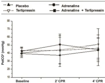
· PetCO2 (mmHg) measured at baseline (before induction
of CA), two minutes of CPR (before the injection of drugs), and 4 min of CPR (2 min after the injection of drugs and immediately before ventricular defibrillation); there were no statistically significant differences between the groups;
· VCO2 (mL/min) measured at baseline (before induction
of CA), two minutes of CPR (before the injection of drugs), and 4 min of CPR (2 min after the injection of drugs and immediately before ventricular defibrillation); although there was a statistically significant decrease in VCO2 at 2 min and 4
min of CPR in relation to baseline, there were no statistically significant differences between the groups at any moment;
· CorPP (mmHg) measured at baseline (before induction of CA), two minutes of CPR (before the injection of drugs), and 4 min of CPR (2 min after the injection of drugs and immediately before ventricular defibrillation). A statistically significant increase in CorPP between the 2 min of CPR (before the injection of drugs) and 4 min of CPR (2 min after the injection of drugs) was observed in the ADR and ADR+TP groups compared to the placebo and isolated TP groups (which was equal to placebo);
· Baseline rectal temperature (oC): ADR: 39.0±0.7; Placebo:
39±0.5; TP: 39.3±0.6; and ADR+TP: 39.6±0.4 (P=0.2085).
Those same variables are shown in Figures 1, 2 and 3.
DISCUSSION
Several studies, both clinical and trial, have investigated the prognostic value of capnography in CA/CPR. The PetCO2
measured by capnography in intubated patients has been
Fig. 1 – Comparative evolution of the variables assessed at baseline (0), 2 min of CPR (2) and 4 min of CPR in the groups studied. Average value and standard deviation of PetCO2 in each moment and group.
Fig. 2 – Comparative evolution of the variables assessed at baseline (0), 2 min of CPR (2) and 4 min of CPR in the groups studied. Average value and standard deviation of VCO2 in each moment and group.
correlated with the quality of CPR maneuvers and with ROSC, considering that it is directly related to DC, which in turn is directly related to pulmonary blood flow, PetCO2 being its reflection[22].
However, no experimental model under invasive mechanical ventilation (IMV), with PEEP=0 cmH2O and FiO2=0.21, has
assessed PetCO2, VCO2, and CorPP in the prediction of success of
ROSC on unassisted CPR for 10 min using terlipressin (a pro-drug, a vasopressin synthetic analogue, with a longer half-life than
Table 1. Descriptive analysis and comparisons of the variables assessed at baseline (0), 2 min of CPR (2), and 4 min of CPR (4) within and among groups.
ADR + TP (n=11) ADR (n=10) Placebo (n=10) TP (n=11) P
PetCO2 0* 42.5±5.2 39.4±3.5 43.4±3.5 40.4±6 *
VCO2 0¥ 121.2±20.5 133.4±53.7 110.2±21.2 119.3±40.8 ¥
CorPP 0§ 88.9±21 89.7±19.5 91.0±18.3 85.5±23.2 §
PetCO2 2* 50.7±13.1 37.4±14 43.4±16.3 44.2±17.7 *
VCO2 2¥ 48.7±27.5 57.5±30.2 47.5±21.2 45.3±24.5 ¥
CorPP 2§ 21.3±10.4 12.6±12.1 22.7±13.5 20.2±15.5 §
PetCO2 4* 44.3±15 43.6±19.3 46.3±12.4 51.0±19.4 *
VCO2 4¥ 46.4±31.1 49.6±32.9 49.2±21.3 35.5±25.5 ¥
CorPP 4§ 44.6±13.1 53.1±15.2 13.7±12 7.0±10.5 §
ADR=adrenaline; TP=terlipressin; PetCO2=end-tidal carbon dioxide tension (mmHg); VCO2=carbon dioxide excretion;
CorPP=coronary perfusion pressure
* Results of the ANOVA for repeated measurements with transformation for posts: Effect of group, P=0.5427; effect of time, P=0.1317; group/time interaction, P=0.3043.
¥ Results of the ANOVA for repeated measurements with transformation for posts:
Effect of group, P=0.7530; effect of time, P=<0.0001; group/time interaction, P=0.8622. Differences between times (profile test for contrast):
Basal ´ 2’CPR, P<0.0001; basal ´ 4’CPR, P<0.0001; 2’CPR ´ 4’CPR, P=0.2286.
§ Results of the ANOVA for repeated measurements with transformation for posts:
Effect of group, P=0.0012; effect of time, P<0.0001; group/time interaction, P<0.0001. Fixed group and time comparison (profile test for contrast):
Placebo group, P<0.0001; Basal fg 2’CPR, P<0.0001; basal fg 4’CPR, P<0.0001; 2’CPR fg 4’CPR, P=0.06. Adrenaline group, P<0.0001; Basal fg 2’CPR, P<0.0001; Basal fg 4’CPR, P<0.0014; 2’CPR fg 4’CPR, P<0.0001. Terlipressin group, P<0.0001; Basal fg 2’CPR, P<0.0001; Basal fg 4’CPR, P<0.0001; 2’CPR fg 4’CPR, P=0.0072. ADR+TP group, P<0.0001; Basal fg 2’CPR, P<0.0001; Basal fg 4’CPR, P=0.0003; 2’CPR fg 4’CPR, P<0.0001. Setting time and comparing groups (Tukey test):
Basal, without differences, P=0.9369; 2’CPR, without differences, P=0.2341; 4’CPR, with differences in Placebo and Adrenaline; Adrenaline and Terlipressin; ADR+TP and Placebo; Terlipressin and ADR+TP, P<0.0001.
Table 2. Descriptive analysis and comparison of the percentage of survivors among the groups.
Surviving groups ADR+TP (n=11) ADR (n=10) Placebo (n=10) TP (n=11) Total (n=42)
No n (%) 2 (18) 2 (20) 8 (80) 10 (91) 22 (52)
Yes n (%) 9 (82) 8 (80) 2 (20) 1 (9) 20 (48)
P<0.0001 (Fisher’s test)
vasopressin), adrenaline and their combination as vasopressor agents, in addition to placebo.
In several published studies, PetCO2 has proved to be a useful
variable in assessing the effectiveness of CPR maneuvers and results in different models of CA.
In the 1980s, Sanders et al.[23] monitored the elimination of
CO2 (VCO2) in experimental studies of CA/CPR. The elimination
and the study reported that all types of maneuvers increased PetCO2. Little more than a decade later, Blumenthal et al.[24],
also in an experimental study, measured PetCO2 and VCO2 and
concluded that higher values of PetCO2 during and after CPR
were associated with a better prognosis.
In our study, there was no statistically significant association between the times and the registered values for PetCO2 with
ROSC, although a sudden and immediate increase in PetCO2 at
the beginning of the CPR was observed, which expresses the pulmonary blood flow, and thus, DC. The data indicates that PetCO2, both at 2 min and 4 min of CPR (more importantly,
because it was after the administration of the drugs), is not correlated with ROSC rates, i.e., it was not different among the four groups. Nevertheless, we have to consider that in our study the CA/CPR model was performed on very young (immature) animals, being an extreme CA model, for 10 min without any assistance, and, after this period, the animals were ventilated with an initial FiO2 (0.21). We should also highlight the fact that we did
not alternate between ventilation and compression, which may have compromised the effectiveness of the alveolar ventilation.
In relation to VCO2 at 2 and 4 min of CPR, it did not differ
statistically between the four groups and, therefore, it was not indicative of ROSC. On the other hand, CorPP proved to be significantly higher in the ADR and ADR+TP groups compared to the Placebo and Terlipressin groups at 4 minutes of CPR, which indicates that CorPP was a predictive factor for ROSC.
Human studies use other variables to assess the successful prediction of ROSC. Different criteria are used, but capnography is also the main focus of the prognostic assessment after CA. One of the first studies in the 1980s by Garnett et al.[25] assessed
patients under IMV who presented CA, and they concluded that the monitoring of PetCO2 during CPR can be useful and serve as
a guide during resuscitation maneuvers.
Another clinical study, conducted by Sanders et al.[26],
assessed PetCO2 in patients subjected to CPR. The authors
reported that all patients listed and those who presented ROSC showed an average PetCO2 ≥ 10 mmHg. None of the patients
with an average PetCO2 ≤ 10 mmHg presented ROSC. The data
in this prospective clinical test indicate that the monitoring of PetCO2 during CPR can be useful in the prediction of ROSC in
CPR in humans.
PetCO2 values have been correlated with CorPP and ROSC
rate. Thus, in an animal model (dogs) of CA/CPR, Sanders et al.[27],
in another study, found a significant correlation (P<0.01) between PetCO2 and CorPP. The data in this study were confirmed by
the same group, in the same year, with small variations in the method. However, the actual physiological relationship between CorPP and PetCO2 during CPR remains uncertain[23].
In our study, we have observed a significant increase in CorPP (P<0.0001) in the 4th minute of CPR (after drugs) in relation to the
2nd min (before drugs) of CPR in the ADR and ADR+TP groups.
However, the same behavior was no observed in PetCO2 and VCO2.
The use of vasopressor agents is suggested in the first cycles of CPR[25]. For different types of CA, vasopressor, adrenaline or
vasopressin can be administered in order to increase myocardial and cerebral blood flow. In the study of Ovalle et al.[20], the use
of some vasopressor agents during CPR was assessed. It was
observed that ADR and its combination with terlipressin, but not isolated terlipressin, were effective in increasing CorPP and ROSC. Furthermore, the ADR+TP combination provided greater hemodynamic stability after ROSC in the surviving animals, suggesting that TP can be a useful drug in handling hypotension after CPR[28].
In most published studies, both clinical and experimental ones, the initial, final, maximum and minimum readings show higher values of PetCO2 in patients who presented ROSC. These
authors highlight that clinical studies receive influences from diseases already present in patients, and this factor should be considered[18].
The PetCO2 values assessed in many (clinical and
experimental) studies can assist in verifying the effectiveness of CPR maneuvers in order to guide, with due caution, the actual results of resuscitation, since very low PetCO2 values may indicate
that there is no more reason to continue the efforts of CPR[29].
In short, PetCO2 and VCO2 values obtained by volumetric
capnography have differed from the findings in some published studies. In addition, those values are not correlated with hemodynamic variables (CorPP) or with ROSC rates in our experimental model with immature swine, in which CA was induced by ventricular fibrillation and the animals remained without assistance for 10 min with subsequent CPR. It should be noted that the animals were under IMV, with FiO2=0.21 and
PEEP=0 cmH2O, and we used vasopressor agents (ADR and TP;
isolated or in association).
CONCLUSION
From the results obtained in the study, both PetCO2 and
VCO2 showed no correlation with ROSC, although VCO2 was 50%
lower during the CPR maneuvers (would it be more sensitive in the detection of a decrease in pulmonary blood flow?). Although we cannot affirm with certainty, some hypotheses were made to explain this fact, namely: FiO2=0.21 during the entire experiment,
immaturity of the animals, time of non-assistance after CA (10 minutes), and positive pressure (IMV) in the CPR, which may have led to an even sharper decrease in the right preload. Thus, further studies are needed to verify the value of volumetric capnography (PetCO2 and VCO2) to guide the effectiveness of CPR maneuvers,
especially with the use of adjuvant vasopressor agents.
ACKNOWLEDGMENTS
We are grateful to Fundação de Amparo à Pesquisa do Estado de São Paulo (Fapesp) (process Nº. 07/08315-0) and to Fundo de Apoio ao Ensino, Pesquisa e Extensão (Faepex)-Unicamp (process Nº 17809) for their financial support; to Ferring Laboratories-Brazil-Ltda, for their kind donation of two 1 mg terlipressin vials for the pilot studies; to the biologists Ana C. Moraes and William A. Silva, from Núcleo de Medicina e Cirurgia Experimental-FCM-Unicamp, for their technical assistance during experimental procedures; and to the statisticians from the Research Commission-FCM-Unicamp, for their technical assistance during data analysis.
REFERENCES
1. Gravenstein JS, Jaffe M, Paulus DA. Capnography: clinical aspects. Carbon dioxide over time and volume. Second edition,. Cambridge: Cambridge University Press; 2011.
2. Toneloto MG, Moreira MM, Bustorff-Silva JM, Souza GF, Martins LC, Dragosavac D, et al. Adjustable inspiratory occlusion valve in experimental bronchopleural fistula. A new therapeutic perspective. Acta Cir Bras. 2015;30(8):561-7.
3. Antonelli RQ, Moreira MM, Martins LC, Negro MS, Baldasso TA, Tincani AJ. Evaluation of the efficiency of the atraumatic endotracheal tube in the pulmonary-gas exchange: an experimental study. Braz J Cardiovasc Surg. 2015;30(6):668-72.
4. Oliveira DG, Toneloto MG, Moreira MM, Bustorff-Silva JM, Souza GF, Martins LC, et al. Hemodynamic, ventilatory and gasometric evaluation of an experimental bronchopleural fistula. Acta Cir Bras. 2015;30(1):1-5. 5. Pereira DJ, Moreira MM, Paschoal IA, Martins LC, Metze K, Moreno
Junior H. Near-fatal pulmonary embolism in an experimental model: hemodynamic, gasometric and capnographic variables. Rev Bras Cir Cardiovasc. 2011;26(3):462-8.
6. Ferreira JH, Terzi RG, Paschoal IA, Silva WA, Moraes AC, Moreira MM. Mechanisms underlying gas exchange alterations in an experimental model of pulmonary embolism. Braz J Med Biol Res. 2006;39(9):1197-204. 7. Silva SM, Paschoal IA, De Capitani EM, Moreira MM, Palhares LC, Pereira MC. COPD phenotypes on computed tomography and its correlation with selected lung function variables in severe patients. Int J Chron Obstruct Pulmon Dis. 2016;11:503-13.
8. Oliveira PM, Moreira MM. Capnography: a feasible tool in clinical and experimental settings. Respir Care. 2015;60(11):1711-3.
9. Veronez L, Pereira MC, Silva SM, Barcaui LA, De Capitani EM, Moreira MM, et al. Volumetric capnography for the evaluation of chronic airways diseases. Int J Chron Obstruct Pulmon Dis. 2014;23(9):983-9. 10. Ribeiro MA, Silva MT, Ribeiro JD, Moreira MM, Almeida CC,
Almeida-Junior AA, et al. Volumetric capnography as a tool to detect early Authors’ roles & responsibilities
ACLM
LCM
IAP
CCISO
SA
MMM
Analysis and/or data interpretation; manuscript writing or critical review of its content; final manuscript approval Analysis and/or data interpretation; manuscript writing or critical review of its content; final manuscript approval Analysis and/or data interpretation; manuscript writing or critical review of its content; final manuscript approval; Conception and design study; realization of operations and/ or trials; analysis and/or data interpretation; manuscript writing or critical review of its content
Conception and study design; execution of operations and/ or trials; analysis and/or data interpretation; manuscript writing or critical review of its content; final manuscript approval;
Execution of operations and/or trials; analysis and/or data interpretation; manuscript writing or critical review of its content; final manuscript approval
peripheral lung obstruction in cystic fibrosis patients. J Pediatr (Rio J). 2012;88(6):509-17.
11. Veronez L, Moreira MM, Soares ST, Pereira MC, Ribeiro MA, Ribeiro JD, et al. Volumetric capnography for the evaluation of pulmonary disease in adult patients with cystic fibrosis and noncystic fibrosis bronchiectasis. Lung. 2010;188(3):263-8.
12. Moreira MM, Terzi RG, Paschoal IA, Martins LC, Oliveira EP, Falcão AL. Thrombolysis in massive pulmonary embolism based on the volumetric capnography. Arq Bras Cardiol. 2010;95(4):e97-9.
13. Welch A. Capnography, mainstream and sidestream modules. Beaverton; 2003.
14. Blumenthal SR, Voorhess WD. The relationship between airway carbon dioxide excretion and cardiac output during cardiopulmonary resuscitation. Resuscitation. 1997;34(3):263-70.
15. Kalenda Z. The capnogram as a guide to the efficacy of cardiac massage. Resuscitation. 1978;6(4):259-63.
16. Moreira MM, Terzi RGG, Ferreira ELA, Moraes AC, Silva WA. Correlação entre a pressão expiratória final de CO2 e o débito cardíaco no choque hemorrágico experimental. RBTI. 2005;17(3):188-13.
17. Sehra R, Underwood K, Checchia P. End tidal CO2 is a quantitative measure of cardiac arrest. Pacing Clin Electrophysiol. 2003;26(1 Pt 2):515-7. 18. Blumenthal SR, Voorhess WD. The relationship between airway
carbon dioxide excretion and cardiac output during cardiopulmonary resuscitation. Resuscitation. 1997;34(3):263-70.
19. Asplin BR, White RD. Prognostic value of end-tidal carbon dioxide pressures during out-of-hospital cardiac arrest. Ann Emerg Med. 1995;25(6):756-61.
20. Ovalle CCIS, Moreira MM, Martins LC, Araujo S. A eficácia da terlipressina versus adrenalina na ressuscitação cardiopulmonar em suínos. Rev Bras Anestesiol. 2011;61(6):728-35.
21. Paradis NA, Martin GB, Rivers EP, Goetting MG, Appleton TJ, Feingold M, et al. Coronary perfusion pressure and the return of spontaneous circulation in human cardiopulmonary resuscitation. JAMA. 1990;263(8):1106-13. 22. Gudipati CV, Weil MH, Bisera J, Deshmukh HG, Rackow EC. Expired
carbon dioxide: a noninvasive monitor of cardiopulmonary resuscitation. Circulation. 1988;77(1):234-9.
23. Sanders AB, Ewy GA, Bragg S, Atlas M, Kern KB. Expired PCO2 as a prognostic indicator of successful resuscitation from cardiac arrest. Ann Emerg Med. 1985;14(10):948-52.
24. Blumenthal SR, Voorhees WD. The relationship of carbon dioxide excretion during cardiopulmonary resuscitation to regional blood flow and survival. Resuscitation. 1997;35(2):135-43.
25. Garnett AR, Ornato JP, Gonzalez ER, Johnson EB. End-tidal carbon dioxide monitoring during cardiopulmonary resuscitation. JAMA. 1987;257(4):512-5.
26. Sanders AB, Kern KB, Otto CW, Milander MM, Ewy GA. End-tidal carbon dioxide monitoring during cardiopulmonary resuscitation. A prognostic indicator for survival. JAMA. 1989;262(10):1347-51.
27. Sanders AB, Atlas M, Ewy GA, Kern KB, Bragg S. Expired PCO2 as an index of coronary perfusion pressure. Am J Emerg Med. 1985;3(2):147-9. 28. Gonzalez MM, Timerman S, Gianotto-Oliveira R, Polastri TF, Canesin MF, Schimidt A, et al; Sociedade Brasileira de Cardiologia. First guidelines of the Brazilian Society of Cardiology on Cardiopulmonary Resuscitation and Cardiovascular Emergency Care. Arq Bras Cardiol. 2013;101(2 Suppl 3):1-221.