1Curso de Pós-graduação em Ciências Médicas, Doutorado em Neurologia;2Laboratório Hidro-Salino, Núcleo de Medicina e Ciru rg i a
Experimental, Departamento de Clínica Médica;3N e u rologia e4Anatomia Patológica, Faculdade de Ciências Médicas, Universidade
Estadual de Campinas (UNICAMP), São Paulo, SP, Brazil. This study was supported by grants from CNPq (500868/91-3) and (302189/2004-1), PRONEX (0134/97), CAPES and FAPESP (00/12216-8).
Received 14 April 2005, received in final form 6 July 2005. Accepted 26 August 2005.
Dr. Manoel Baldoino Leal Filho - Rua Thomaz Tajra 1222/300 - 64048-380 Teresina PI - Brasil. E-mail: [email protected]
HEMODYNAMIC PARAMETERS AND
NEUROGENIC PULMONARY EDEMA
FOLLOWING SPINAL CORD INJURY
An experimental model
Manoel Baldoino Leal Filho
1, Rosana C. Morandin
2,
Amanda R. de Almeida
2, Elizabeth C. Cambiucci
2, Konradin Metze
4,
Guilherme Borges
3, José Antonio Rocha Gontijo
2.
ABSTRACT - Neurogenic pulmonary edema is a serious and always life-threatening complication following several lesions of the central nervous system. We re p o rt an experiment with 58 Wi s t a r-Hanover adult male rats. Two gro u p s w e re formed: control (n=4) and experimental (n=54). The experimental group sustained acute midthoracic spinal c o rdinjury by Fogart y ’s balloon-compression technique containing 20µL of saline for 5, 15, 30 or 60 seconds. The rats w e reanesthetized by intraperitoneal (i.p.) sodium pentobarbital (s.p.) 60 mg/Kg. The quantitative neurological out-come was presented at 4, 24 and 48 hours from compression to characterize the injury graduation in diff e rent gro u p s . Poor outcome occurred with 60 seconds of compression. Six animals died suddenly with pulmonary edema. Using the pro c e d u re to investigate the pulmonary edema during 60 seconds of compression, followed by decompre s s i o n and time-course of 60 seconds, 20 rats were randomly asigned to one of the following groups: control (1, n=4, anes-thetized by i.p. s.p., 60 mg/Kg but without compression) and experimental (2, n=7, anesanes-thetized by i.p. xylazine 10 mg/Kg and ketamine 75 mg/Kg) and (3, n=9, anesthetized by i.p. s.p., 60 mg/Kg). The pulmonary index (100 x wet lung weight / body weight) was 0.395 ± 0.018 in control group, rose to 0.499 ± 0.060 in group 2, and was 0.639 ± 0.14 in group 3. Histologic examination of the spinal cord showed parenchymal ru p t u res and acute hemorrh a g e . Comparison of the pulmonary index with morphometric evaluation of edema fluid-filled alveoli by light micro s c o p y showed that relevant intra-alveolar edema occurred only for index values above 0.55. The results suggest that the p u l m o n a ryedema induced by spinal compression is of neurogenic nature and that the type of anesthesia used might be important for the genesis of lung edema.
KEY WORDS: spinal cord injury, neurogenic pulmonary edema, central nervous system lesions.
P a r â m e t ros hemodinâmicos e edema pulmonar neurogênico após traumatismo raquimedular: mode-lo experimental
RESUMO - Edema pulmonar neurogênico é complicação séria e aumenta o risco de vida em pacientes com várias lesões do sistema nervoso central. Apresentamos uma experiência com 58 ratos Wistar machos e adultos. Foram for-mados dois grupos: controle (n=4) e experimental (n=54). O grupo experimental sofreu trauma raquimedular torá-cico médio com o cateter-balão de Fogarty contendo 20µL de salina por 5, 15, 30 ou 60 segundos de compressão. Os ratos foram anestesiados com pentobarbital sódico (p.s.), 60 mg/Kg intraperitoneal (i.p.). Foi investigada a re l a ç ã o e n t re a lesão medular e o tempo de compressão. A evolução neurológica foi quantificada e apresentada com 4, 24 e 48 horas da compressão para caracterizar a graduação da lesão nos diferentes grupos. A pior evolução ocorre u com 60 segundos de compressão. Seis animais morreram subitamente com edema pulmonar. Vinte ratos foram ran-domicamente distribuídos em um dos seguintes grupos: controle (1, n=4, anestesiados com p.s. i.p., 60 mg/Kg, mas sem compressão) e experimental (2, n=7, anestesiados com xilasina 10 mg/Kg e ketamina 75 mg/Kg) e (3, n=9, aneste-siados com p.s. i.p., 60 mg/Kg). O índice pulmonar (100 x peso pulmonar / peso corporal) foi 0,395 ± 0,018 no gru p o c o n t role, 0,499 ± 0,060 no grupo 2, e 0,639 ± 0,14 no grupo 3. O exame histológico da medula espinhal mostrou ru p-turas no parênquima e hemorragia aguda. Comparando-se o índice pulmonar com o índice morfométrico através da microscopia óptica, evidenciou-se que ocorreu edema intra-alveolar relevante para índices acima de 0,55. A pre-sente experiência sugere que o edema pulmonar induzido pela compressão medular é de natureza neurogênica e que o tipo de anestesia utilizada no experimento poderia ter participação na gênesis do edema pulmonar.
N e u rogenic pulmonary edema is characterized as an acute, protein-rich lung edema occurring s h o rt-ly after spinal cord injury. Yet, the precise pathoge-netic mechanisms, incidence and clinical significan-ce of development of elicited neurogenic
pulmo-n a ry edema remaipulmo-n upulmo-nclear1. The pulmonary
alveo-l a r- c a p i alveo-l alveo-l a ry barrier needs to satisfy two confalveo-licting re q u i rements: it must be extremely thin for eff i-cient gas exchange and also strong enough to wi-thstand the extremely high stress in the capillary
wall when capillary pre s s u rerises2. The strengh of
the pulmonary blood-gas barrier on the thin side is attributable to the type IV collagen in the
base-ment membranes2 - 4. However, when the wall s t re s s
rises to very high levels, ultrastructural changes o c c u r
in the barr i e r, a condition known as stress failure2.
Pathophysiological conditions such as high-altitu-de pulmonary ehigh-altitu-dema, neurogenic pulmonary ehigh-altitu-de- ede-ma, severe left ventricular failure, mitral stenosis, overinflation of the lung and others result in stre s s
f a i l u re3. Recent experimental findings suggest that
rapid changes in gene expression for extracellular matrix proteins and growth factors occur in re
s-ponse to increases in capillary wall stress2.
In patients with spinal cord injuries re s p i r a t o-ry complications are still an important cofator of morbidity and mort a l i t y. Fontes et al.5p u b l i s h e d
cases of acute neurogenic pulmonary edema after several several seizure episodes followed by crani-oplasty and subarachnoid hemorrhage due to a c a v e rnoma. Additionally, these authors pre s e n t e d a literature review since 1990 of human cases of n e u-rogenic pulmonary edema, which resulted in
four-teen reports (21 cases) to be analysed5.
Although diff e rent experimental animal mod-els that simulate the traumatic spinal lesions seen in human have been described, in the present com-munication we have assessed the time-dependent re p e rcussion of hemodynamic parameters, sensory and motor activities and pulmonary edema in an original experimental model of spinal cord injury in rats and also perf o rmed an extensive review of the literature about the subject.
METHOD
Fifty-eight male Wi s t a r-Hannover rats, weighing between 300 and 350 g, were obtained from the Univer-s i t y ’Univer-s breeding center. The general guidelineUniver-s eUniver-stabliUniver-shed by the Brazilian College for Animal Experimentation ( C O-BEA) were followed throughout this study. The experi-ment design was previously approved by the Institutional Committee for Ethics in Animal Research. The rats were anesthetized with intraperitoneal sodium
pentobarbi-tal (60mg/kg body weight, i.p.) and the level of anesthe-sia was controlled by corneal reflex monitoring. In ord e r to evaluate the effect of spinal cord injury on systolic ( S B P ) / diastolic (DBP) blood pre s s u re (in mmHg) and heart ra-te (HR, in bpm) by means of a BESE polygraph, the right c a rotid art e ry of all animals was cannulated and they were randomly assigned to one of these groups:
1) Control (sham) group:four anesthetized rats we-re submitted to ligamentum flavum we-removal followed by insertion of Fogart ycatheter balloon (12-062-2F) into
the dorsal epidural space 1 cm cranially to midthoracic levelwithout insufflations of the catheter and spinal cord c o m p ression (SC).
2) Experimental SC group 1:23 anesthetized rats were submitted to the same surgical pro c e d u re as group 1 and s u ff e red additional SC at midthoracic level by insuff l a-tions of 20µL of saline solution into the balloon during 5, 15, 30 and 60 seconds, being evaluated 4-h after SC.
3) Experimental SC group 2:17 anesthetized rats were submitted to the same surgical pro c e d u re as group 1 and s u ff e red additional SC at midthoracic level by insuff l a-tions of 20µl of saline solution into the balloon during 5, 15, 30 and 60 seconds, being evaluated 12-h after SC.
4) Experimental SC group 3:14 anesthetized rats sub-mitted to the same surgical pro c e d u re as group 1 and s u ff e red additional SC at midthoracic level by insuff l a-tions of 20µL of saline solution into the balloon during 5, 15, 30 and 60 seconds, being evaluated 24-h after SC. Subsequently the experimental group was submit-ted to time-course extradural compression. The animals w e re observed in a flat, barr i e r- f ree environment for spontaneous motor activity of the hind limbs accord i n g to Khan et al., which was scored as: (0) no movement in the hind limbs, no weight bearing; (1) barely perc e p t ib l e movement of hind limbs, no weight bearing; (2) freq u e n t movement of hind limbs, no weight bearing; (3) a b i l i t y to support weight on hind limbs, may take one or two steps; (4) ability to walk with mild deficit; and (5) nor-mal walking6. Also, the neural sensorial test was per-f o rmed accordingly and scored as per-follows: (0) no re a c-tion to foot pinch; (1) foot jerks without consistent with-drawal toward the body; (2) normal, rapid withwith-drawal of hind limb to body; and (3) hyperflexion of the hind limb, spastic shaking6.
At the end of each blood pre s s u re and heart rate re c o rding, the rats were perfused by the left carotid a rt e ry saline containing heparin (2%) for 5 min under cons-tant pre s s u re. This was followed by perfusion with 0.1 M phosphate buffer (PB; pH 7.4) containing 4% (w/v) p a r a f o rmaldehyde and 0.1 M sucrose with overnight fi-xation by freezing 24 hours later the spinal cord and the lung were removed and analysed.
ti-me-course of 60 seconds. The animals were randomly asigned to one of the following groups: control (1, n=4, anesthetized by i.p. s.p., 60mg/Kg but without compre s-sion) and experimental (2, n=7, anesthetized by i.p. xyla-sine 10mg/Kg and ketamine 75mg/Kg) and (3, n=9, anes-thetized by i.p. s.p., 60mg/Kg). After the experiment the animals were sacrificed using an overdose of anesthe-sia and the lungs were harvested and the pulmonary in-dex was calculed. The lungs were fixed by immersion for 24 hours in 4% formaldehyde, dehydrated and em-bedded in paraffin and 5µm haematoxylin-eosin stained sections were examined by light microscopy.
For quantitative evaluation of the pulmonary sec-tions there were randomly selected 10 microscopic fields (objective magnification 40 x) per case and counted the p e rcentage of alveoli which were at least partially filled with eosinophilic edema fluid. The morphometric index of edema fluid-filled alveoli was evaluated by light mi-croscopy in paraffin sections.
Statistical analysis –All data are re p o rted as means ± SEM. Data obtained over time were analyzed using appro-priate ANOVA.Post hoccomparisons between selected means were done with Bonferro n i ’s contrast test when initial ANOVA indicated statistical diff e rences between experimental groups. Comparisons involving only two means within or between groups were done using Stu-d e n t ’sttest. Apvalue < 0.05 was considered significant.
RESULTS
The basal (systolic and diastolic) blood pre s s u re and heart rate levels in the control and all experi-mental groups were similar. In the control sham-operated group, the heart rate was constant dur-ing the whole experiment, whereas the systolic and diastolic blood pre s s u redecreased significantly dur-ing the experiment (SBP from: 129.2 ± 4.7 mmHg to 115 ± 5.0 mmHg, p=0.03; and DBP from: 100.0 ± 1.0 mmHg to 87.5 ± 5.0 mmHg, p=0.001). In the
pento-barbital anesthetized rats with SC, peak heart rate rose from 212.0 ± 19.0 bpm to 258.0 ± 22.0 bpm (Fig 1) and fell afterw a rds below baseline level (160.0 ± 45.0 bpm, p=0.0002). In these experiments, systolic a rterial pre s s u re doubled the baseline pre s s u re (116.0 ± 14.0 mmHg) during spinal cord compression (232.0 ± 33.0 mmHg) (see Fig 1) and decreased afterw a rd s to still elevated levels (148.0 ± 14.0 mmHg, p=0.0001). The diastolic pre s s u re showed similar statistically sig-nificant changes, and increased from baseline lev-els of 91.4 ± 10.7 mmHg to 131.4 ± 8.0 mmHg dur-ing SC compression, droppdur-ing to 116.0 ± 6.0 mmHg after compression (p=0.0001).
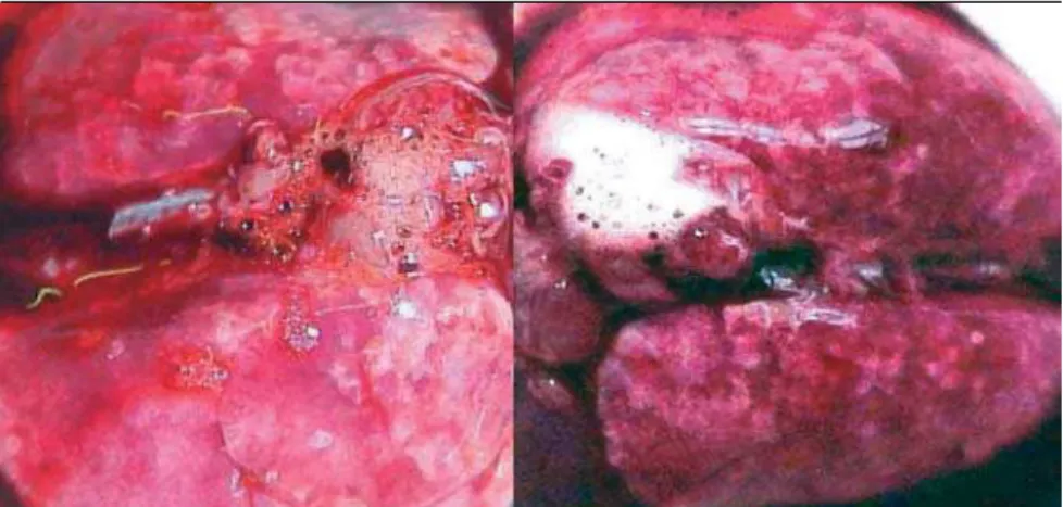
F i g u re2 and 3 exemplify spinal cord injury and lung specimens from the control and 4-hour post-SC experimental group after perfusion with 0.1M phosphate buffer containing 4% paraform a l d e-hyde and 0.1 M sucrose and overnight fixation in freezing.
The time-response rated scores obtained in neu-ral, sensorial and motor activity tests during evalua-tion perf o rmed by open-field testing had a nor-mal perf o rmance in control (sham) non-SC gro u p . I n c reasing spinal cord compression time-course f ro m five to 60-sec resulted in graded time-dependent d e c reases, in sensitive (neurosensorial scores 4-h post-SC - 5 sec: 2 ± 0.5; 15 sec: 1.7 ± 0.9; 30 sec: 1 ± 0.8 and 60 sec: 0.7 ± 0.6; group 24-h post-SC - 5 s e c : 2 ± 0.5; 15 sec 1.8 ± 0.9; 30 sec: 1 ± 0.9 and 60 sec: 0.7 ± 0.6 and group 48-h post-SC - 5 sec: 2 ± 0.5; 1 5 sec: 1.3 ± 0.8; 30 sec: 1 ± 0.8 and 60 sec: 0.6 ± 0.4) and motor activity test scores (motor activity 4-h post-SC - 5 sec: 5 ± 0.1; 15 sec: 3.7 ± 0.4; 30 sec: 2.5 ± 0.5 and 60 sec: 1.6 ± 0.8; group 24-h post-SC - 5 sec: 4.7 ± 0.2; 15 sec: 4.4 ± 0.3; 30 sec: 3.5 ± 0.4 and 60 sec: 2.2 ± 0.5 and group 48-h post-SC - 5 sec: 4.5
± 0.3; 15 sec: 3.6 ± 0.4; 30 sec: 2.5 ± 0.5 and 60 sec: 1 ± 0.8). Although there was significant and gradu-al time-dependent increase in motor (p = 0.001) and neurosensorial (p = 0.02) deficits, these abnor-malities were unaffected when evaluated at 4, 24 and 48 hours after SC (motor activity scores, p = 0 . 5 and neurosensorial scores, p = 0.1).
Histologic examination of the spinal cord sho-wed in the area of compression parenchymal ru p-t u res accompanied by acup-te hemorrhage. Lighp-t m i-c rosi-copii-c examination of pulmonary tissue show e d vascular congestion with perivascular edema and a v a rying amount of intra-alveolar accumulation of p roteinaceous fluid. Comparison of pulmonary in-dex and morphometric data obtained by light mi-c rosmi-copy in paraffin semi-ctions showed that re l e v a n t intra-alveolar edema occured only for index valu-es above 0.55. The pulmonary edema was invvalu-esti- investi-gated by the pulmonary index that was 0.395 ±
0.018 in control group, rose to 0.499 ± 0.060 in g ro u p 2, and was 0.639 ± 0.14; p=0.0018 in group 3.
DISCUSSION
Since the diagnosis of neurogenic pulmonary edema is often based on exclusion and as many c a-ses may be subclinical, the incidence of neuro g e n i c p u l m o n a ry edema may be underestimated. In the mammalian lung, alveolar gas and blood are sepa-reted by an extremely thin membrane, despite the fact that mechanical failure could be catastro p hi c for gas exchange. Stress failure causes increased p e r-meability with protein leakage or frank hemorrh a-ge, and probably has a role in several types of lung disease. In anesthesized rabbits, at capillary trans-mural pre s s u res gre a t e r- t h a n - o r-equal-to 40 m m H g , d i s ruption of the capillary endothelium and
alve-olar epithelium was seen in some locations4. N e u
ro-genic pulmonary edema is associated with a
varieFig 2. Macroscopic aspects of the pulmonary edema in rats submitted to spinal cord com -pression study during 60 seconds.
se the amount of edema development for a given d e g ree of pulmonary hypert e n s i o n1 2.
Widdicombe re p o rted that the trachea-bro n-chial vasculature is controlled by adre n e rgic,
cholin-e rgic and pcholin-eptidcholin-ergic ncholin-ervous mcholin-echanisms1 3. This
author show that sympathetic nerves release nor-epinephrine and neuropeptide Y (both of which a re constrictor agents) while parasympathetic ner-ves release acetylcholine and usually vasoative in-testinal polypeptide (both of which are vasodila-tors). Activation of pulmonary C-fiber receptors may cause a powerful vasodilatation, mainly via sympa-thetic motor nerves, and cardiac and chemore c e
p-tor reflexes also influence airway vascular tone1 3.
S e n s o ry nerves in the airway mucosa are re s p o n s i-ble for local axon reflexes and these nerves contain n e u ropeptides such as substance P, neurokinins A and B, and calcitonin gene-related peptide (all these
n e u ropeptides are powerful vasodilators)1 3.
The neuropeptide Y is believed to be an impor-tant mediator in neurogenic pulmonary edema, re s-ponsible for the increased pulmonary vascular per-meability and diff e rences in receptor modulation. This might be an explanation for the fact that neu-rogenic pulmonary edema could be more pro-nounced due to administration of sodium
pento-barbital when this anesthesic is used1 4and justify
the pulmonary edema verified in the present ex-periment, where the xylazine could modulate no-r a d no-re n e no-rgic no-receptono-rs and ketamine, neuno-ro p e p y i d e Y receptors, diff e rently from sodium pentobarbital.
Injection of ibotenic acid, a glutamate agonist, into the ventral medullary raphe (VMR), especial-ly the nucleus raphe magnus, of the rat pro d u c e d re s p i r a t o ry failure and death following a pre d i c-table course of events, but pre t reatment with PPP, a sigma receptor agonist, or scopolamine, a musca-rinic cholinergic antagonist, prevented pulmonary f a i l u reand death1 5. Based on these results, it is s u
g-gested that the VMR has a role in regulation of p u l m o n a ry blood flow and that the pre l i m i n a ry p h a rmacological studies suggest that disru p t i o n of glutamatergic and cholinergic mechanisms
me-diates the lethal pulmonary phenomenon1 5.
Imai-zumi et al. had always investigated the role of the
caudal ventrolateral medulla (CVL) in rats10.
The use of neuro - e x c i t a t o ry (L-glutamate) and n e u ro - i n h i b i t o ry (muscimol) agents into the de-p ressor region of the CVL of anesthetized rabbits indicated that the sympathoinhibitory neurons in the CVL medulla tonically suppress the activity of sympathetic preganglionic neurons controlling myo-ty of central nervous system lesions. Some authors
explain that a massive centrally mediated
sympa-thetic discharge occurs as a result of the lesion7 - 1 1.
In the present experimental model, the behavi-oral tests after 60 seconds of spinal cord compre s-sion showed a striking influence of this pro c e d u re in the neurological perf o rmance. Six animals that w e re submitted to a spinal cord compression for 60 seconds (Fig 3) developed pulmonary edema, all of them in the 4-h group (Fig 2). Additionally, a s demonstrated in the Figure 1, the spinal cord com-p ression during 60 seconds also resulted in eleva-tion of the arterial pre s s u reand drop in the heart rate. This evidence corroborates the hypothesis of massive centrally mediated sympathetic discharge.
Nathan and Reis observed that bilateral electro-lytic lesions of the anterior hypothalamus in unre-strained rats resulted within 2 hours in the devel-opment of arterial hypertension, tachycardia, hy-p e rt e rmia and increased locomotor activity, often leading to pulmonary edema and death. Similar lesions in paralyzed, artificially ventilated rats pro-duced comparable changes. They observed that a rterial hypertension, elevated peripheral re s i s t a n-ce, and diminished cardiac output were reversed t o n o rmal by alpha-receptor blockade with phentola-mine. Also, these authors demonstrated that bilat-eral adre n a l e c t o m y, adrenal demedullation or a d renal denervation perf o rmed prior to lesion in-ducement prevented the development of art e r i a l h y p e rtension and pulmonary edema, as well as the changes in peripheral resistance, cardiac
out-put, and body temperature7. Glazer and Ross
ob-s e rved noradre n e rgic (NE) bulb-ob-spinal innerv a t i o n to midthoracic sympathetic preganglionic nuclei in the rat thoracic cord by immunocytochemical l o-calization of dopamine-beta-hydroxylase, a
speci-fic NE antigen8.
The rapid development of pulmonary edema that may occur in the rabbit after the intracistern a l injection of fibrinogen and thrombin has classical-ly been considered to result from a cholinergic me-diated increase in vascular perm e a b i l i t y. Experimen-tal work tested this hypothesis by evaluating the relationship between the degree of pulmonary hy-p e rtension and hy-postmortem extravascular lung w-ater content (EVLW) in both non-vagotomized and vagotomized rabbits, which had been administre d t h rombin and fibrinogen intracistern a l l y1 2. The
a-c a rdial a-contraa-ctility as well as peripheral vasomotor tone, and that dysfunction of these medullary neu-rons could underlie some forms of experimental h y p e rt e n s i o n1 6. It was proposed that neurogenic
pul-m o n a ry edepul-ma is a functional disturbance pro v o k e d by adverse stimuli from outside the lungs and that in the rat the primary aff e rent fiber is essential to the production of this entity based on the effect of neonatal capsaicin treatment (to destroy unmyeli-nated C-fibers) on neurogenic pulmonary - e d e m a
f rom fliud-percussion brain injury in the adult-rat1 7.
It was suggested that when endotheliumde-rived relaxing factor (EDRF) release is inhibited during massive sympathetic nervous system activi-t y, pulmonary vascular resisactivi-tance is markedly in-creased, which causes the right ventricule to fail. The reduced right ventricular output maintains p u l m o n a rymicrovascular pre s s u rebelow levels
re-q u i red for edema development1 8. Another study
revealed that intracranial hypertension elicits vaso-constriction of the systemic and pulmonary re s i s t-ance and capacitt-ance vessels and the major cause of volume and pre s s u reloading in the pulmonary c i rculation is acute left ventricular failure re s u l
t-ing in a dramatic decrease in aortic flow19.
The intrathecal (i.t.) injection of endothelins to conscious rats was found to cause re s p i r a t o ry a rrest. The increase of pulmonary vascular perm e-ability and edema induced by i.t. endothelin-1 are due to an intense pulmonary vasoconstriction me-diated by alpha-adrenoceptors, following the re-lease of catecholamines in response to the activa-tion of endothelin receptor in the spinal cord. This central phenomenon seems to be reflexogenic, in-cluding the involvement of primary afferent
C-fi-bers and spinal cord ascending fiC-fi-bers to the brain2 0.
Hamdy et al. used fibrinogen and thrombin in-jected into the rat’s cisterna magna to induce neu-rogenic pulmonary edema. They observed that n e u ropeptide Y has a relationship to the high pro-tein concentration ratio or to increased pulmonary vascular perm e a b i l i t y, which consequently may c o n-tribute to the development of neurogenic
pul-m o n a ry edepul-ma in rats2 1. Sympathetic
hyperactivi-ty during sudden intracranial hypertension leads to cardiovascular instability, myocardial dysfunc-tion and neurogenic pulmonary edema. One study o b s e rved that intrathecal lidocaine prevents car-diovascular collapse and neurogenic pulmonary edema in a rat model of acute intracranial hyper-tension22.
Other re s e a rchbrought new data on the spinal mechanisms of autonomic dysreflexia and card i o-vascular dysfunctions, such as visceral stimulation of rats with chronic spinal cord injury that activate a d renal sympathetic preganglionic neurons, which could induce release of catecholamines by the
a d renal medulla2 3. The effects of nitric oxide (NO)
in the central nervous system on incidence and se-verity in the fibrin-induced pulmonary edema mod-el were evaluated in rats that were left unilater-ally vagotomized 1, 2 and 4 weeks before injec-tions of fibrinogen and thrombin into the cister-na magcister-na, after sectioning of the right vagus ner-ve. In the present study, the brain NO synthase lev-el in the medulla oblongata was lev-elevated in the 2-week group, compared to the control, but de-c reased in the 4-week group. Inde-cidende-ces of pulmo-nary edema were 100% in the control group, dec reasing to 78% in the 1week group, 17% in the 2 -week group and back to 72% in the 4--week gro u p . The lung water ratio, a parameter of severity, de-monstrated a similar pettern of change as the inci-dence. The lowered incidence and severity obtai-ned in the 2-week group were reversed by intra-c i s t e rnal injeintra-ction of N-omega-nitro - L - a rginine me-thyl ester (L-NAME). These results sustain the idea that an increase in nitric oxide, possibly in the nu-cleus tractus solitarius 2 weeks after left vagoto-m y, vagoto-may have an inhibitory action on the
develop-ment of neurogenic pulmonary edema in rats24.
T h e re is evidence that the motor cortex is in-volved in cardiovascular adjustments associated with somatic motor activity, as it has functional c o n-nections with the CVL medulla, a brainstem re g i o n critically involved in the control of blood pre s s u re and the regulation of plasma catecholamine lev-els.The CVL medulla sends projections to the spinal i n t e rmediolateral nucleus, where pre g a n g l i o n i c n e u rons take control of heart and blood vessels
(T2 segment) and adrenal medulla (T8 segment)2 5.
In the present experiment the compression was done in the middle thoracic spine cord and this f a c t could probably justify the occurence of hemody-namics disturbances and pulmonary edema.
whe-re the mean fwhe-requency was 27.8 ± 8.6 and 13.6 ±1.4 (SE) breaks/mm for endothelium, re s p e c t i v e l y2 6.
Morphological characteristics of changes in the spine and lungs of patients with spine injuries who died in hospital were: spinal edemas, morphologi-cal changes in the lungs characterized by a phase-wise process, depended on the volume of injury, d u-ration of hospitalization and medical care, manif e s-ted by disorders of blood content of the organ, d
e-velopment of tissue edema, and pneumonia27.
In the present study the histologic evidence of p a renchymal ru p t u res and acute hemorrhage car-acterized the spinal cord injury and it was coinci-dent with the pulmonary edema in the group of sodium pentobarbital under 60 seconds balloon-c o m p ression. The histologiballoon-cs evidenballoon-ce desballoon-cribed a n d the hemodynamic parameters evaluated corro b-orate with the idea that the pulmonary edema h a d an important participation of the anesthetic dru g in the genesis of the neurogenic pulmonary edem a .
Some experiences in the literature are re v i e w e d and related, for example: acute neurogenic pulmo-n a ry edema ipulmo-n a 28-year-old womapulmo-n who pre s e pulmo-n t-ed ru p t u reof an internal carotid art e ry aneury s m
and subarachnoid hemorrh a g e2 8; two patients with
n e u rogenic pulmonary edema, one with head
in-j u ry and another with intracerebral hemorrh a g e2 9;
n e u rogenic pulmonary edema after injuries of the
c e rvical spine3 0; evidence of lung injury during
re-c o n s t rure-ctive thorare-columbosare-cral surg e ry for adult spinal deformities with pulmonary art e ry pre s s u re
m o n i t o r i n g3 1; sudden unexpected and unexplained
death in autopsied epilepsy patients, most of w h o m had pulmonary and/or cerebral edema as the cause of death32.
A c c o rding to the literature, two therapeutic points are the most important in the control of t h e p roblem: the first is re s p i r a t o ry support and the second is the hemodynamic improvement.
This knowledge is of great importance due to the need to learn more about the subject and how to solve the life-threatening complication in pa-tients with neurogenic pulmonary edema.
REFERENCES
1. Schwarz S, Schwab S, Keller E, Bertram M, Hacke W. Neurogenic im-pairment of cardiopulmonary function in acute cerebral lesions. Ner-venarzt 1997;68:956-962.
2. West JB, Mathieu-Costello O. Stru c t u re, strenght, failure, and re m o d-eling of the pulmonary blood-gas barrier. Ann Rev Physiol 1999;61: 543-572.
3. West JB, Mathieu-Costello O. Stress-induced injury of pulmonary cap-illaries. Proc Assoc Am Phys 1998;110:506-512.
4. West JB, Tsukimoto K, Mathieu-Costello O, Prediletto R. Stress failure in pulmonary capillaries. J Appl Physiol. 1991;70:1731-1742.
5. Fontes RBV, Aguiar PH, Zanetti MV, Andrade F, Mandel M, Te i x e i r a MJ. Acute neurogenic pulmonary edema: case reports and literature review. J Neurosurg Anesthesiol 2003;15:144-150.
6. Khan T, Havey RM, Sayers ST, Patwardhan A, King WW. Animal mod-els of spinal cord contusion injuries. Lab Anim 1999;49:161-172. 7. Nathan MA, Reis DJ. Fulminating arterial hypertension with pulmonary
edema from release of adrenomedullary catecholamines after lesions of the anterior hypothalamus in the rat. Cir Res 1975;37:226-235. 8 . Glazer EJ, Ross LL. Localization of noradre n e rgic terminals in
sympathe-tic preganglionic nuclei of the rat: demonstration by immunocytochemi-cal loimmunocytochemi-calization of dopamine-beta-hydroxylase. Brain Res 1980;185:39-49. 9. Kiker JD, Woodside JR, Jelinek GE. Neurogenic pulmonary edema
associated with autonomic dysreflexia. J Urol 1982;128:1038-1039. 10. Imaizumi T, Granata AR, Benarroch EE, Sved A F, Reis DJ. Contributions
of arginine vasopressin and the sympathetic nervous system to fulmina-ting hypertension after destruction of neurons of caudal ventro l a t e r a l medulla in the rat. J Hypertens 1985;3:491-501.
11. Barbeito L, Fernandez C, Silveira R, Dajas F. Evidences of a sympatho-a d rensympatho-al dysfunction sympatho-after lesion of the centrsympatho-al norsympatho-adre n e rgic psympatho-athwsympatho-ays in rats. J Neural Transm 1986;67:205-214.
12. Bosso FJ, Lang SA, Maron MB. Role of hemodynamics and vagus nerves in development of fibrin-induced pulmonary-edema. J Appl Physiol 1990;69:2227-2232.
13. Widdicombe JG. Neural control of airway vasculature and edema. A m Rev Resp Dis 1991;143(Suppl):S18-S21.
14. Leal MB Filho, Morandin RC, Almeida AR, et al. Importance of anesthe-sia for the genesis of neurogenic pulmonary edema in spinal cord injury. Neurosci Lett 2005;373:165-170.
15. C a r ruth MK, Fowler AA, Fairman RP, Mayer DJ, Leichnetz GR. Res-p i r a t o r y - f a i l u re without Res-pulmonary-edema following injection of gluta-mate agonist into the ventral medullary paphe of the rat. Brain Res Bull 1992;28:365-378.
1 6 . D rolet G, Chalmers J, Blessing W. Va s o d e p ressor neurons in medulla alter c a rdiac contractility and cardiac output. Hypertension 1993;21:210-215. 17. Levasseur JE, Patterson JL, Garcia CI, Moskowitz MA, Choi SC, Kontos
HA. Effect of neonatal capsaicin treatment on neurogenic pulmonary-edema from fluid-percussion brain injury in the adult-rat. J Neuro s u rg 1993;78:610-618.
18. Pilati CF, Maron MB, Bosso FJ. Role of EDRF in the cardiopulmonary dysfunction produced by massive sympathetic activation. J A p p l Physiol 1995;78:1642-1650.
19. Chen HI. Hemodynamic mechanisms of neurogenic pulmonary ede-ma. Biol Sig 1995;4:186-192.
20. Poulat P, Couture R. Increased pulmonary vascular permeability and oedema induced by intrathecally injected endothelins in rat. Eur J Phar-macol 1998;344:251-259.
21. Hamdy O, Nishiwaki K, Yajima M, et al. Presence and quantification of neuropeptide Y in pulmonary edema fluids in rats. Exp Lung Res 2000;26:137-147.
2 2 . Hall SRR, Wang L, Milne B, Ford S, Hong M. Intrathecal lidocaine pre-vents cardiovascular collapse and neurogenic pulmonary edema in a rat model of acute intracranial hypertension. Anesth Analg 2002;94:948-953. 23. Leman S, Sequeira H. Activation of adrenal preganglionic neurons dur-ing autonomic dysreflexia in the chronic spinal cord injured rat. A u t o n Neurosci 2002;98:94-98.
24. Feng GG, Nishiwaki K, Kondo H, Shimada Y, Ishikawa N. Inhibition of fibrin-induced neurogenic pulmonary edema by previous unilate-ral left-vagotomy correlates with increased levels of brain nitric oxide synthase in the nucleus tractus solitarii of rats. Autonomic Neuro s c i e n c e Basic and Clinical 2002;102:1-7.
25. Viltart O, Mullier O, Bernet F, Poulain P, Ba-M’Hamed S, Sequeira H. Motor cortical control of cardiovascular bulbar neurones projecting to spinal autonomic areas. J Neurosci Res 2003;73:122-135.
26. Tsukimoto K, Mathieu-Costello O, Prediletto R, Elliott AR, West JB. Ul-t r a s Ul-t rucUl-tural appearances of pulmonary capillaries aUl-t high Ul- transmur-al pressures. J Appl Physiol 1991;71:573-582.
27. Solokhin EV, Belova TS, Naumov MR, et al. Morphological changes in lungs of patients with spinal injuries died in hospital. Sud Med Ekspert 2000;43:17-19.
28. Brito JCD, Diniz MCA, Rosas RR, Da Silva JAG. Acute neurogenic pul-monary edema: case report. Arq Neuropsiquiatr 1995;53:288-293. 29. Dragosavac D, Falcão ALE, Araújo S, Terzi RGG. Neurogenic
pulmo-nary edema: report of two cases. A rq Neuropsiquiatr 1997;55:305-309. 30. Stocker R, Burgi U. Respiratory problems after injuries of the cervical
spine. Schweiz Med Wochenschr 1998;128:1462-1466.
31. Urban MK, Urquhart B, Boachie-Adjei O. Evidence of lung injury dur-ing re c o n s t ructive surgery for adult spinal deformities with pilmonary artery pressure monitoring. Spine 2001;26:387-390.