581 1. São José do Rio Preto Pediatric Cardiovascular Surgery Service
-Hospital de
Base - São José do Rio Preto Medical School, SP, Brazil.
Address for correspondence: Ulisses Alexandre Croti
Hospital de Base – FAMERP – Avenida Brigadeiro Faria Lima, 5544
Ulisses Alexandre CROTI1, Domingo Marcolino BRAILE1, Adriana Érica YAMAMOTO1, Ana Carolina Leiroz Ferreira Botelho Maisano KOZAK1
Rev Bras Cir Cardiovasc 2008; 23(4): 581-582
CLINICAL-SURGICAL CORRELATION
RBCCV 44205-1038
Comunicação interatrial ostium secundum: pouca clínica e muita repercussão hemodinâmica
Ostium secundum-type communication: few clinical
symptoms and great hemodynamic repercussion
CEP 15090-000 – São José do Rio Preto – SP – Brasil Phone (Fax): 17 – 97726560 / 3201 5025
E-mail: [email protected]
Article receveid on October 26th, 2008 Article accepted on November 21st, 2008 CLINICAL DATA
9-year-and-6-month child, 37.4 kg, female, from Colômbia, São Paulo State. She did not present signs or symptoms -aside from the ignored complaint to the family of a “sensation of chest tightness” that improved with deep breathing. In a routine medical consultation (after patient came down with tonsillitis) irregular cardiac auscultation was revealed, and an echocardiogram was requested, revealing congenital heart disease with pulmonary hyperflow.
Her physical examination upon admission into our practice was relatively normal; she presented with good general condition, eutrophic, afebrile and acyanotic. We noticed only regular heart rhythm without murmurs, with fixed doubling of second heart sound (more audible at a pulmonary focus).
ELECTROCARDIOGRAM
Sinus rhythm, heart beat of 88 bpm, SÂP 0°, SÂQRS +120°, PR 160s, QRS 80s. Right ventricle overload (RV), right QRS axis deviation at V1 and notched R-wave with 13mm amplitude (Figure 1).
RADIOGRAM
Visceral situs solitus with levocardia. Normal heart area with normal cardiothoracic index of 0.53. Pulmonary vascular network unchanged, free diaphragmatic domes (Figure 2).
ECHOCARDIOGRAM
Situs solitus with levocardia. The venoatrial,
atrioventricular and ventriculo-arterial connections were all normal. Doppler detected interatrial septum with minimal flow entering into the right atrium (RA) compatible with
Fig. 1 – Electrocardiogram showing little interatrial communication
582
CROTI, UA ET AL - Ostium Secundum-type interatrial communication: few clinical symptoms and great hemodynamic repercussion
Rev Bras Cir Cardiovasc 2008; 23(4): 581-582
ostium secundum-type interatrial communication and turbulent systolic flow in the RA, indicating slight tricuspid insufficiency. The RV systolic pressure was 40 mmHg. The pulmonary to systemic blood flow ratio (Qp/Qs) estimation was 3,3:1, with interatrial communication measuring up to 20mm and severe hemodynamic repercussion.
DIAGNOSIS
It is common for children in our clinics to not present signs and symptoms of heart disease. Often, these children practice physical activities and usually their parents have problems believing in the pre-existence of the disease.
Few clinical symptoms is often characteristic of interatrial communication without murmurs, and the electrocardiogram and chest rediography seem clear. The investigation must be appropriate and the operation must be indicated according to hemodynamic repercussion (QP/QS 1.5:1 or more) preferably between the ages of 3 and 4, in order to avoid several injuries to the heart and lungs [1,2 ].
OPERATION
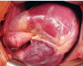
Median transsternal thoracotomy, opening of the pericardium; we observed distention of the right chambers and dilation of pulmonary trunk (Fig. 3). We initiated conventional cardiopulmonary bypass (CPB) circuit using cannulas in the aorta and vena cava.
While still at normal body temperature, the aorta was clamped, the RA was opened, and aspiration for decompression of cavities was performed, as well as single anterograde hypothermic blood cardioplegia at 4ºC.
The edges of the interatrial septal defect (Figure 4) are identified. Sutures were used at the inferior edge (near the inferior vena cava). We used a 5-0 polypropylene continuous suture, closing up the orifice of the defect with a bovine pericardium patch (Figure 5).
Fig. 4 – Opened right atrium showing large ostium secundum-type interatrial communication
Fig. 3 – Right chamber distension after pericardium opening
The patient was monitored in the ICU for 48 hours. Drains and central catheter were removed and the patient was referred to the recovery ward. She was discharged from the hospital on the fourth postoperative day, with a control echocardiography showing the correction of the defect.
Fig. 5 – Opened right atrium with ostium secundum-type interatrial communication corrected with bovine pericardium patch
REFERENCES
1. Christensen DD, Vincent RN, Campbell RM. Presentation of atrial septal defect in the pediatric population. Pediatr Cardiol. 2005;26(6):812-4.