3 2 8 Arq Bras Oftalmol. 2013;76(5):328-9
Cartas ao Editor |
Letterstothe editorUnilateral central retinal artery occlusion as the sole presenting sign
of Susac syndrome in a young man: case report
Oclusão unilateral da artéria central da retina como único sinal de apresentação da síndrome de Susac
em jovem do sexo masculino: relato de caso
RobeRt Alex egAn1
Submitted for publication: August 21, 2013 Accepted for publication: August 25, 2013
1Physician, Oregon Neurology, Tualatin, Unite States.
Funding: No specific financial support was available for this study.
Disclosure of potential conflicts of interest: R.A.Egan, None.
Corresponding author: Robert Alex Egan. Director of Neuro-Ophthalmology. Oregon Neurology - 19260 SW 65th Ave, Ste 280 - Tualatin, OR, United States, 97062 - E-mail: [email protected]
Submitted for publication: September 16, 2013 Accepted for publication: October 1, 2013
1Physician, Medical School, University of São Paulo, USP - São Paulo (SP), Brazil.
Funding: No specific financial support was available for this study.
Disclosure of potential conflicts of interest: M.L.R.Monteiro, None.
Correspondence address: Mário Luiz Ribeiro Monteiro. NeuroOphthalmology Service. Escola de Me -dicina da Universidade de São Paulo. Av. Angélica, 1.757 - Conj. 61 - São Paulo (SP) - 01227-200 - Brazil - E-mail: [email protected]
I read with interest the report of “Unilateral central retinal artery oc-clusion as the sole presenting sign of Susac syndrome in a young man: case report” by Apóstolos-Pereira et al.(1) Central retinal artery occlusion is very rare in Susac syndrome as evidenced by the paucity of reports in the clinical literature. The corpus callosum findings in this case are pathognomonic for the disease with the central callosal lesion and the 2 small spoke lesions in the posterior callosum. However, the fundus appearance is somewhat odd with the significant platelet-like embolic material in the arterioles. These are not Gass plaques as has been des-cribed previously(2). Did they disappear after his initial presentation? There are also characteristic fluorescein angiographic findings des-cribed previously(3). Was the typical autofluorescense pattern located
remotely from the retinal artery occlusion? I am particularly interested in learning if the right eye showed this pattern prior to infarction; if so, can this be published? Were anti-en dothelial cell antibodies sent?
REFERENCES
1. Apostolos-Pereira SL dos, Kara-Jose LB, Marchiori PE, Monteiro ML. Unilateral central retinal artery occlusion as the sole presenting sign of Susac syndrome in a young man: case report. Arq Bras Oftalmol. 2013;76(3):192-4.
2. Egan RA, Ha Nguyen T, Gass JD, Rizzo JF 3rd, Tivnan J, Susac JO. Retinal artery wall plaques in Susac Syndrome. Am J Ophthalmol. 2003;135(4):483-6.
3. Egan RA, Hills WL, Susac JO. Gass plaques and fluorescein leakage in Susac Syndrome. J Neurol Sci. 2010;299(1-2):97-100.
We thank Dr. Egan for his comments regarding our paper(1) and for the opportunity to further discuss Susac syndrome (SS), a unique condition which has held our greatest interest for many years. Soon after the first two cases reported by Susac et al.(2), we examined four cases reported in two sequential publications(3,4) that allowed its
cha-racterization as “A microangiopathic syndrome of encephalopathy, hearing loss, and retinal arteriolar occlusions”(4). We would like to point out that, as a fellow of Dr. W. F. Hoyt, we shared cases with him and sig-nificantly influenced him in naming the condition SS at a Symposium in 1986 when another case was presented to him for discussion by
Reply:
Fibrin-like material in the retinal arterioles and fluorescein angiography findings
in Susac syndrome
Material semelhante à ibrina em arteríolas retínicas e achados de angiograia luoresceínica
na síndrome de Susac
Cartas ao Editor | Letters to the editor
3 2 9
Arq Bras Oftalmol. 2013;76(5):328-9 Susac(5). Interestingly, very little has changed in our understanding of
the disease for more than three decades. Dr. Egan discussed topics in which our knowledge has advanced, the main one being the findings on magnetic resonance imaging (MRI). While MRI abnormalities were initially observed(4), subsequent advances in tech nology allowed to identify typical lesions in the corpus callosum(6), which are extremely important in the differential diagnosis and of which our recent case is an example(1). He also shares our point that central retinal artery occlu-sion is in very unusual and a more recently observed occurrence as the disease preferentially affects arterioles in the periphery of the retina(3,4).
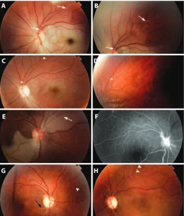
Figure 1. Fundus photograph of the left eye (A and B) and the right eye (E) at presentation; of the left eye three weeks later (C and D) and of both eyes two years later (G and H). Note that the blood column relex of several arterioles was partially or totally substituted by long segments of ibrin-like white material in the acute phase (white arrows in A, B and E) that signiicantly reduced or even disappeared at follow up (white arrowheads in C, D, G and H) while some arterioles developed a “silver streak” appearance (black arrow in G). Fluorescein angiography of the right eye at presentation shows branch artery occlusions and areas of segmental arterioles staining (F).
A
C
E
G
B
D
F
H
As for the observation regarding the material presented in the arteries of our case (Figure 1A, B and E), although we admit that it was somewhat larger than usual, we do not think it argues against SS. In fact, early on we observed that “the blood column reflex of several arterioles was partially or totally substituted by long segments of fibrin-like white material” (see figure 2 in Monteiro et al.(4)). We have seen similar findings in other cases and it is our understanding that these segments of fibrin-like material are also characteristic of SS and differ from platelet emboli mainly because they are located along the arteriole and not at its bifurcation. These fibrin-like material frequently decreases in size or disappear during follow-up, as observed in our case (Figure 1C, D, F and G), sometimes resembling the small Gass plaques reported by Egan et al.(7), and suggesting that the two findings lie within a spectrum of the same phenomenon in SS.
We agree that fluorescein angiography is important not only to show arteriolar occlusions but also to show segmental arteriolar staining(8), as observed in our current case (Figure 1 F). This finding, supports our initial suggestion that SS was an autoimmune disease against some unknown antigen present in the endothelium which has a blood-tissue barrier mechanism, as observed in the retina, cochlea and brain(3,4). Finally, we did not observe abnormalities in the initially unaffected eye of our case but unfortunately a fluorescein angiography was not performed until the second eye was involved; nor did we test the patient for recently described anti-endothelial cell antibodies. On the other hand, we do share Dr. Egan’s interest in knowing whether arteriolar staining on fluorescein angiography or Gass plaques on fundus observation could precede retinal arterial occlusions and certainly hope that further studies will be able to clarify this issue.
REFERENCES
1. Apostolos-Pereira SL dos, Kara-Jose LB, Marchiori PE, Monteiro ML. Unilateral central retinal artery occlusion as the sole presenting sign of Susac syndrome in a young man: case report. Arq Bras Oftalmol.2013;76(3):192-4.
2. Susac JO, Hardman JM, Selhorst JB. Microangiopathy of the brain and retina. Neuro-logy.1979;29(3):313-6.
3. Coppeto JR, Currie JN, Monteiro ML, Lessell S. A syndrome of arterial-occlusive reti-nopathy and encephalopathy. Am J Ophthalmol.1984;98(2):189-202.
4. Monteiro ML, Swanson RA, Coppeto JR, Cuneo RA, DeArmond SJ, Prusiner SB. A microangiopathic syndrome of encephalopathy, hearing loss, and retinal arteriolar occlusions. Neurology. 1985;35(8):1113-21.
5. Susac JO. Susac’s syndrome: the triad of microangiopathy of the brain and retina with hearing loss in young women. Neurology. 1994;44(4):591-3.
6. Susac JO, Murtagh FR, Egan RA, Berger JR, Bakshi R, Lincoff N, et al. MRI findings in Susac’s syndrome. Neurology. 2003;61(12):1783-7. Comment in: Neurology. 2004; 63(4):761; author reply 761.
7. Egan RA, Ha Nguyen T, Gass JD, Rizzo JF 3rd, Tivnan J, Susac JO. Retinal arterial wall plaques in Susac syndrome. Am J Ophthalmol.2003;135(4):483-6.