Contents lists available at
ScienceDirect
Toxicology Letters
j o u r n a l h o m e p a g e :
w w w . e l s e v i e r . c o m / l o c a t e / t o x l e t
Reduced cardiovascular alterations of tartar emetic administered in
long-circulating liposomes in rats
Naira R. Maciel
a
, Priscila G. Reis
a
, Kelly C. Kato
a
, Alessandra T. Vidal
a
, Homero N. Guimarães
b
,
Frederic Frézard
c
, Neila M. Silva-Barcellos
a
, Andrea Grabe-Guimarães
a
,
∗
aDepartamento de Farmácia, EF, Universidade Federal de Ouro Preto (UFOP), Ouro Preto, Minas Gerais, Brazil
bDepartamento de Engenharia Elétrica, EE, Universidade Federal de Minas Gerais (UFMG), Belo Horizonte, Minas Gerais, Brazil cDepartamento de Fisiologia e Biofísica, ICB, Universidade Federal de Minas Gerais (UFMG), Belo Horizonte, Minas Gerais, Brazil
a r t i c l e
i n f o
Article history: Received 20 May 2010
Received in revised form 3 September 2010 Accepted 6 September 2010
Available online 15 September 2010
Keywords: Antimonial tartrate Long-circulating liposomes ECG
Arterial pressure Cardiotoxicity
a b s t r a c t
Trivalent antimonial drugs, including tartar emetic (TA), are known to induce important cardiotoxicity observed by electrocardiographic abnormalities. Liposome encapsulation was found to reduce the overall acute toxicity of TA. The present work investigated the cardiovascular parameters alterations of rats submitted to the treatment with free and encapsulated TA in long-circulating liposomes. Liposomes were made using lipids DSPC, DSPE-PEG and cholesterol. The cardiovascular signals, electrocardiogram (ECG) and arterial blood pressure (AP), were recorded from anaesthetized Wistar rats after intravenous (IV) administration of a single specially high dose (17 mg/kg) of TA in liposomes and in free form. The IV administration of TA solution caused significant increase of QT interval of ECG and significant reduction of AP when compared to the control group. These alterations were not observed when liposomes TA were administered and the profile of ECG and AP data was quite similar to the control groups. In conclusion, a liposomal formulation of TA showed a reduced cardiotoxic profile for TA when compared to the free form.
© 2010 Elsevier Ireland Ltd. All rights reserved.
1. Introduction
Toxic effects on the cardiovascular system are routinely
described for many drugs, including anti-parasitic ones (
Crumb and
Cavero, 1999; Batey and Coker, 2002
). Non-cardiac drugs
induc-ing rhythm disturbances, like QT interval prolongation, which have
the electrocardiogram (ECG) features of ‘torsade de points’, were
described in the 1960s (
Shah, 2007
). The determination of cardiac
toxicity using QT interval prolongation continued to be
stud-ied (
Morganroth, 1993
) and it is a necessity for decision-making
during drug development (
EMEA, 1997, 2005; Haverkamp et al.,
2000
).
The trivalent antimonial drugs, including tartar emetic (TA),
were the first class of compounds employed in the clinical
treat-ment of schistosomiasis (
Cioli et al., 1995
), and TA was also used to
treat leishmaniasis in the 1912–1960 (
Frézard et al., 2005
).
How-ever, their use was discontinued because of their low therapeutic
index and side effects like trombocytopenia, other dyscrasias, and
∗Corresponding author at: Universidade Federal de Ouro Preto, Escola de Farmá-cia, Rua Costa Sena, 171, Centro, Ouro Preto, 35400-000 Minas Gerais, Brazil. Tel.: +55 31 3559 1628; fax: +55 31 3559 1628.
E-mail addresses:grabe@ef.ufop.br,agguimar@gmail.com(A. Grabe-Guimarães).
electrocardiographic disturbances. The cardiovascular alterations
induced by antimonial compounds include ECG alterations such
as ST segment inversion and QT interval prolongation, and,
conse-quently, ‘torsade de points’ arrhythmias and sudden cardiac arrest
(
Lacerda-Junior et al., 1965; Chulay et al., 1985
).
Brazil is considered one of the main area of schistosomiasis
distribution in the world (
Seubert et al., 1977
), and the
chemother-apy plays an important role in reducing schistosomiasis morbidity
(
WHO, 1993
). However, it has always been limited due to the
diffi-culty of discovering drugs presenting high efficacy and reasonable
tolerance (
Seubert et al., 1977; Andrews et al., 1983
). In this
con-text, efforts have been devoted to the improvement of old drugs by
development of novel formulations. Importantly, TA encapsulated
in long-circulating pegylated liposomes was more effective against
established Shistosoma mansoni infection during the late stages
of infection, than free TA or TA encapsulated in conventional
lipo-somes (
De-Melo et al., 2003
). Furthermore, liposome encapsulation
was found to reduce the overall acute toxicity of TA (
De-Melo et al.,
2003
). However, the impact of liposome encapsulation, specifically
in reducing cardiotoxicity of TA has not yet been determined. Thus,
the aim of the present work was to compare the cardiovascular
parameters, mainly the cardiotoxic markers, in rats submitted to
the treatment with free TA and TA encapsulated in long-circulating
liposomes.
199 (2010) 234–238 235
2. Materials and methods
2.1. Drugs and reagents
TA was purchased from Sigma (USA),l-␣-distearoylphosphatidylcholine (DSPC)
and PEG (2000)-distearoylphosphatidyl-ethanolamine (DSPE-PEG) were supplied by Lipoid GmBh (Ludwigshafen, Germany), and cholesterol was purchased from Sigma (USA). The solvents were of analytical grade and all other chemicals were commercially available. Water was purified by reverse osmosis (Symplicity System 185, Millipore, USA).
2.2. Preparation of tartar emetic containing pegylated liposomes
Liposomes were made from DSPC, cholesterol and DSPE-PEG at a molar ratio of 5:4:0.3. The encapsulation of TA (80 g/l in water) or PBS (150 mM NaCl, 10 mM phosphate, pH 7.2) was carried out in freeze–thawed multilamellar vesicles (Mayer et al., 1985). The multilamellar vesicles (lipid concentration of 120 g/l) were repeat-edly extruded through two stacked polycarbonate membranes of 200 nm pore size (Nayar et al., 1989), and finally submitted to dialysis against saline (150 mM NaCl) to purify TA containing liposomes and to remove non-encapsulated TA.
2.3. Liposome characterization
The concentration of encapsulated antimony was determined after mixing an aliquot of the suspension with 0.5 ml of a 20% (w/v) triton X-100 solution to solubilize the liposomes. Antimony was determined photometrically, using the chromogen bromopyrogallol red (BPR) (Frézard et al., 2001). According to this method, the absorbance of BPR at 560 nm decreases proportionally to the amount of antimony, as a consequence of the formation of the 1:1 BPR–Sb(III) complex. A calibration curve was established using TA as the source of antimony. The mean hydrodynamic diameter and polydispersity index of the vesicles suspensions were measured using the 3000 HS Zetasizer equipment (Malvern Instruments, Worces-tershire, England).
2.4. Experimental animals
Male Wistar rats (250±30 g) were randomly distributed into four experimental groups: the first received 17 mg of Sb/kg (3.4 mg Sb/ml in PBS) of TA intravenously (IV); a second received 17 mg of Sb/kg (4.36 mg Sb/ml) of liposomal TA IV. The other two groups received control solutions containing only vehicles: PBS or empty liposomes. The final volume administered was 0.8±0.2 ml. The animals were anaes-thetized with 60 mg/kg of sodium thiopental administered intraperitoneally. When the anesthesia reached the appropriate depth, the animals were tracheotomized to facilitate breathing. The femoral artery and vein were catheterized to recording of arterial blood pressure (AP) and IV drug administration, respectively. The catheters were previously filled with 1% heparin in 0.9% NaCl sterile solution. Stainless steel needle electrodes were inserted subcutaneously to record ECG.
All procedures related to the use of animals in these studies were reviewed and conform to the Ethical Principles of Animal Experimentation (Brazilian College of Animal Experimentation) and were approved by the UFOP Ethics Committee under number 99/2007.
2.5. Determination of cardiovascular parameters and protocols
AP was continuously recorded using a disposable pressure transducer (TruWave, Edwards Life Sciences) connected to a signal conditioning system. Limb lead II ECG was continuously recorded using subcutaneous stainless steel needle electrodes connected by a shielded cable to a biopotential amplifier. The AP signal conditioning system and the biopotential amplifier were designed and built in our laboratory, and all the care related with the frequency response of amplifiers (0.5–100 Hz to the biopotential amplifier and 0–30 Hz to AP amplifier) and the non-utilization of 60 Hz notch filters was taken to avoid distortion on recorded signals (Vale-Cardoso and Guimarães, 2010). The output signals of these systems were sampled at 1200 Hz by a 16-bits A/D conversion board (DaqBoard/2000, IOtech, USA) and stored on a PC hard disk.
The ECG and AP signals were recorded for 5 and 20 min before and after injec-tion of the different formulainjec-tions, respectively. Thereafter, segmented data records of 30 s were performed every 10 min up to 1 h after the injection of the different formulations. The stored records were analyzed off-line. From the stored records were extracted 2 s segments (raw data), containing 6 to 12 heart beats depending on the heart rate, and all the cardiovascular parameters were calculate as a mean value of these segments (filtered data). The cardiac parameters extracted from ECG records were QT (interval between the beginning of the Q-wave and the end of the T-wave), RR (interval between two successive R-waves and used to obtain the heart rate: HR = 60/RR), PR (interval between the beginning of the P-wave and the end of the R-wave) and QRS (interval from the beginning of the Q-wave to the end of the S-wave) intervals. Several mathematical formulae have been proposed to min-imize the QT interval dependence on heart rate (Simonson et al., 1962), deriving a heart rate corrected QT interval (QTc). The best known are Bazett’s (QTc = QT/RR1/2) (1920) and Fridericia’s (QTc = QT/RR1/3) (1920) formulae. We choose the Fridericia’s
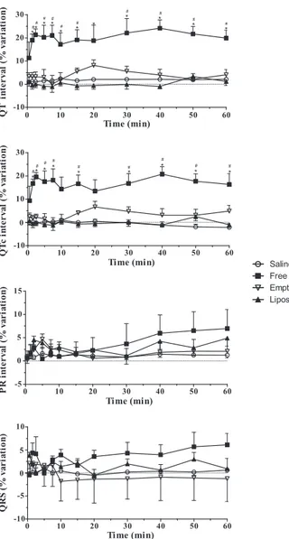
Fig. 1.Percentual variation of lead II ECG parameters obtained in anaesthetized rats after IV administration of free TA or in liposomes (17 mg Sb/kg) or control solutions. #Pvalue < 0.05 related to the saline group and*Pvalue < 0.05 related to the TA in liposome group.
formula, based on the work ofAbernethy et al. (2001), who suggest this correction criteria for the QTc for the cases of RR < 500 ms, as was consistently observed in our experiments (Vidal et al., 2010). The cardiovascular parameters extracted from AP records were systolic (SAP) and diastolic blood pressure (DAP) signals.
The percentage variations of each parameter, used inFigs. 1 and 2, were expressed as the mean value after drug injection minus the mean value before drug injection, divided by the mean value before drug injection and multiplied by 100.
2.6. Sb(III) determination in blood
Two other groups, that also received 17 mg of Sb/kg of free or of liposomal TA, were performed in order to collect blood samples at 10, 30 and 60 min after IV administration. The blood sample was storaged at−20◦C until the dosage proce-dure. Antimony was determined in serum and total blood by electrothermal atomic absorption spectrometry (ETAAS) using a Perkin-Elmer AA600 graphite furnace atomic absorption spectrometer, as previously described (Costantini et al., 1985). Samples were diluted 40 times in water containing 0.2% (v/v) HNO3and Sb was determined using pyrolise and atomization temperatures of 1300◦C and 2100◦C and Pd(NO3)2and Mg(NO3)2as matrix modifiers.
2.7. Statistics
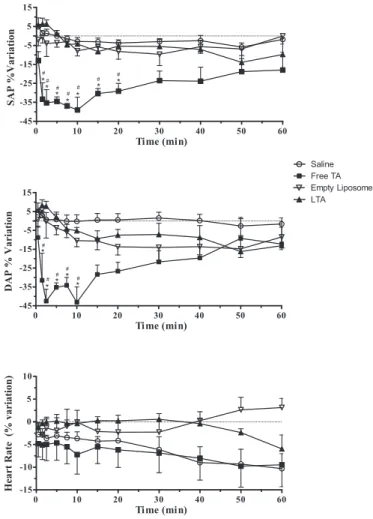
Fig. 2.Percentual variation of arterial pressure and heart rate obtained in anaes-thetized rats, after IV administration of free TA or in liposomes (17 mg Sb/kg) or control solutions.#Pvalue < 0.05 related to the saline group and*Pvalue < 0.05 related to the TA in liposome group.
3. Results
3.1. Liposome characterization
The mean hydrodynamics diameter of the vesicles was equal
to 149 nm (polydispersity 0.014) for liposomal TA and 199 nm
(polydispersity 0.016) for empty liposomes. Encapsulation of TA
was achieved with a trapping efficiency of 14.5% and a final
anti-mony/lipid ratio of 0.054 (w/w). The encapsulation of TA was found
to be stable since trapping efficiency was similar after 10 days
stor-age at 4
◦C compared to the initial value (
Table 1
).
Table 1
Percentual of encapsulated antimony.
Concentration of Sb(III) (mg/ml)
% of encap-sulation Soon after preparation 4.36 14.6 After 10 days of preparation 4.15 13.8
3.2. Determination of cardiovascular parameters
The IV administration of 17 mg of Sb/kg of TA solution in
Wis-tar rats caused significant increase of QT and QTc intervals of ECG,
that was observed since 1 min after its administration (
Fig. 1
), as it
was expected to occur (
Chulay et al., 1985; Thakur, 1998
), and was
maintained until the end of the experiment (1 h after
administra-tion). The increase of QT and QTc intervals was not observed when
the liposomal TA was administered, neither soon after its IV
admin-istration nor until the end of the experiment, and this protective
effect was similar among all the animals evaluated. No significant
alterations were observed in QRS and PR intervals after TA in free
ou liposomal forms. The free form of TA was also able to
signifi-cantly reduce the blood pressure (SAP and DAP), mainly between
1 and 20 min after its administration (
Fig. 2
). The filtered values
presented in
Table 2
show that very low levels of AP were reached
after administration of the free form of TA. These alterations were
not observed when TA in liposomes was injected (
Fig. 2
), indicating
that liposome encapsulation can prevent cardiovascular toxicity. In
resume, the results obtained from the animals that received TA in
liposomes were quite similar to those observed in control groups
(saline and empty liposomes), all along the time of the experiment
(
Figs. 1 and 2
, and
Table 2
). Also, there was no significant
alter-ation of ECG parameters in control groups which received either
empty liposomes or PBS (
Figs. 1 and 2
).
Table 2
reports in details
the filtered values of cardiovascular parameters measured before
and after the administration of each formulation of TA.
3.3. Sb determination in blood
The Sb(III) determination showed that a large amount of Sb
is maintained in blood compartment until 1 h after liposomal TA
administration. In contrary, when free TA was administered the
concentration of Sb(III) in blood, since 10 min after, was lower than
Sb from liposomal TA (
Fig. 3
).
4. Discussion
Tartar emetic, as other antimony compounds, is a drug that
can induce cardiotoxicity (
Honey, 1960; Thakur, 1998; Kuryshev
et al., 2006
). In this case, the most important ECG abnormalities
are ST segment inversion, QT interval prolongation, and therefore
Table 2
Filtered values of arterial blood pressure, heart rate and electrocardiographic parameters, measured before and after IV injection of 17 mg/kg free TA or liposomal TA at different times. The values represent the mean±S.E.M;*Pvalue < 0.05 compared to the time 0 (control period).
Heart rate (bpm)
Systolic AP (mm Hg)
Diastolic AP (mm Hg)
QT interval (ms)
QTc interval (ms)
PR interval (ms)
QRS interval (ms) TA
Time (min) 0 334±17.7 114±9.7 86±9.8 65±1.6 116±1.8 57±2.1 21±0.8 5 324±12.9 75±7.0* 55±6.0* 80±3.1* 136±4.9* 57±1.9 21±0.8 15 322±13.5 79±7.4* 61±6.8* 79±3.4* 135±5.4* 58±2.3 21±0.9 30 316±10.9 88±10.2 66±7.9 81±3.8* 135±4.1* 59±2.4 22±0.9 60 306±14.2 93±12.5 74±9.0 80±2.9* 135±5.8* 62±2.6 22±0.8 Liposomal TA
199 (2010) 234–238 237
10 30 60
1000 10000 100000 1000000
Free TA Liposomal TA
[Sb] (mg/l)
Time (min)
Fig. 3.Sb (III) blood concentration 10, 30 and 60 min after 17 mg of Sb/kg of free or liposomal IV administration.
‘torsades de points’ and sudden heart attack (
Lacerda-Junior et al.,
1965; Chulay et al., 1985
). Its ability to induce QT interval
prolon-gation has already been reported in anaesthetized guinea-pigs and
rabbits (
Alvarez et al., 2005
). In the present study it was showed
that QT interval prolongation and decreased blood pressure occur
after free form of TA administration, evidencing its cardiovascular
toxicity. There are some possible mechanisms to explain the
anti-monial toxicity. Antimony compounds can be genotoxic (
De Boeck
et al., 2003
), and can induce cytotoxicity and apoptosis (
Lecureur
et al., 2002; Mann et al., 2006
), effects that are important for its
use as anti-tumoral, but also could contribute to its cardiotoxicity.
Considering that both trivalent antimony and arsenic compounds
are metalloids belonging to group V of the periodic table, they
share many chemical properties and are thought to interfere with
biological processes in a similar manner; just like that it is
possi-ble to suggest a similar mechanism for TA toxicity as a rationale.
The QT interval prolongation, among other electrocardiographic
abnormalities, was also observed in patients treated with arsenic
trioxide (As
2O
3) for acute promyelocytic leukemia (
Ohnishi et al.,
2000
). In addition to prolongation of the action potential duration,
some studies have shown cellular Ca
2+overload, lipid peroxidation
caused by reactive oxygen species generation and decreased
intra-cellular ATP concentration (
Yamazaki et al., 2006
). It was reported
by
Drolet et al. (2004)
that As
2O
3is a potent blocker of both
I
Krand
I
Ksin hERG and KCNQ1 + KCNE1 transfected CHO cells.
Other-wise, it was reported that As
2O
3(
Ficker et al., 2004
) and antimonial
compounds (
Kuryshev et al., 2006
) reduce hERG/
I
Kr(a specific
car-diac ion channel that carries the rapidly activating delayed rectifier
potassium current,
I
Kr) currents, not by direct block, but by
inhibi-tion of hERG/
I
Krtrafficking to the cell surface (
Dennis et al., 2007
).
It should be pointed out that these experiments were carried out
with hERG (human ERG) transfected cells, and there are
signifi-cant differences in ERG protein expression between the species.
In rats, rERG (rat homologue of hERG) protein and functional
I
Krexpression are higher in atria than ventricles, whereas in mouse and
human, ERG (mERG and hERG) expression is higher in the
ventri-cles (
Pond et al., 2000
). Also, it was previously shown that antimony
potassium can induce a lethal oxidative stress in cardiac myocytes
(
Tirmenstein et al., 1995
), which may arise due to a deficiency of
antioxidant defenses rather than direct reduction of molecular
oxy-gen (
Tirmenstein et al., 1997
). The intracellular calcium increase
by antimony can also induced cardiac myocyte death (
Wey et al.,
1997; Ohnishi et al., 2000
). All these effects together could explain
the malfunction of the heart muscle caused by myocyte injury, and
could influence the arterial pressure control, beyond other
unde-termined mechanisms.
In fact, the main objective of our work was the study of the
toxic effects of TA encapsulated in liposomes on the
cardiovascu-lar system, assuming that the mechanism of action of TA has not
been changed. The specific investigation of ECG and blood pressure
changes caused by TA in the rat model is first reported here. It was
known that non-lethal doses of the TA reduce the ability of cardiac
myocytes to mobilize calcium during excitation–contraction phase
of cardiac cycle (
Toraason et al., 1997
), and that in neonatal rat
cul-tured cardiac myocytes, the non-lethal concentration of antimony
potassium tartrate was able to increase glutathione levels and the
synthesis of stress proteins, which may be responsible for
protec-tion against the toxicity caused by high doses used in treatments
(
Snawder et al., 1999
). In this way, the TA in liposomes is probably
less able to induce cardiotoxicity because the control release of the
high dose of TA used in this study.
Another important result reported here is the reduced effect
on blood pressure of TA in liposomes, preventing hypotension,
which is a common adverse reaction induced by the free form of
TA. However, this effect only represents a major advantage of
lipo-somes over the free form, when high doses are necessary. To treat
schistosomiasis is necessary to use multiple IV doses of TA every
day, for a month or more (
Cioli et al., 1995
), and it was reported
some options of doses, from 26.0 to 28.5 mg Sb/kg IV, until a total
of 2 g/day of TA, for 18–30 days (
Honey, 1960
). A previous study,
showed that intraperitoneal or subcutaneous administration of 11
and 27 mg Sb/kg TA in pegylated liposome reduced significantly
the worm burden of mice with Shistosoma mansoni infection and
they all survived, even when treated with the higher dose, in
con-trast with free TA that was 100% lethal with the dose of 27 mg/kg
(
De-Melo et al., 2003
). Additionally, the time-course of antimony
release from liposomes used in the present work has already been
determined by
De-Melo et al. (2003)
in mice serum and the results
showed that about 7% of encapsulated antimony was found to be
released within 24 h and 9 days was the time estimated to release
50% of Sb from liposomes. The distribution profile of free TA was
also determined before by
Ness et al. (1947)
and it was shown that
in 24 h only about 10% of Sb remained in blood. In the present work,
17 mg Sb/kg of TA in free or liposomal form was used, keeping the
animal alive during the experiments. A higher dose of TA free form
(18 mg/kg) in rats induced 100% death. To show the ability of
lipo-somes to avoid the cardiovascular toxicity of TA, a near to lethal
dose was used to demonstrate the advantage of TA encapsulation
in liposomes.
The polydispersity index and the average size of this suspension
of liposomes were very similar to those previously prepared
lipo-somal TA (
De-Melo et al., 2003
), demonstrating the reproducibility
of the preparation methodology.
The present work shows a reduction of TA cardiotoxicity
result-ing from the properties of liposomes, which probably induced
to a lower concentration of TA available for association with the
cardiac tissue as compared with the administration of the free
drug. It is noteworthy that such a reduction of cardiotoxicity has
been previously described, in the case of doxorubicin following its
encapsulation in long-circulating liposomes (
Papahadjopoulos et
al., 1991
), but this observation not necessarily indicates that this
ability of liposomes could be observed for all cardiotoxic drugs.
The present work is the first to demonstrate the prevention of
cardiotoxicity of an antimonial drug encapsulated in liposomes.
5. Conclusion
Conflict of interest statement
Authors declare there are no conflict of interest.
Acknowledgements
We wish to thank FAPEMIG (Rede Nanobiomg) and CNPq for its
grants, the scholarship from CNPq and CAPES, and our Universities
UFOP, UFMG, for the complete support.
References
Abernethy, D.R., David, L., Wesche, J.T., Ohrt, B., Mohanty, S.C., Pezzullo, J.C., Schuster, B.G., 2001. Stereoselective halofantrine disposition and effect: concentration-related QTc prolongation. Br. J. Clin. Pharmacol. 51 (3), 231–237.
Alvarez, M., Malécot, C.O., Gannier, F., Lignon, J.M., 2005. Antimony-induced car-diomyopathy in guinea-pig and protection byl-carnitine. Br. J. Pharmacol. 144 (1), 17–27.
Andrews, P., Thomas, H., Pohlke, R., Seubert, J., 1983. Praziquantel. Med. Res. Rev. 3 (2), 147–200.
Batey, A.J., Coker, S.J., 2002. Proarrhythmic potential of halofantrine, terfenadine and clofilium in a modified in vivo model of torsade de pointes. Br. J. Pharmacol. 135 (4), 1003–1012.
Chulay, J.D., Spencer, H.C., Mugambi, M., 1985. Electrocardiographic changes during treatment of leishmaniasis with pentavalent antimony (sodium stibogluconate). Am. J. Trop. Med. Hyg. 34 (4), 702–709.
Cioli, D., Pica-Mattoccia, L., Archer, S., 1995. Antischistosomal drugs: past, present and future? Pharmacol. Ther. 68 (1), 35–85.
Costantini, S., Giordano, R., Rizzica, M., Benedetti, F., 1985. Applicability of anodic-stripping voltammetry and graphite furnace atomic-absorption spectrometry to the determination of antimony in biological matrices: a comparative study. Analyst 110, 1355–1359.
Crumb, W., Cavero, I.I., 1999. QT interval prolongation by non-cardiovascular drugs: issues and solutions for novel drug development. Pharm. Sci. Technol. Today 2 (7), 270–280.
De Boeck, M., Kirsch-Volders, M., Lison, D., 2003. Cobalt and antimony: genotoxicity and carcinogenicity. Mutat. Res. 533, 135–152.
De-Melo, A.L., Silva-Barcellos, N.M., Demicheli, C., Frézard, F., 2003. Enhanced schis-tosomicidal efficacy of tartar emetic encapsulated in pegylated liposomes. Int. J. Pharm. 255 (1–2), 227–230.
Dennis, A., Wang, L., Ficker, E., 2007. Herg channel trafficking: novel targets in drug-induced long QT syndrome. Biochem. Soc. Trans. 35 (5), 1060–1063. Drolet, B., Simard, C., Roden, D.M., 2004. Unusual effects of a QT-prolonging drug,
arsenic trioxide, on cardiac potassium currents. Circulation 109 (1), 26–29. European Medicines Agency, 1997. Points to consider: the assessment of the
potential for QT interval prolongation by non-cardiovascular medicinal prod-ucts. Doc. n CPMP/986/96. Avaiable from: http://ferran.torres.name/links/ links CPMP ICH.htm# Toc68179441.
European Medicines Agency, 2005. ICH note for guidance on the clinical evaluation of QT/QTc interval prolongation and proarrhythmic potential for non-antiarrhythmic drugs (ICH E14). Doc. n CHMP/ICH/2/04. Avaiable from:http:// www.emea.eu.int/pdfs/human/ich/000204en.pdf.
Ficker, E., Kuryshev, Y.A., Dennis, A.T., Obejero-Paz, C., Wang, L., Hawryluk, P., Wible, B.A., Brown, A.M., 2004. Mechanisms of arsenic-induced prolongation of cardiac repolarization. Mol. Pharmacol. 66 (1), 33–44.
Frézard, F., Demicheli, C., Ferreira, C.S., Costa, M.A., 2001. Glutathione-induced conversion of pentavalent antimony to trivalent antimony in meglumine anti-moniate. Antimicrob. Agents Chemother. 45 (3), 913–916.
Frézard, F., Schettini, D.A., Rocha, O.G.F., Demicheli, C., 2005. Lipossomas: pro-priedades físicoquímicas e farmacológicas, aplicac¸ões na quimioterapia à base de antimônio. Quim. Nova. 28 (3), 511–518.
Haverkamp, W., Breithardt, G., Camm, A.J., Janse, M.J., Rosen, M.R., Antzelevitch, C., Escande, D., Franz, M., Malik, M., Moss, A., Shah, R., 2000. The potential for QT prolongation and proarrhythmia by non-antiarrhythmic drugs: clincal and regulatory implications. Report on a policy conference of the European Society of Cardiology. Eur. Heart J. 21, 1216–1231.
Honey, M., 1960. The effects of sodium antimony tartrate on the myocardium. Br. Heart J. 22 (5), 601–616.
Kuryshev, Y.A., Wang, L., Wible, B.A., Wan, X., Ficker, E., 2006. Antimony-based antileishmanial compounds prolong the cardiac action potential by an increase in cardiac calcium currents. Mol. Pharmacol. 69 (4), 1216–1225.
Lacerda-Junior, F.S., Ferminiani, H., Mota, C.C.S., Baranski, M.C., 1965. Comparative study of electrocardiographic changes determined by treatment with tri- and pentavalent antimony. Rev. Inst. Med. Trop. Sao Paulo. 7 (4), 210–217. Lecureur, V., Le Thiec, A., Le Meur, A., Amiot, L., Drenou, B., Bernard, M., Lamy, T.,
Fauchet, R., Fardel, O., 2002. Potassium antimonyl tartrate induces caspase and reactive oxygen species-dependent apoptosis in lymphoid tumoral cells. Br. J. Haematol. 119, 608–615.
Mann, K.K., Davison, K., Colombo, M., Colosimo, A.L., Diaz, Z., Padovani, A.M.S., Guo, Q., Scrivens, P.J., Gao, W., Mader, S., Miller Jr., W.H., 2006. Antimony trioxide apoptosis is dependent on SEK1/JNK signaling. Toxicol. Lett. 160 (158), 170. Mayer, D., Hope, M.J., Cullis, P.R., Janoff, A.S., 1985. Solute distributions and trapping
efficiencies observed in freeze-thawed multilamellar vesicles. Biochim. Biophys. Acta. 817 (1), 193–196.
Morganroth, J., 1993. QTc interval prolongation: is it beneficial or harmful? Sympo-sium proceedings. Am. J. Cardiol. 72, 1–59B.
Nayar, R., Hope, M.J., Cullis, P.R., 1989. Generation of large unilamellar vesicles from long-chain saturated phosphatidylcholines by extrusion technique. Biochim. Biophys. Acta. 986 (2), 206–1206.
Ness, A.T., Brady, F.J., Cowie, D.B., Lawton, A.H., 1947. Anomalous distribution of anti-mony in white rats following the administration of tartar emetic. J. Pharmacol. Exp. Ther. 90 (2), 174–180.
Ohnishi, K., Yoshida, H., Shigeno, K., Nakamura, S., Fujisawa, S., Naito, K., Shinjo, K., Fujita, Y., Matsui, H., Takeshita, A., Sugiyama, S., Satoh, H., Terada, H., Ohno, R., 2000. Prolongation of the QT interval and ventricular tachycardia in patients treated with arsenic trioxide for acute promyelocytic leukemia. Ann. Intern. Med. 133 (11), 881–885.
Papahadjopoulos, D., Allen, T.M., Gabizon, A., Mayhew, E., Matthay, K., Huang, S.K., Lee, K.D., Woodle, M.C., Lasic, D.D., Redemann, C., Martin, F.J., 1991. Sterically stabilized liposomes: improvements in pharmacokinetics and antitumor thera-peutic efficacy. Proc. Natl. Acad. Sci. U.S.A. 88 (24), 11460–11464.
Pond, A.L., Scheve, B.K., Benedict, A.T., Petrecca, K., Van-Wagoner, D.R., Shrier, A., Ner-bonne, J.M., 2000. Expression of distinct ERG proteins in rat, mouse, and human heart—Relation to functional IKrchannels. J. Biol. Chem. 275 (8), 5997–6006. Seubert, J., Pohlke, R., Loebich, F., 1977. Synthesis and properties of praziquantel, a
novel broad spectrum anthelmintic with excellent activity against Schistosomes and cestodes. Experientia 33 (8), 1036–1037.
Shah, R.R., 2007. Cardiac repolarization and drug regulation: assessing cardiac safety 10 years after the CPMP guidance. Drug Saf. 30 (12), 1093–1110.
Simonson, E., Cady, L.D., Woodbury, M., 1962. The normal Q-T interval. Am. Heart J. 63 (6), 747–753.
Snawder, J.E., Tirmenstein, M.A., Mathias, P.I., Toraason, M., 1999. Induction of stress proteins in rat cardiac myocytes by antimony. Toxicol. Appl. Pharmacol. 159 (2), 91–97.
Thakur, C.P., 1998. Sodium antimony gluconate, amphotericin and myocardial dam-age. Lancet 351, 1928–1929.
Tirmenstein, M.A., Mathias, P.I., Snawder, J.E., Wey, H.E., Toraason, M., 1997. Antimony-induced alterations in thiol homeostasis and adenine nucleotide sta-tus in cultured cardiac myocytes. Toxicology 119 (3), 203–211.
Tirmenstein, M.A., Plews, P.I., Walker, C.V., Woolery, M.D., Wey, H.E., Toraason, M.A., 1995. Antimony-induced oxidative stress and toxicity in cultured cardiac myocytes. Toxicol. Appl. Pharmacol. 130 (1), 41–47.
Toraason, M., Wey, H.E., Richards, D.E., Mathias, P.I., Krieg, E., 1997. Altered Ca2+mobilization during excitation–contraction in cultured cardiac myocytes exposed to antimony. Toxicol. Appl. Pharmacol. 146 (1), 104–115.
Vale-Cardoso, A.S., Guimarães, H.N., 2010. The effect of 50/60 Hz notch filter appli-cation on human and rat ECG recordings. Physiol. Meas. 31 (1), 45–58. Vidal, A.T., Guimarães, H.N., Paula, D.C.C., Frézard, F., Silva-Barcellos, N.M.,
Grabe-Guimarães, A., 2010. Prolonged cardioprotective effect of pyridostigmine encapsulated in liposomes. Life Sci. 86 (1–2), 17–23.
Wey, H.E., Richards, D., Tirmenstein, M.A., Mathias, P.I., Toraason, M., 1997. The role of intracellular calcium in antimony-induced toxicity in cultured cardiac myocytes. Toxicol. Appl. Pharmacol. 145 (1), 202–210.
World Health Organization, 1993. The Control of Schistosomiasis. Second Report of WHO Expert Committee. Technical Report Series (830), Geneva, 86p. Yamazaki, K., Terada, H., Satoh, H., Naito, K., Takeshita, A., Uehara, A., Katoh, H.,