UNIVERSIDADE FEDERAL DO CEARÁ
DEPARTAMENTO DE SAÚDE MATERNO INFANTIL
MESTRADO PROFISSIONAL EM SAÚDE DA MULHER E DA CRIANÇA
CLARA TAÍNA SILVA LIMA
ASSOCIAÇÃO DA EPISIOTOMIA E LACERAÇÃO PERINEAL GRAVE COM DISFUNÇÃO DO ASSOALHO PÉLVICO NO PÓS-PARTO REMOTO
CLARA TAÍNA SILVA LIMA
ASSOCIAÇÃO DA EPISIOTOMIA E LACERAÇÃO PERINEAL GRAVE COM DISFUNÇÃO DO ASSOALHO PÉLVICO NO PÓS-PARTO REMOTO
Dissertação apresentada ao Programa de Pós-Graduação do Departamento de Saúde Materno Infantil da Universidade Federal do Ceará, para obtenção do título de Mestre em Saúde da Mulher e da Criança. Área de concentração: Atenção Integrada e Multidisciplinar à Saúde Materno-Infantil.
Linha de Pesquisa: Atenção à Saúde Materna e Perinatal.
Orientadora: Profa. Dra. Simony Lira do Nascimento.
Coorientador: Prof. Dr. Leonardo Robson Pinheiro Sobreira Bezerra.
CLARA TAÍNA SILVA LIMA
ASSOCIAÇÃO DA EPISIOTOMIA E LACERAÇÃO PERINEAL GRAVE COM DISFUNÇÃO DO ASSOALHO PÉLVICO NO PÓS-PARTO REMOTO
Dissertação apresentada ao Programa de Pós-Graduação do Departamento de Saúde Materno Infantil da Universidade Federal do Ceará, para obtenção do título de Mestre em Saúde da Mulher e da Criança. Área de concentração: Atenção Integrada e Multidisciplinar à Saúde Materno-Infantil.
Aprovada em: ___/___/______
BANCA EXAMINADORA
______________________________________________________________________ Profa. Dra. Simony Lira do Nascimento (Orientador)
Universidade Federal do Ceará (UFC)
______________________________________________________________________ Prof. Dr. Leonardo Robson Pinheiro Sobreira Bezerra
Universidade Federal do Ceará (UFC)
______________________________________________________________________ Dra. Simone Botelho Pereira
Universidade Federal de Alfenas (UNIFAL-MG)
______________________________________________________________________ Prof. Dr. Francisco Herlânio Costa Carvalho
RESUMO
Objetivo: Avaliar a associação entre episiotomia e laceração perineal grave (terceiro e quarto graus) com a presença da incontinência urinária (IU), função contrátil e achados ultrassonográficos dos músculos do assoalho pélvico (MAP) no pós-parto remoto. Materiais e Métodos: Produto 1 - foi realizada uma Revisão Sistemática e Meta-análise de estudos observacionais que avaliaram a associação entre lesões perineais do parto e as alterações morfológicas dos MAP avaliadas pelo ultrassom (US). Produto 2
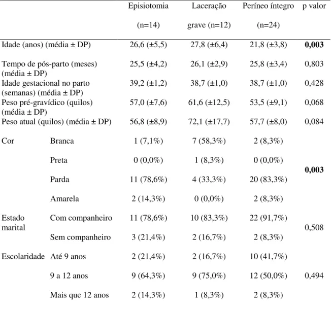
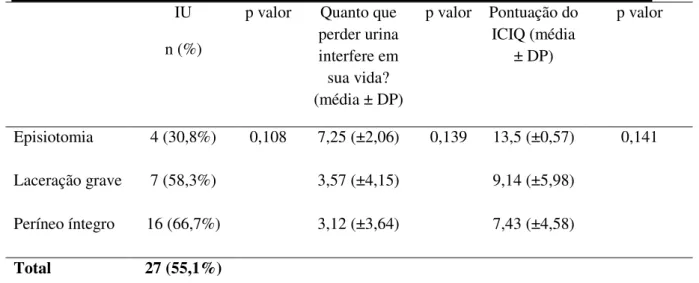
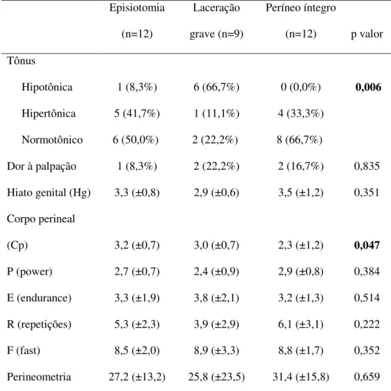
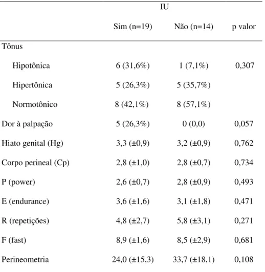
– estudo observacional de corte transversal. Realizado com mulheres primíparas oriundas da Maternidade Escola Assis Chateaubriand (MEAC), que estavam no período de 12 a 36 meses após parto vaginal a termo. Foram excluídas mulheres com infecção no trato urogenital, histórico de câncer ginecológico ou retal, cirurgias prévias nos MAP, doenças desmielinizantes da medula espinhal ou que engravidaram novamente. Os prontuários de todas as mulheres que tiveram parto vaginal entre maio a novembro de 2015 foram analisados, e a partir dessa análise, foram identificadas as mulheres que sofreram laceração perineal grave e as que realizaram episiotomia. As mulheres foram selecionadas para compor três grupos: 1) mulheres que foram submetidas à episiotomia (n=12); 2) mulheres que sofreram laceração perineal grave (n=9) e 3) mulheres que mantiveram o períneo íntegro (n=12). A avaliação inicial foi composta por um questionário clínico e sociodemográfico, e International Consultation on Incontinence Questionnaire - Short Form (ICIQ-SF). A avaliação física foi composta por medidas do Pelvic Organ Prolapse Quantification System (POP-Q), palpação digital dos MAP e perineometria. As análises estatísticas foram realizadas com os testes Qui-quadrado e U de Mann-Whitney. Resultados: Produto 1 - Tanto a episiotomia quanto a laceração perineal grave estão associadas à maior probabilidade de avulsão do músculo levantador do ânus (OR=1,77, 95% CI 1,25-2,51) (OR=4,31, 95% CI 2,34-7,91), respectivamente; defeitos dos esfíncteres do ânus (OR 2,82, 95% CI 1,71-4,67) e aumento da área do hiato urogenital em comparação com mulheres que não sofreram essas lesões obstétricas. Produto 2 - não se observou relação entre a presença da IU com os traumas perineais ou com a função muscular (tônus, dor, hiato genital, corpo perineal, força e resistência). Porém, foi possível observar diferença estatística na alteração do tônus muscular (p=0,006) e no tamanho do corpo perineal (p=0,047) entre os grupos. A presença de IU antes (p=0,021) e durante (p=0,025) a gestação tiveram relação com o desenvolvimento de IU no pós-parto. Conclusões: A episiotomia e a laceração perineal grave se relacionam com a presença de lesões nos MAP visualizadas pelo US, porém clinicamente não observamos relação com a presença da IU. Logo, há uma incerteza com relação as alterações visualizadas pelo US serem determinantes para ter IU.
ABSTRACT
Objective: To evaluate the association between episiotomy and severe perineal laceration (third and fourth degreen) with the presence of urinary incontinence (UI), contractile function and ultrasonographic findings of the pelvic floor muscles (PFM) in the postpartum remote. Materials and Methods: Product 1 – A Systematic Review and Meta-Analysis of observational studies evaluating the association between perineal childbirth lesions and morphological alterations pf PFM evaluated by ultrasound was performed. Product 2 – cross-sectional observational study. It was performed with primiparous women from the Maternity School Assis Chateaubriand (MEAC), who were in the period from 12 to 36 months after term vaginal delivery. Women with urogenital tract infection, history of gynecological or rectal cancer, previous surgeries in the PFM, carriers of demyelinating diseases of the spinal cor dor who became pregnant again were excluded. The records of all women who had vaginal delivery between May and November 2015 were analyzed, and from this analysis, women who suffered severe perineal laceration and those who underwent episiotomy were identified. The women were selected to composse three groups: 1) women who underwent episiotomy (n=12); 2) women who suffered severe perineal laceration (n=9) and 3) women who maintained intact perineum (n=12). The initial evaluation consisted of a clinical and sociodemographic questionnaire, and the International Consulation on Incontinence Questionnaire – Short Form (ICIQ-SF). The physical evaluation was composed of Pelvic Organ Prolapse Quantification System (POP-Q) measurements, digital MAP palpation and perineometry. Statistical analyzes were performed using the Chi-square and U Mann-Whitney tests. Results: Product 1– Both episiotomy and severe perineal laceration are associated with a greater likelihood of avulsion of the anus levator muscle (OR = 1,77, 95% CI 1,25-2,51) (OR = 4,31, 95% CI 2,34-7,91), respectively; defects of the sphincters (OR = 2,82, 95% CI 1,71-4,67) and increased urogenital hiatos área compared to women who did not experience these obstetric lesions. Product 2 – there was no relationship between the presence of UI and perineal traumas or muscle function (tone, pain, genital hiatos, perineal body, strength and resistance). However, it was possible to observe a statistical difference in muscle tone (p = 0.006) and perineal body size (p = 0.047) between groups. The presence of UI before (p = 0.021) and during (p = 0.025) gestation were related to the development of UI in the postpatum period. Conclusions: Episiotomy and severe perineal laceration are related to the presence of lesions in the MAP visualized by the US, but clinically we did not observe a relation with the presence of UI. Therefore, there is uncertainty regarding the changes seen by the US being decisive for having UI.
SUMÁRIO
1.INTRODUÇÃO ... 8
1.1. Efeito da gravidez e parto sobre o assoalho pélvico ... 8
1.2. Relação entre lesões obstétricas com incontinência urinária e função dos músculos do assoalho pélvico... 9
1.3. Relação entre lesões obstétricas e alterações morfológicas identificadas pelo ultrassom ... 10
2. JUSTIFICATIVA ... 12
3. OBJETIVOS ... 12
3.1. Objetivo Geral ... 12
3.2. Objetivos Específicos ... 13
4. HIPÓTESES ... 13
5. MATERIAIS E MÉTODOS ... 13
5.1. Local e tipo de estudo ... 13
5.2. População e amostra ... 13
5.3. Seleção dos sujeitos ... 14
5.3.1. Critérios de inclusão ... 14
5.3.2. Critérios de exclusão ... 14
5.4. Variáveis ... 14
5.4.1. Variáveis independentes ... 14
5.4.2. Variáveis dependentes ... 14
5.4.3. Variáveis de controle ... 15
5.5. Lista de instrumentos de coleta de dados ... 15
5.6. Seleção e treinamento de pessoal ... 16
5.7. Plano de coleta de dados ... 17
5.8. Análise estatística ... 17
5.9. Aspectos éticos ... 18
5.10. Financiamento ... 18
6. RESULTADOS ... 18
Produto 1 – Revisão Sistemática e Meta-Análise ... 19
Produto 2 – Artigo Original ... 40
7. CONCLUSÕES ... 58 REFERÊNCIAS ... 59 APÊNDICE A –LISTA DE VERIFICAÇÃO ... 63
APÊNDICE B – QUESTIONÁRIO CLÍNICO E SOCIODEMOGRÁFICO 64
APÊNDICE C – TERMO DE FIEL DEPOSITÁRIO ... 66
APÊNDICE D – TERMO DE CONSENTIMENTO LIVRE E
ESCLARECIDO ... 67 ANEXO A – International Consulation on Incontinence Questionnaire –
1. INTRODUÇÃO
1.1Efeito da gravidez e parto sobre o assoalho pélvico
Os músculos do assoalho pélvico (MAP) são responsáveis por suportar as vísceras e manter a continência (ASHTON-MILLER et al., 2001; HERSCHORN, 2004; RAIZADA, MITTAL, 2008; YAVAGAL et al., 2011). Anatomicamente dividem-se em duas camadas musculares ou diafragmas: 1) musculatura profunda do assoalho pélvico, que é composta pelos músculos pubovisceral, puborretal e íleococcígeo, que juntos formam o músculo levantador do ânus; e 2) musculatura superficial, que é composta pelos músculos isquiocavernoso, bulboesponjoso e transverso superficial do períneo (HERSCHORN, 2004; BO; SHERBURN, 2005; RAIZADA; MITTAL, 2008).
A gestação e o parto vaginal são considerados fatores de risco para as disfunções do assoalho pélvico (DAPs). A pressão crescente do útero e do peso fetal nos MAP, juntamente com as alterações hormonais relacionadas à gravidez podem levar à redução da força dos MAP, assim como alteração na sua função de apoio. Isso provocará mobilidade no colo vesical e na uretra, levando a incompetência do esfíncter uretral (SANGSAWANG; SANGSAWANG, 2013). Durante o parto vaginal ocorre estiramento e compressão dos músculos, nervos e tecidos conjuntivo, podendo ocorrer desnervação parcial ou total dos MAP (SNOOKS et al, 1990; DIETZ; WILSON, 2005) e avulsão do músculo levantador do ânus (van DELFT et al, 2014; 2015). Lesões obstétricas como laceração perineal e episiotomia são agravantes, e vêm sendo insistentemente estudados.
A episiotomia, procedimento obstétrico amplamente utilizado, consiste em realizar uma incisão nos MAP que pode ser em dois sentidos: mediana ou médio-lateral, e tem como justificativa a prevenção de lesões perineais mais graves, retais e dos esfíncteres anais interno e externo (AYTAN et al, 2005). Uma das principais indicações para a realização da episiotomia é a iminência de laceração de 3º e 4º graus, percebida subjetivamente pela rigidez perineal durante o trabalho de parto. Porém ainda não é clara a real proteção que a episiotomia pode oferecer, e as evidências apontam que seu uso deve ser restrito e não rotineiro. Em uma meta-análise, que incluiu 8 estudos (5.541 mulheres), foi observado que o uso restrito mostra menor risco para trauma perineal severo (RR 0,67, IC 95% 0,49-0,91), trauma perineal posterior (RR 0,88, IC 95% 0,84-0,92), necessidade de suturar o trauma perineal (RR 0,71, IC 95% 0,61-0,81) e complicações de cicatrização após sete dias de pós-parto (RR 0,69, IC 95% 0,56-0,85); sem diferenças para dispareunia e incontinência urinária. Questões como as reais indicações para a realização de episiotomia restrita e a sua associação com as DAPs ainda necessitam ser esclarecidas à luz das evidências científicas (CARROLI; MIGNINI, 2009).
1.2. Relação entre lesões obstétricas com incontinência urinária e função dos músculos do assoalho pélvico
Estima-se que a prevalência de IU na gestação varia de 30% a 38,7% (VALETON; AMARAL, 2011; DALY; CLARKE; BEGLEY, 2018). Porém, o parto vaginal, especialmente o primeiro parto, é reconhecido como potencialmente traumático para os MAP, quando comparado com a cesariana, podendo levar à IU de esforço em até 31% das mulheres no pós-parto (WESNES et al, 2009; BOYLES et al, 2009; DIEZ-ITZA et al, 2010; TAHTINEN et al, 2016).
Acredita-se que as lesões diretas e indiretas das fáscias e músculos durante o parto vaginal comprometem a função de suporte da bexiga e do esfíncter urinário, sendo estes os principais mecanismos etiológicos que justificam o aumento do risco de IU de esforço em três vezes [3,4 (0,9–6,6)] após o parto vaginal espontâneo, e em cinco vezes [5,7 (2,6-9,7)] após episiotomia. Essa IU pode permanecer até 12 meses após o parto de primíparas (KOKABI; YAZDANPANAH,2017).
comparadas com as que sofreram lacerações perineais graves, avaliadas pelo questionário de qualidade de vida King’s Health Questionnaire (KHQ). Neste caso, esses autores acreditam que a episiotomia pode servir como um fator de proteção para as disfunções dos MAPs.
Correlações entre laceração perineal e episiotomia com a função muscular do assoalho pélvico também estão sendo estudadas. Sartore et al, (2004) estudou os efeitos da episiotomia médio-lateral na função dos MAP. Para tanto, comparou os MAP de mulheres que foram e que não foram submetidas a incisão. Observaram que as mulheres submetidas a episiotomia apresentaram maior incidência de dispareunia e pior desempenho nos testes de manometria anal e perineometria, mas não foi observado diferença entre os grupos na incidência de prolapso de órgãos pélvicos (POP). Concluíram ainda que a episiotomia não protege contra a IU, incontinência fecal e POP. Em outro estudo randomizado controlado, realizado por Dannecker et al, (2005), avaliou-se a pressão uretral, manometria anal, força dos MAP, e a presença de IU, fecal e dispaurenia em dois grupos de mulheres: as que realizaram e as que não realizaram episiotomia a partir de uma laceração iminente no momento do parto vaginal. Observaram que não houve diferença estatística entre os grupos. O estudo de Bharucha et al, (2012) avaliou por ressonância magnética mulheres com e sem incontinência fecal, e percebeu que a episiotomia foi o único fator obstétrico associado com o risco de qualquer lesão dos MAP.
Um estudo realizado por Leeman et al, (2007) comparou a função muscular de mulheres que suturaram uma laceração perineal de segundo grau após o parto com as que não suturaram e com mulheres que não tiveram nenhum grau de laceração. Observou-se que não houve diferença estatística entre os grupos para função sexual, continência e força dos MAP.
Handa et al, (2012) realizou a avaliação de 449 mulheres no período de 5 a 10 anos após o último parto, e observaram que a episiotomia não foi associada a qualquer desordem dos MAP (IU de esforço, bexiga hiperativa, POP ou incontinência fecal). Já as mulheres com história de mais de uma laceração perineal espontânea foram significativamente mais propensas a ter POP.
Além dos métodos mais tradicionais de avaliação da função do assoalho pélvico, como o exame clínico através da palpação, perineometria e manometria, atualmente pesquisadores têm mostrado maior interesse em estudos com ultrassom (US) transperineal. Esse possibilita a investigação da estrutura e função muscular sem penetração vaginal. Segundo Ubukata et al, (2015), métodos não invasivos para avaliação dos MAP evitam desconfortos e constrangimentos à paciente. Esse constrangimento pode mascarar as contrações dessa musculatura em um primeiro momento do exame entre terapeuta e paciente.
Até recentemente a ressonância magnética era o único método possível de avaliar as modificações nas estruturas anatômicas dos MAP. Ao contrário da ressonância magnética, o US transperineal tem menor custo, sua realização é mais viável clinicamente, além de ser uma técnica segura e confiável para ser utilizado em mulheres grávidas e no pós-parto (VAN VEELEN et al, 2013; STÆR-JENSEN et al, 2013; FALKERT et al, 2013). Também se mostrou de alta confiabilidade para medir as dimensões do hiato genital e traumas no músculo levantador do ânus (VAN VEELEN et al, 2013; VAN VEELEN et al, 2014).
Correlações entre as lesões obstétricas e alterações morfológicas visualizadas pelo US estão sendo bastante estudadas. No estudo de Falkert et al, (2010) foi observado que o uso da episiotomia ou presença de lacerações perineais não tiveram influência sobre as alterações morfológicas visualizadas pelo US transperineal, realizado dois dias após o parto. Do contrário, o estudo de Shek et al, (2016) mostra que as lacerações perineais graves estão sim associadas com a avulsão do músculo levantador do ânus, visualizadas pelo mesmo método. O estudo de Cassadó et al, (2014), ao avaliar a relação entre a episiotomia e as alterações morfológicas no músculo levantador do ânus após o parto vaginal, percebeu que não houve relação entre essas variáveis.
Apesar da importante morbidade a longo prazo, relacionada às lesões do assoalho pélvico decorrentes do parto vaginal, afetarem a qualidade de vida das mulheres, os resultados dos estudos ainda são contraditórios. Desta forma, fica claro a necessidade de mais pesquisas voltadas para essa temática.
Portanto, o objetivo desse estudo é realizar uma revisão sistemática da literatura sobre o impacto da episiotomia e da laceração perineal grave sobre as lesões do assoalho pélvico secundárias ao parto vaginal, avaliado por US, assim como avaliar a associação entre a episiotomia e a laceração perineal grave com a presença da IU e função dos MAP em mulheres no pós-parto remoto.
2. JUSTIFICATIVA
Apesar da morbidade a longo prazo, relacionada às lesões no assoalho pélvico durante o parto vaginal, que preocupa e afeta a qualidade de vida das mulheres, os achados literários que avaliam a relação entre os traumas obstétricos com a IU dentro de um período maior que um ano após o parto são poucos e contraditórios. Assim, fica claro a importância da realização de um novo estudo que avalie as repercussões da episiotomia e da laceração perineal grave nos MAP de mulheres no pós-parto remoto torna-se relevante.
A identificação da influência da episiotomia e da laceração perineal grave sobre o assoalho pélvico e o diagnóstico precoce das disfunções dos MAP no pós-parto poderá contribuir para melhorar a assistência dada pelos profissionais de saúde a essa população. Além disso, o US transperineal é um método de baixo custo e de alta confiabilidade, e por ser um método não invasivo, torna-se fundamental em circunstâncias onde uma técnica invasiva seja inapropriada.
3. OBJETIVOS
3.1. Objetivo Geral
laceração perineal grave com a presença da IU e função dos MAP em mulheres no pós-parto remoto.
3.2. Objetivos Específicos
● Investigar, por revisão sistemática da literatura, o impacto da episiotomia e da laceração perineal grave sobre os MAP após o parto vaginal, avaliado por US. ● Investigar a associação entre a episiotomia e a laceração perineal grave com a
presença da IU em mulheres.
● Avaliar a associação entre a episiotomia e a laceração perineal grave com a função muscular do assoalho pélvico, percebida pela palpação digital e perineometria de pressão, em mulheres.
4. HIPÓTESES
● Episiotomia e laceração perineal grave estão relacionadas com as alterações morfológicas visualizadas pelo US, como avulsão do músculo levantador do ânus.
● Episiotomia e laceração perineal grave estão associadas com a presença da IU em mulheres.
● Episiotomia e laceração perineal grave estão associadas com redução da força dos MAP de mulheres no pós-parto remoto.
5. MATERIAIS E MÉTODOS
5.1. Local e Tipo de Estudo
Este estudo apresenta um delineamento do tipo observacional de corte transversal e foi executado na Maternidade Escola Assis Chateaubriand (MEAC).
5.2. População e Amostra
5.3. Seleção dos Sujeitos 5.3.1. Critérios de inclusão
● Mulheres no pós-parto ● Pós-parto de 12 a 36 meses ● Parto vaginal
● Parto a termo ● Primíparas
5.3.2. Critérios de exclusão ● Nova gestação
● Infecção atual no trato urogenital ● Câncer ginecológico ou retal ● Cirurgia nos MAP
● Tratamentos fisioterápicos dos MAP entre o parto e o convite para o estudo ● Doenças desmielinizantes traumáticas ou não traumáticas da medula espinhal ● Demências
5.4. Variáveis
5.4.1. Variáveis Independentes
● Laceração perineal grave: quando ocorre a lesão parcial ou completa do esfíncter anal, sem o envolvimento do epitélio anal (terceiro grau) ou também com a lesão do epitélio anal (quarto grau) (RCOG, 2015).
● Episiotomia: procedimento obstétrico que consiste em realizar uma incisão nos MAP, podendo ser em dois sentidos: mediana ou médio-lateral (AYTAN et al, 2005).
● Períneo íntegro: quando não houver lesão no períneo decorrente do parto, ou com laceração de primeiro grau (lesão de pele perineal e/ou mucosa vaginal) (RCOG, 2015).
5.4.2. Variáveis Dependentes
● Função contrátil dos MAP: capacidade do assoalho pélvico de contrair e relaxar a partir de comandos verbais, avaliada através da Escala Modificada de Oxford (VAN DELFT; THAKAR; SULTAN, 2015) e da perineometria de pressão (BO; SHERBURN, 2005; HUNDLEY; WU; VISCO, 2005).
5.4.3. Variáveis de Controle:
● Idade: tempo transcorrido desde o nascimento até o dia do último parto. ● Peso atual: último peso corporal medido.
● Uso de fórceps: parto onde um instrumento de preensão e tração é utilizado para a extração da cabeça fetal.
● Tipo de gestação: gestação única, quando a mulher esteve grávida de um único feto.
● Prolapso de Órgãos Pélvicos: descida dos órgãos pélvicos pela vagina associada à presença de sintomatologia, como sensação de peso ou de “bola” na vagina. Os POPs podem ser de parede anterior ou parede posterior da vagina, ou apical. ● IU antes da gestação: queixa de qualquer perda involuntária de urina iniciada
antes da gestação.
● IU durante a gestação: queixa de qualquer perda involuntária de urina iniciada durante a gestação.
5.5. Lista de Instrumentos de coleta de dados
● Lista de verificação (Apêndice A): composta pelos critérios de inclusão e exclusão da pesquisa.
● Questionário clínico e sociodemográfico (Apêndice B): composto por perguntas referentes aos dados pessoais, história da última gestação, desfechos perinatais, dados do recém-nascido, presença de IU, avaliação antropométrica, medições do Pelvic Organ Prolapse Quantification System (POP-Q), e avaliação dos músculos do assoalho pélvico através da Escala Modificada de Oxford e perineometia, detalhados a seguir.
impacto da IU, além de um conjunto de oito itens de autodiagnóstico, relacionados às causas ou a situações de IU vivenciadas pelos pacientes. Sua pontuação é dada através da soma das questões 3, 4 e 5, e quanto maior a pontuação, maior o impacto da IU na qualidade de vida dos avaliados. Esse questionário foi traduzido e validado para o português (TAMANINI et al, 2004). ● Pelvic Organ Prolapse Quantification System (POP-Q): esse sistema é
específico para descrever e quantificar o POP em mulheres, sendo um sistema padrão e de confiabilidade comprovada. Vários pontos de referência são considerados para realizar as medições, que são verificadas com uma régua. Anteriormente tem-se três pontos (Aa, Ba e C) e três posteriormente (Ap, Bp, D). Três outras medidas são tomadas: comprimento vaginal em repouso; o comprimento do hiato genital (gh) a partir do meio do meato uretral para o anel himenal posterior, e o comprimento do corpo perineal (pb) a partir do aspecto posterior do hiato genital para a abertura anal (PERSU et al, 2011). Dois parâmetros embasados no POP-Q foram utilizados nesse estudo: hiato genital e corpo perineal.
● Escala Modificada de Oxford: consiste numa escala de 0 a 5, avaliada através da palpação vaginal digital, sendo 0 – ausência de contração; 1 – contração tênue e trêmula; 2 – contração fraca; 3 – contração boa; 4 – contração moderada e 5 – contração forte (VAN DELFT; THAKAR; SULTAN, 2015).
● Perineometria de pressão: será realizada através do equipamento Peritron®. Essa é uma forma de avaliação confiável e que mostra a pressão vaginal, em milímetros de mercúrio, como medida de força dos MAP (BØ; SHERBURN 2005; HUNDLEY; WU; VISCO, 2005).
5.6. Seleção e treinamento de pessoal
participaram desse estudo, como descrito a seguir. Porém, não atendiam aos critérios de inclusão.
5.7. Plano de Coleta de dados
Os prontuários de todas as mulheres que tiveram parto vaginal no período entre maio a novembro de 2015 foram analisados, e a partir dessa análise foram identificadas as mulheres que sofreram laceração perineal grave e as que foram submetidas à episiotomia. As mulheres foram selecionadas para compor três grupos: 1) mulheres que foram submetidas à episiotomia; 2) mulheres que sofreram laceração perineal grave e 3) mulheres que mantiveram o períneo íntegro.
Os pesquisadores entraram em contato por telefone com as pacientes para realizar a avaliação inicial, composta por um questionário clínico e sociodemográfico (dados pessoais, história obstétrica, complicações na última gestação, sintomas de IU) e pelo ICIQ-SF. Os dados perinatais e do recém-nascido foram retirados dos prontuários. Ao fim da ligação, a avaliação física era agendada.
Durante avaliação física a paciente foi posicionada em litotomia e inicialmente foram realizadas as medições do hiato genital e corpo perineal, baseadas no POP-Q. Em seguida a palpação digital foi realizada para identificar tônus muscular, dor ao toque vaginal e força dos MAP através da Escala Modificada de Oxford. Por fim, a perineometria era realizada introduzindo a sonda vaginal, recoberta por um preservativo não lubrificado, na cavidade vaginal. Para tal, a paciente realizou a contração máxima dos MAP três vezes, para que pudéssemos fazer a média. As contrações foram associadas à respiração e sempre realizadas no início da expiração, e o tempo de um minuto entre uma contração e outra foi respeitado, evitando a fadiga da musculatura. Por fim, a participante era encaminhada ao banheiro para vestir-se, finalizando os procedimentos de avaliação.
5.8. Análise Estatística
teste U de Mann-Whitney para as variáveis contínuas. O valor de p<0,05 foi considerado estatisticamente significante. Os dados foram processados no SPSS 20.0.
5.9. Aspectos Éticos
Este projeto foi encaminhado ao Comitê de Ética em Pesquisa da MEAC, de acordo com a resolução 466/12 do Conselho Nacional de Saúde, que estabelece os preceitos éticos para a pesquisa envolvendo seres humanos (BRASIL, 2012), e aprovado com parecer de número (1.620.173), além de respeitar o Código de Ética Profissional do Fisioterapeuta. O consentimento foi obtido pelo pesquisador antes da realização dos testes. O termo de fiel depositário foi entregue na MEAC (Apêndice C). O termo de consentimento livre e esclarecido (TCLE) (Apêndice D) foi lido pela participante e as dúvidas foram esclarecidas pelo pesquisador responsável.
5.10. Financiamento
Essa pesquisa foi aprovada para receber financiamento do Edital Universal CNPQ 2016, com número de processo: 430064/2016-0.
6. RESULTADOS
Produto 1
–
Revisão Sistemática e Meta-análise
Ultrasonographic measurements of pelvic floor muscles after episiotomy and perineal laceration: systematic review and meta-analysis
Lima, CTS; Brito, GA; Karbage, SAL; Bilhar, APM; Grande, AJ; Carvalho, FHC; Bezerra, LRPS; Nascimento, SL.
Abstract
Objectives: evaluate the impact of episiotomy and severe perineal tear on pelvic floor muscle (PFM) through ultrasonographic evaluation, in primiparous women in the postpartum. Methods: The databases used were MEDLINE via PubMed, LILACS via BVS, Embase via Elsevier and Cochrane Library. The eligibility criteria were primiparous women evaluated in the period between three to 24 months postpartum, who underwent episiotomy or had severe perineal tear, and in which the outcome evaluated included an evaluation of one or more morphological aspects of the PFM evaluated by ultrasonography. Two independent reviewers initially selected the studies based on titles and abstracts. The full texts of the potentially relevant articles were subsequently retrieved for a final evaluation, and their references were checked. Were collect data on the study design and objective, sample size, population studied, postpartum period, control group, parity, age, and assessed outcome. Results: The final selection was composed of 18 articles for the systematic review, and 10 for the meta-analysis. Women with levator ani muscle (LAM) avulsion were 1.77 times more likely to have undergone episiotomy (OR = 1,77, IC 95% 1,25-2,51), 4.31 times more likely to have severe perineal tear (OR = 4,31, IC 95% 2,34-7,91) and those who suffered severe laceration were 1.54 times more likely to have LAM avulsion after delivery (OR=1.54, 95%CI 0.27-8.72). Women with defects in the anal sphincters, were 2.82 times more likely to have suffered severe perineal tear (OR=2.82, 95%CI 1.71-4.67). Conclusions: that episiotomy and severe perineal tear are independent risk factors for anatomical changes to the PFM, and this can be useful for identifying women who are at greater risk of developing pelvic floor dysfunctions. Systematic review registration number: CRD42017075750.
key words:Pelvic floor.Episiotomy. Perineal tear. Vaginal birth. Postpartum. Ultrasonographic.
Introduction
Episiotomy and severe perineal tear are important etiological obstetric factors of trauma to the pelvic floor muscle (PFM)1. Episiotomies are suggested as a protective factor of PFM, and have been routinely performedwith the intention of avoiding severe perineal lacerations; despite this they havebeen introducedwithout strong scientific evidence2. Currently the routine use of episiotomy is discouraged, although it is still widely used in some countries. Therefore, it is still controversial and requires research that identifies the real effects of this procedure2,3. The prevalence of its routine use varies widely, for example 9.7% in Sweden, 20.1% in Nigeria and 100% in Taiwan4.
Severe perineal tear is considered when there is partial or complete lesion of the anal sphincter without the involvement of the anal epithelium (third-degree tear) or with laceration of the anal epithelium (fourth-degree tear)5, can be clinically identified at the time of delivery in 9% of women,and it has been related to the presence of intestinal symptoms,although there is a weak correlation between the extent of the lesion and the severity of clinical symptoms6
.
Some authors have reported on the relationship between these obstetric factorsand the presence of urinary incontinence (UI), anal incontinence (AI),dyspareunia and pelvic organ prolapse (POPs) in postpartum7-9. However, other studies have denied this relationship10,11, evidencing the need to investigate the impact of these two factors on PFM.
Anatomical and functional alterations of PFM can occur due to vaginal delivery traumas such as muscular hyperdistension or pudendal nerve injury, and certain alterations such as levator ani muscle (LAM)avulsion, increased genital hiatus areaand sphincter defects can only be identified by magnetic resonance imaging or transperineal ultrasound (US)9. Studies that correlate these anatomical changes with episiotomy and severe perineal tear have increased in the last decade, however they still present contradictory results1,12-14.
The objective of this systematic review is to evaluate the impact of episiotomy and severe perineal tear on PFM through ultrasonographic evaluation, in primiparous women in the postpartum.
Methods
The search in the electronic databases was performed by two independent researchers (CTSL and GAB), in the period from december 2016 to november 2017.The databases used were MEDLINE via PubMed, LILACS via BVS, Embase via Elsevier and Cochrane Library, using the following keywords: vaginal birth, postpartum period, episiotomy, obstetric anal sphincter injury, perineal tear, perineal laceration, birth injuries, anus levator avulsion, pelvic floor disorders, pelvic floor function, ultrasound. Comprehensive data on the search strategy in the databases are available in the supporting information (Appendix 1).
Study selection and data extraction
Studies performed on primiparous women evaluated in the period between three to 24 months postpartum (population), who underwent episiotomy or had severe perineal tear (exposure),and in which the outcome evaluated included an evaluation of one or more morphological aspects of the PFM evaluated by ultrasonography (primary outcome) were selected. Observational studies were included, while review studies, expert opinion and/or magazine reviews were excluded.
The two independent reviewers (CTSL and GAB) initially selected the studies based on titles and abstracts to identify those that met the inclusion criteria, excluding those that clearly did not relate to the subject of this review. The full texts of the potentially relevant articles were subsequentlyretrieved for a final evaluationand reading of the full articles by two people independently (CTSL and GAB), and their references were checked to identify potential relevant studies that were not identified in the electronic search. Possible disagreements during the process were resolved by athird evaluator experienced in systematic reviews.
Data collection and analysis
The full texts of the articles were evaluated to collect data on the study design and objective, sample size, population studied,postpartum period, control group, parity, age, and assessed outcome (pelvic floor evaluation methods).
level). A forest plot was generated for each analysis and used to assess the relative strength of the intervention effect. A random effect model was used. Inconsistency (I2) was explored as an indicator of statistical heterogeneity. A random effect was used when I² was greater than 75%, otherwise a fixed effect was applied. All analyzes were performed in the Review Manager software (REVMAN) (Copenhague: The Nordic Cochrane Center, The Cochrane Collaboration 2011).The methodological quality of the studies included in the systematic review was evaluated by the Newscastle-Ottawa Scale (NOS).Articles were not excluded based on this assessment.
This systematic review was previous registered at the site PROSPERO – International Register of Systematic Reviews (http://www.crd.york.ac.uk/prospero/) CRD42017075750. The guidelines for meta-analyzes and systematic reviews of patterns of observational studies (http://www.prisma-statement.org/) and MOOSE (Meta-analysis of observational studies in epidemiology) were adhered to.
Results
A flow chart according to PRISMA Statement describes steps for studies inclusion. The electronic search resulted in a total of 978 references. Eighty-four (84) complete articles were evaluated after the exclusion of duplicate articles and of titles and abstracts clearly unrelated to the topic. The final selection was composed of 18 articles for the systematic review, and 10 for the meta-analysis. Three of the selected studies are abstracts published in event annals. One article was selected after evaluating the lists of references (Figure 1).
All studies included in the review are of the observational type, with evaluations performed from 3 to 18 months postpartum,and only 4 studies also performed the assessments during the gestational period15-18. However, data regarding the evaluations performed during the gestational and immediate puerperium were not considered for this review (Table 1).
The studied sample had an average age ranging from 24.4 - 32.9 years. Three studies did not include this information13,15,19. Regarding parity, the sample was recruited as nulliparous or primiparous (Table 1).
The studies were developed with a population from the United States14,24,26, Australia1,13,16,18, Europe11,12,15,19-22or Asia25. One study was conducted based on Australian and Chilean data17. It was not possible to identify the study location of two studies.
The obstetric episiotomy factors and severe perineal tear were correlated with the morphological aspects of the US, and presented varied results. Ten (10) studies evaluated the outcome of LAM avulsion. Episiotomy and severe laceration were risk factors for avulsion in six studies1,17,18,20,21,23;however, only 3 showed statistical significance in the relationship between episiotomy and avulsion in the univariate analysis1,17,18. Three studies showed no relationship between episiotomy and severe laceration with avulsion11,12,22, and one study25 only showed a relationship with severe laceration, and not with episiotomy.Regarding the defects in the anal sphincters, three studies showed a relationship between laceration and/or episiotomy13,26,27, while another study14 showed no relationship between severe laceration and greater probability of injuryin the internal anal sphincter. Episiotomy had a relationship with the largest area of the urogenital hiatus in two studies19,24. No relationship between the evaluated obstetric factors and the perineal body mobility, anorectal junction and mobility of the urethrovesical junction were observed15,16 (Table 2).
Meta-analysis
Of the ten studies included in the meta-analysis, five1,11,20,22,23 evaluated the outcome LAM avulsion,observing that women with LAM avulsion were 1.77 times more likely to have undergone episiotomy (OR=1.77, 95%CI 1.25-2.51) (Figure 2),and 4.31 times more likely to have severe perineal tear (OR=4.31, 95%CI 2.34-7.91) (Figure 3). Two studies12,25 showed that women who suffered severe laceration were 1.54 times more likely to have LAM avulsion after delivery (OR=1.54, 95%CI 0.27-8.72) (Figure 4). Three studies13,14,26 evaluated anal sphincter defects,and found that women with these defects were 2.82 times more likely to have suffered severe perineal tear (OR=2.82, 95%CI 1.71-4.67) (Figure 5).
Figure 3 – Forest plot of comparison: women with Levator Ani Muscle (LAM) avulsion versus women without LAM avulsion, outcome: severe perineal tear.
Figure 4 - Forest plot of comparison: women with severe perineal tear (S.P.T.) versus women without (S.P.T.), outcome: levator ani muscle avulsion.
Figure 5 – Forest plot of comparison: women with severe perineal tear (S.P.T.) versus women without S.P.T., outcome: external anal sphincter lesion.
Risk of bias assessment
exposed)13,17,19. There were no data on the comparability of the cohorts in some of the studies, as reported in the literature12,14,15,16,19,22,26 based on the design or analysis. Regarding the outcome domain, all the studies presented an adequate follow-up period of at least three months, with small follow-up losses.
Figure 6 -Newcastle-Ottawa risk of bias tool.
DISCUSSION
Based on the data from this systematic review and meta-analysis, we found that both episiotomy and severe perineal tear are associated with increased likelihood of LAM avulsion,sphincter defectsand increased urogenital hiatus areacompared with women who did not suffer episiotomy or severe laceration.
Another reported limitation was regarding the design. Three studies were carried out with a retrospective design1,17,18. These studies only showed a relationship between episiotomy and LAM avulsion in the univariate analysis. In the multivariate analysis, only severe laceration was a risk factor1,18. This demonstrates that other factors may also be related to the LAM avulsionsuch as head circumference, birth weight, and a prolonged second stage of labor.
Two studies showed a relationship between episiotomy and increased urogenital hiatus area19,24. Rostaminia et al.24 also studied the relationship between laceration and the hiatus area, however with no statistical significance.It is believed that LAM avulsion also has an effect on the dimensions of the urogenital hiatus area, which may predispose to genital prolapse10. However, it was not possible to combine the results of the studies with this outcome in the meta-analysis.
Four studies evaluated the relationship between obstetric factors and sphincter defects assessed through US. Three studies showed an association between severe lacerations and internal and external anal sphincter defects13,26,27. Bradley et al.27 also studied the relationship between episiotomy and internal anal sphincter injury, demonstrating weaker associations. Among all women who presented severe perineal tear at delivery, those with episiotomy were more likely to have a persistent sphincter defect shown by the US. In this study, more than 90% of the episiotomies were performed midline,therefore all types of episiotomy were grouped as a single potential risk factor. Rojas et al.13 only found a statistically significant relationship between episiotomy and external anal sphincter lesion in the univariate analysis.
Despite the repair of the severe laceration, the reason there is a persistence of anal sphincters injury visualized by the US after deliveryis still not clear. It is believed that it may be due to poor performance of the repair technique, impaired healingor incorrect clinical classification of laceration during labor and delivery13,26,27. However, it may also consist of a person's own anatomical alteration, non-obstetric traumas, or false-positive ultrasound findings which explain sphincter injuries in women who had vaginal births without laceration or those with cesarean sections14,26.
women who had no severe laceration, and which the authors attributed to the formation of scar tissue at the site or to adequate repair of laceration during delivery.
Richter et al.26 and Mariwether et al.14 highlight the absence of evaluating the PFM with US prior to delivery and immediately after the repair of the perineal laceration as a limitation of the study.
Limitations of the systematic review and meta-analysis
The included studies differ regarding the design and standardization of the transperineal and/or endoanal US technique, the outcome measures evaluated, and the performed statistical analysis,which prevented all studies included in the systematic review from being combined in the conducted meta-analysis.
The results of this systematic review only apply to primiparous women.This was necessary because most studies excluded multiparous women in an attempt to reduce confounding factors by different outcomes in deliveries.However, it is known that multiparity is also considered a risk factor for lesions and dysfunctions of the PFM28.
Clinical Implications
The relationship between POP, LAM avulsion and increased area of urogenital hiatus29,30, as well as the relationship between anal incontinence (AI) and sphincter defects31 are well known.This systematic review with meta-analysis shows thatepisiotomy and severe perineal tear are independent risk factors for these anatomical changes to the PFM,and this can be useful for identifying women who are at greater risk of developing pelvic floor dysfunctions,as well as to reinforce the need for measures to avoid routine episiotomy and to prevent severe lacerations. There is evidence that prenatal physiotherapeutic interventions can prevent obstetric injuries (episiotomy and perineal laceration)32.
Table 1 – Summary of the data regarding the characteristics of the study sample included in the Systematic Review. Study (Author,
Year)
Title and location Inclusion criteria Sample (n) Evaluation
period Average age (years) Parity Caudwell-Hall
et al., 2017 Intrapartum predictors of maternal levator ani injury. Australia Nulliparous, with no previous pregnancy ≥ 20 weeks, planned vaginal delivery, single pregnancy without complications, between 34 and 36 weeks of gestation and maternal age ≥18 years.
Initial: 1148
Final: 844 3 to 4 months postpartum 29.0 Nulliparous
González et al.,
2017 Is obstetric anal sphincter injury a risk factor for levator ani muscle avulsion in vaginal delivery? Spain
Primparous diagnosed with
severelaceration at the time of first vaginal delivery, with no history offecal
incontinence, pelvic surgeries or any condition that could distort the LAM anatomy.
Final: 80 6 to 12 months
postpartum GC: 30.9 CG: 31.9 Primiparous
Rostaminia et al., 2016
New measures for predicting birth-related pelvic floor trauma. The United States
Single pregnancy, maternal age above 18 years, gestational history not over 20 weeks, who can read and understand English.
Initial: 187 Final: 173
3 months after delivery
29.6 Primiparous
Garcia-Mejido
et al., 2016 Levator ani muscle injuries associated with vaginal vacuum assisted delivery determined by 3/4D transperineal ultrasound.
Nulliparous, full-term gestation (37-42 weeks), without prior corrective PFM surgery, in the active phase of labor, with fetus in cephalic presentation who signed the free and informed consent.
Initial: 151
Final: 146 6 months after delivery GC: 30.4 CG: 28.1 Primiparous
Shek et al.,
2016 Perineal and vaginal tears are clinical markers for occult levator ani muscle trauma: a retrospective observational study. Australia
Nulliparous women, between 35-38 weeks of single gestation, who were planning for a vaginal delivery.
Initial: 1048
Final: 774 3 to 6 months postpartum 28.0 Nulliparous
Valsky et al.,
2016 Third- or Fourth-Degree Intrapartum Anal Sphincter Tears Are Associated With Levator Ani Avulsion in Primiparous women. Israel
Primiparous women with a confirmed intrapartum clinical diagnosis of third or fourth degree lacerations, according to the surgical evaluation and the Royal College of Obstetricians and Gynecologists categories, which were immediately repaired by the overlap technique.
Initial: 708
Laterza et al.,
2015 Pelvic floor dysfunction after levator trauma 1-year postpartum: a prospective case-control study. Germany
Group of cases: primiparous with vaginal births, with LAM trauma shown by 3D US on the 2nd or 3rd day postpartum and 1 year later.
Control group: primiparous with vaginal births in the same period, with intact LAM shown by 3D US in the initial postpartum period and after 1 year.
Final: 40
GC: 20 CG: 20 48 to 72 hours after deliveryand 12 months postpartum
GC: 30
CG: 31.5 Primiparous
Van Delft et al., 2015
The natural history of levator avulsion one year following childbirth: a prospective study. UK
Single pregnancy, maternal age > 18 years, gestational history not over 20 weeks, who can read and understand English.
Initial: 269 Final: 147 3 months postpartum and 12 months postpartum
31.2 Primiparous
Cassadó et al.,
2014 Does Episiotomy Protect Against Injury of the Levator Ani Muscle in Normal Vaginal Delivery? Spain
Primiparous with vaginal delivery. Initial: 298
Final: 194 5 to 12 months after delivery GC: 101 CG: 93 Primiparous
Meriwether et
al., 2014 Postpartum translabial 2D and 3D ultrasound measurements of the anal sphincter complex in primiparous women delivering by vaginal birth versus cesarean delivery. The United States
Group of cases: healthy nulliparous women who underwent prenatal care with midwives.
Control group: nulliparous women who did not enter the second stage of labor and progressed to a cesarean section.
Initial: 696 GC: 448; CG: 248
Final: 433 GC: 299; CG: 143
6 months
postpartum GC: 24.4 CG: 26.7 Primiparous
Van Delft et al.,
2014 Levator ani muscle avulsion during childbirth: a risk prediction model. UK
Single pregnancy, maternal age > 18 years, gestational history not over 20 weeks, who can read and understand English.
Initial: 269 Final: 191 Sub-groups: No avulsion: 161; Smaller avulsion: 7; Greater avulsion: 23 3 months
postpartum No avulsion: 30.7 Smaller avulsion: 25 Greater avulsion: 32.9 Primiparous
Nardi et al.,
2013 Pelvic floor ultrasound in puerperium. ABSTRACT. Italy Not reported. Final: 84 2 to 6 months after delivery Not reported Primiparous Rojas et al.,
2012
The prevalence of anal sphincter injury in primiparous women. ABSTRACT Australia
Not reported. Initial: 343
Final: 320
3 to 6 months after delivery
Not reported
Chantarasorn et
al., 2012 Mobility of the perineal body and anorectal junction before and after childbirth. Australia
Single pregnancy, age> 18 years and no
previous pregnanciesover 20 weeks. Initial: 200 Final: 157 36 to 38 weeks of gestation and 3 to 6 months postpartum
26.2 Nulliparous
Lopez et al., 2012
Does episiotomy damage the pelvic floor? ABSTRACT. Australia and Chile
Not reported. Initial: 639
Final: 490
36 to 38 weeks of gestation and 3 to 6 months postpartum
29.5 Primiparous
Bradley et al.,
2007 Risk factors for sonographic internal anal sphincter gaps 6-12 months after delivery complicated by anal sphincter tear.
Women identified while hospitalized after giving birth to a single fetus, with third or fourth degree of perineal laceration
Initial: 106
Final: 106 6 to 12 months postpartum 27.7 Primiparous
Richter et al., 2006
Endoanal Ultrasound Findings and Fecal Incontinence Symptoms in Women With and Without Recognized Anal Sphincter Tears. The United States
Primiparous identified during
hospitalization after delivery of a single fetus
Initial: 256 Final: 251 Group 1 Severe tear: 106 Group 2 (vaginal without severe tear): 106 Group
3(cesarean): 39
6 to 12 months postpartum Group 1: 27.5 Group 2: 26.1 Group 3: 28.3 Primiparous
Wijma et al.,
2003 Anatomical and functional changes in the lower urinary tract following spontaneous vaginal delivery. The Netherlands
Nulliparous women, with a single
gestation, without history of incontinence, pelvic operations or neurological disease before the pregnancy.
Initial: 117
Final: 62 38 weeks of gestation, 6 weeks and 6 months postpartum
Not
reported Nulliparous
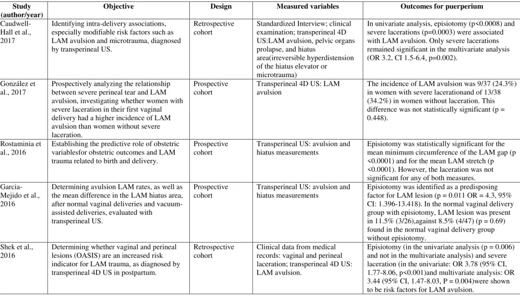
Table 2 - Description of the objectives, variables measured, and outcomes of the studies included in the Systematic Review. Study
(author/year) Objective Design Measured variables Outcomes for puerperium
Caudwell-Hall et al., 2017
Identifying intra-delivery associations, especially modifiable risk factors such as LAM avulsion and microtrauma, diagnosed by transperineal US.
Retrospective
cohort Standardized Interview; clinical examination; transperineal 4D US:LAM avulsion, pelvic organs prolapse, and hiatus
area(irreversible hyperdistension of the hiatus elevator or
microtrauma)
In univariate analysis, episiotomy (p<0.0008) and severe lacerations (p=0.0003) were associated with LAM avulsion. Only severe lacerations remained significant in the multivariate analysis (OR 3.2, CI 1.5-6.4, p=0.002).
González et
al., 2017 Prospectively analyzing the relationship between severe perineal tear and LAM avulsion, investigating whether women with severe laceration in their first vaginal delivery had a higher incidence of LAM avulsion than women without severe laceration.
Prospective
cohort Transperineal 4D US: LAM avulsion The incidence of LAM avulsion was 9/37 (24.3%) in women with severe lacerationand of 13/38 (34.2%) in women without laceration. This difference was not statistically significant (p = 0.448).
Rostaminia et
al., 2016 Establishing the predictive role of obstetric variablesfor obstetric outcomes and LAM trauma related to birth and delivery.
Prospective
cohort Transperineal US: avulsion and hiatus measurements Episiotomy was statistically significant for the mean minimum circumference of the LAM gap (p <0.0001) and for the mean LAM stretch (p <0.0001). However, the laceration was not significant for any of both measures.
Garcia-Mejido et al., 2016
Determining avulsion LAM rates, as well as the mean difference in the LAM hiatus area, after normal vaginal deliveries and vacuum-assisted deliveries, evaluated with
transperineal US.
Prospective
cohort Transperineal US: avulsion and hiatus measurements Episiotomy was identified as a predisposing factor for LAM lesion (p = 0.011 OR = 4.3, 95% CI: 1.396-13.418). In the normal vaginal delivery group with episiotomy, LAM lesion was present in 11.5% (3/26),against 8.5% (4/47) (p = 0.69) found in the normal vaginal delivery group without episiotomy.
Shek et al.,
2016 Determining whether vaginal and perineal lesions (OASIS) are an increased risk indicator for LAM trauma, as diagnosed by transperineal 4D US in postpartum.
Retrospective
cohort Clinical data from medical records: vaginal and perineal laceration; transperineal 4D US: LAM avulsion.
Valsky et al.,
2016 Assessing the LAM lesion rates in primiparous women with third and fourth degree tear compared with women without lacerations.
Prospective
cohort Transperineal 3D US: LAM avulsion; clinical data from medical records: maternal history, delivery room information and neonatal details.
The groups differed significantly regarding the LAM avulsion rates: 38 of 94 women (40.4%) of the laceration group had LAM avulsion according to the US, compared to 75 of 464 (16.2%) of the comparison group (P <0.001, [OR], 3.53, 95% CI [CI], 2.18-5.7). Both groups did not differ for episiotomy rates.
Laterza et al.,
2015 Evaluating the association between LAM lesion and PFM dysfunction at 12 months postpartumand determining whether LAM trauma reveals anatomical signs of pelvic organ prolapse as early as one year after vaginal delivery.
Prospective
case-control Australian Pelvic Floor Questionnaire:PFM symptoms; Structured interview: UI and AI, constipation, POP symptoms and sexual function; POP-Q; Oxford Method: PFM strength and endurance; transperineal US: LAM avulsion and genital stretch
No differences were observed in the characteristics of patients in both groups regarding episiotomy (p = 0.52) and perineal laceration (p = 0.51)
Van Delft et
al., 2015 Exploring the natural history of LAM avulsions within one year postpartum and correlating these findings with signs and symptoms of PFM dysfunction.
Prospective
cohort Oxford method: PFM strength and endurance; POP-Q; St Mark's incontinence score system: Intestinal function;Questionnaires (ICIQ-SF and ICIQ-VS); Urinary and sexual function; transperineal US: LAM avulsion and genital hiatus
Compared to women with no longer evident avulsion one year postpartum,women with persistent LAM avulsion had more episiotomies (p = 0.022) and more OASIS (p = 0.010).
Cassadó et al.,
2014 Studying the relationship between episiotomy and LAM lesion in vaginal delivery through US.
Prospective
cohort Transperineal US: avulsion and stretch measurements LAM avulsion was detected in 25 women (12.9%): in 11 (10.9%) of the 101 with episiotomy and in 14 (15.1%) of the 93 withoutepisiotomy.The difference was not statistically significant (p = 0.401).The relative risk of avulsion in patients with episiotomy is 0.82 (95% CI: 0.52-1.31). Not reaching statistical significance.
Meriwether et al., 2014
Evaluating the normative measures of transperineal 2D and 3D US for the anal sphincter complex at 6 months postpartum, and comparing these measures among women with vaginal and cesarean delivery.
Prospective cohort
Transperineal US: Thickness of pubovisceral muscle and anal sphincter complex; and endoanal US (missing data): IAS and EAS
Women who suffered third or fourth degree tears were not more likely to have a dysfunction in the proximal IAS (0 of 19 [0%]) vs. 14 of 404 (3%), p
in 402 [5%] p = 0.97) compared to women without severe laceration.
Women who suffered fourth-degree tears were also not more prone to EAS defects (0 of 19 vs 4
of 402 [1%], p = 1.0).
Women who suffered third or fourth degree tears had EAS slightlythicker measurements after 12h. (2.57 ± 0.83 vs 2.11 ± 0.83 mm, p = 0.03).
Van Delft et
al., 2014 Establishing the incidence of LAM avulsion in primiparous women and to developing a clinically applicable risk prediction model for this avulsion.
Observational
cohort Transperineal US: LAM avulsion and genital hiatus Severe laceration was a risk factor for LAM avulsion [OR] 4.4, 95% CI 1.6-12.1
Nardi et al.,
2013 Evaluating the impact of the first vaginal delivery and of a mid-lateral episiotomy, using transperineal 2D US.
Retrospective
cohort Transperineal US: stretch and mobility of the bladder neck measurements
In comparing US measurements between the two LAM stretch groups, it was significantly higher in women undergoing episiotomy, especially during maximum Valsalva, but also during contraction. Rojas et al.,
2012
Determining the prevalence of OASIS in primiparous women.
Retrospective cohort
Transperineal US: sphincter defects and avulsion
LAM avulsion was diagnosed in 44
(13.8%),residual EAS defects according to the US in 69 patients (21.6%); 6 were complete (1.9%). Nine had been diagnosed with third-degree tears, 29 with second-degree tears and 2 with first-degree tears;3 had intact perineums.31 had episiotomy (5 extended for third degree lacerations). Of the 12 diagnosed with third-degree tear, 2 had a complete defect (all 6 slices) at follow-up,7 had a residual defect, 2 had minor abnormalities, and 1 was classified as normal. The relationship between episiotomy and sphincter injury was significant only in the univariate analysis (OR = 3.46, CI 1.89-6.32, p <0.001) Chantarasorn
et al., 2012 Determining changes in the mobility of the perineal body and anorectal junction before and after delivery, using transperineal US.
Observational
cohort Structured interview: urinary function; transperineal US: perineal body and anorectal junction
Lopez et al.,
2012 Evaluating the effect of episiotomy on the LAM morphology, in preparation for a randomized controlled trial.
Retrospective
cohort Transperineal US: hiatal area during Valsalva, neck of the bladder, cystocele and avulsion.
The univariate analysis showed statistical significance in the relation between episiotomy and avulsion (OR 2.07 (1.138-3.765, p = 0.017)), however, it was not significant in the multivariate analysis (OR 0.5-2.2). Severe lacerations
remained significant in the multivariate analysis (OR 4.263 (1.156-11.344, p = 0.0272)). Bradley et al.,
2007 Identifying risk factors for IAS lesion observed in the endoanal US at 6-12 months postpartumin women who underwent repair of a sphincter disruption during delivery.
Prospective
cohort Endoanal US: Internal and external anal sphincter defects. Severe perineal lacerations (OR 15.4, 95% CI, 4.8, 50) and episiotomy (OR 3.3, 95% CI 1.2, 9.1) are associated with sphincter injury.
Richter et al.,
2006 Estimating whether primiparous women with severe lacerations during vaginal delivery increased the ultrasound findings of sphincter lesions, and whether the ultrasound findings between 6-12 months after delivery are associated with symptoms of fecal incontinence.
Observational
cohort Fecal Incontinence Severity Index: AI; endoanal US: Sphincter defects
Women who had a fourth-degree tear had more IAS (p <0.001) and EAS lesions (OR 3.8, 95% CI 1.4-10.0) than those with third-degree tears.The probability of an EAS defect was higher in the group with severe lacerations (51%) than in the vaginal (31%)or cesarean section (28%) control groups (OR 2.3, 95% CI 1.3-4.0).
Wijma et al., 2003
Assessing the incidence of UI during pregnancyand after spontaneous vaginal delivery, and its relationship with changes in static and dynamic PFM function.
Prospective cohort
Pad test: UI; transperineal US: position and mobility of the urethrovesical junction.
None of the obstetric variables (episiotomy or lacerations of varying degrees) were correlated with urethrovesical junction measurements.
REFERENCES
1. Shek KL, Green K, Hall J, Guzman-Rojas R, Dietz H. Perineal and vaginal tears are clinical markers for occult levator ani trauma: a retrospective observational study. Ultrasound in Obstetrics & Gynecology 2016; 47: 224-7.
2. Aytan H, Tapisiz OL, Tuncay G, Avsar FA. Severe perineal lacerations in nulliparous women and episiotomy type, European Journal of Obstetrics & Gynecology and Reproductive Biology 2005; 121: 46-50.
3. Dannecker C, Hillemanns P, Strauss A, Hasbargen U, Hepp H, Anthuber C. Episiotomy and perineal tears presumed to be imminent: the influence on the urethral pressure profile, analmanometric and other pelvic floor findings – follow-up study of a randomized controlled trial. Acta Obstetricia et Gynecologica Scandinavica 2005; 84: 65-71.
4. Graham ID, Carroli G, Davies C, Medves JM. Episiotomy Rates Around the World: An Update. Birth 2005; 32: 219-23.
5. Royal College of Obstetricians & Gynaecologists. The Management of Third- and
Fourth-Degree Perineal Tears. Green-top Guideline, n.29,
2015.https://www.rcog.org.uk/en/guidelines-research-services/guidelines/gtg29/ [3 November 2017].
6. Dudding TC, Vaizey CJ, Kamm MA. Obstetric anal sphincter injury: incidence, risk factors, and management. Annals of Surgery 2008; 247: 224-237.
7. Sartore A, De Seta F, Maso G, Pregazzi R, Grimaldi E, Guaschino S. The Effects of Mediolateral Episiotomy on Pelvic Floor Function After Vaginal Delivery. Obstetrics & Gynecology 2004; 103: 669-73.
8. Scheer I, Andrews V, Thakar R, Sultan AH. Urinary incontinence after obstetric anal sphincter injuries (OASIS)—is there a relationship? Int Urogynecol J 2008; 19: 179-183.
9. Bharucha AE, Fletcher JG, Joseph Melton III L, Zinsmeister AR. Obstetric Trauma, Pelvic Floor Injury and Fecal Incontinence: A Population-Based Case-Control Study. The American Journal of Gastroenterology 2012; 107: 902-911.
10. Falkert A, Willmann A, Endress E, Meint P, Seelbach-Gobel B. Three-dimensional ultrasound of pelvic floor: is there a correlation with delivery mode and persisting pelvic floor disorders 18–24 months after first delivery? Ultrasound in Obstetrics & Gynecology 2013; 41: 204-209.
12. González MS, Garriga JC, Capel CD, Roda OP, Capó JP, Saladich IG. In obstetric anal factor for levator ani muscle avulsion in vaginal delivery? Ultrasound Obstet Gynecol 2017, 49: 257-262.
13. Guzman Rojas RA, Shek K, Langer S, Dietz H. The prevalence of anal sphincter injury in primiparous women. Int Urogynecol J 2012; 23 (Suppl 2):S43–S244.
14. Meriwether KV, Hall RJ, Leeman LM, Migliaccio L, Qualls C, Rogers RG. Postpartum translabial 2D and 3D ultrasound measurements of the anal sphincter complex in primiparous women delivering by vaginal birth versus Cesarean delivery. Int Urogynecol J 2014; 25: 329-336.
15. Wijma J, Potters AEW, Wolf BTHM, Tinga DJ, Aarnoudse JG. Anatomical and functional changes in the lower urinary tract following spontaneous vaginal delivery. International Journal of Obstetrics and Gynaecology 2003; 110: 658-663.
16. Chantarasorn V, Shek KL, Dietz HP. Mobility of the perineal body and anorectal junction before and after childbirth. Int Urogynecol J 2012; 23: 729–733.
17. Lopez VM, Shek KL, Guzman Rojas R, Dietz HP. Does episiotomy damage the pelvic floor? Int Urogynecol J 2012; 23 (Suppl 2):S43–S244.
18. Caudwell-hall J, Atan IK, Martin A, Rojas RG, Langer S, Sher K, Dietz HP. Intrapartum predictors of maternal levator ani injury. Acta Obstet Gynecol Scand 2017;
96: 426-431.
19. Nardi M, Ciattaglia F, Vincenzi R. Pelvic floor ultrasound in puerperium. Tech Coloproctol 2013; 17:133–147.
20. van Delft K, Thakar R, Sultan AH, Schwertner-Tiepelmann N, Kluivers K. Levator ani muscle avulsion during childbirth: a risk prediction model. British Journal of Obstetrics and Gynaecology 2014; 121: 1155-1163.
21. van Delft K, Thakar R, Sultan AH, Inthout J, Kluivers K. The natural history of levator avulsion one year following childbirth: a prospective study. British Journal of Obstetrics and Gynaecology 2015; 122: 1266–1273.
22. Laterza RM, Schrutka L, Umek W, Albrich A, Koelbl H. Pelvic floor dysfunction after levator trauma 1-year postpartum: a prospective case–control study. Int Urogynecol J 2015; 26: 41-47.
25. Valsky DV, Cohen SM, Lipschuetz M, Hochner-Celnikier D, Daum H, Yagel I, Yagel S. Third- or Fourth-Degree Intrapartum Anal Sphincter Tears Are Associated With Levator Ani Avulsion in Primiparas. J Ultrasound Med 2016; 35: 709-715.
26. Richter HE, Fielding JR, Bradley CS,Handa VL, Fine P,FitzGeraldMP, Visco A, Wald A, Hakim C, Wei JT, WeberAM. Endoanal Ultrasound Findings and Fecal Incontinence Symptoms in Women With and Without Recognized Anal Sphincter Tears. American College of Obstetricians and Gynecologists 2006; 108: 1394-1401. 27. Bradley CS, Richter HE,Gutman RE, Brown MB, Whitehead WE, Fine PM, Hakim C, Harford F, WeberAM. Risk factors for sonographic internal anal sphincter gaps 6 to 12 months after delivery complicated by anal sphincter tear. American Journal of Obstetrics and Gynecology 2007; 197: 310.e1-310.e5.
28. Dietz HP, Steensma AB. The prevalence of major abnormalities of the levator ani in urogynaecological patients. An International Journal of Obstetrics & Gynaecology 2006; 113: 225–230.
29. DeLancey JOL, Miller JM, Kearney R, Howard D, Reddy P, Umek W, Guire KE, Margulies RU, Ashton-Miller JA. Vaginal birth and de novo stress incontinence: Relative contributions of urethral dysfunction and mobility. Obstetrics & Gynecology 2007;110: 354–362.
30. Dietz HP, Shek KL, Chantarasorn V, Langer SEM. Do women notice the effect of childbirth-related pelvic floor trauma? Australian and New Zealand Journal of Obstetrics and Gynaecology 2012; 52: 277–281.
31. Cerro CR, Franco EM, Santoro GA, Palau MJ, Wieczorek P, Espuña-Pons M. Residual defects after repair of obstetric anal sphincter injuries and pelvic floor muscle strength are related to anal incontinence symptoms. Int Urogynecol J 2017; 28: 455-460.
32. Leon-Larios F, Corrales-Gutierrez I, Casado-Mejía R, Suarez-Serrano C. Influence of a pelvic floor training programme to prevent perineal trauma: A quasi-randomised controlled trial. Midwifery 2017; 50: 72-77.
33. Salsi G, Cataneo I, Dodaro G, Rizzo N, Pilu G, Sanz Gascón M, Youssef A. Three-dimensional/four-dimensional transperineal ultrasound: clinical utility and future prospects. Int J Women’s Health 2017; 9: 643–656.