374 Radiol Bras. 2011 Nov/Dez;44(6):374–380
Integrating computer-aided diagnosis tools into the picture
archiving and communication system
*
Integrando ferramentas de auxílio ao diagnóstico no sistema de arquivamento e comunicação de imagens
Samuel Covas Salomão1, Paulo Mazzoncini de Azevedo Marques2
Objective: This paper presents a model for integration of computer-aided diagnosis algorithms into the picture archiving and communication systems workflow that has been developed on the basis of the dcm4che2 open source toolkit. Materials and Methods: The proposed integration model consists of an image processing server and communication services. The data management follows the workflow defined by the post-processing workflow profile (PWF) developed by Integrating the Healthcare Enterprise (IHE) and utilizes the DICOM secondary capture functionality. An application for diffuse lung disease has been utilized as proof of concept. Results: Based on a cross validation method, the standard classification algorithm presented 78% accuracy. The integration enables the visualization of processed images as a new series in the original study. Conclusion: The proposed integration model is based on IHE profiles and allows the establishment of standardized procedures. The principles used to integrate the workflow are applicable to any non-interactive post-processing task.
Keywords: Computer-aided image processing; Picture archiving and communication systems; DICOM secondary capture; IHE.
Objetivo: Este artigo apresenta um modelo de integração de algoritmos de diagnóstico auxiliado por computador dentro do fluxo de trabalho dos sistemas de gerenciamento de imagens, desenvolvido com base em um conjunto de ferra-mentas computacionais de código aberto e uso livre chamado dcm4che2. Materiais e Métodos: O modelo de inte-gração é composto por um servidor de processamento de imagens e por serviços de comunicação. O gerenciamento de dados segue o fluxo de trabalho definido pelo perfil de pós-processamento (PWF) do Integrating the Healthcare Enterprise (IHE) e utiliza a funcionalidade de captura secundária do DICOM. Uma aplicação para lesões difusas de pulmão foi utilizada para prova de conceito. Resultados: O algoritmo de classificação de padrões apresentou acurá-cia de 78%, com base em um método de teste de validação cruzada. A integração possibilita a visualização das ima-gens processadas como uma nova série dentro do estudo original. Conclusão: O modelo de integração proposto baseia-se em perfis do IHE e permite o estabelecimento de procedimentos padronizados. Os princípios utilizados para inte-gração do fluxo de trabalho são aplicáveis para qualquer tarefa não interativa de pós-processamento de imagens. Unitermos: Processamento de imagens médicas; Sistemas de arquivamento e comunicação de imagens; Captura secundária do DICOM; Perfis de integração do IHE.
Abstract
Resumo
* Study developed at Centro de Ciências das Imagens e Física Médica do Hospital das Clínicas da Faculdade de Medicina de Ribeirão Preto da Universidade de São Paulo (CCIFM/HC-FMRP-USP), through the Interunits Program of Post-graduation in Bio-engineering (EESC/IQSC/FMRP), Universidade de São Paulo, Ribeirão Preto, SP, Brazil. Financial support: Conselho Nacional de Desenvolvimento Científico e Tecnológico (CNPq).
1. Master of Bioengineering, Independent Advisor of Health Informatics, Ribeirão Preto, SP, Brazil.
2. Private Docent, Associate Professor, Department of Inter-nal Medicine, Faculdade de Medicina de Ribeirão Preto da Uni-versidade de São Paulo (FMRPUSP), Ribeirão Preto, SP, Brazil.
Salomão SC, Azevedo-Marques PM. Integrating computer-aided diagnosis tools into the picture archiving and communication system. Radiol Bras. 2011 Nov/Dez;44(6):374–380.
tion system) concept was developed to meet such a need. It is an archiving and communication system dedicated to imag-ing diagnosis that allows immediate access to medical digital images from any sector in a hospital(2). The PACS has quickly be-come the first option for the management of images in the clinical and hospital envi-ronments and, together with the radiology information system (RIS) and the hospital information system (HIS), comprise the basis for a filmless radiology service. A fundamental aspect of the workflow in a digital (filmless) radiology environment is the guarantee of the consistency of the data transmitted from one component to the cesses in hospitals(1). Until then, every
equipment was considered as being an iso-lated system, connected only to its own workstation and a printer. However, the increased utilization of data in digital for-mat has led to the need for development of a computational structure which allowed a consistent and automatic exchange of im-ages data in the hospital environment. The PACS (picture archiving and communica-INTRODUCTION
The utilization of information systems for the management of clinical images and data started being more effectively studied in the late 1980’s, with the increase in the utilization of digital data acquisition
pro-Mailing Address: Dr. Paulo Mazzoncini de Azevedo Marques. Departamento de Clínica Médica, FMRPUSP. Avenida Bandeiran-tes, 3900, Monte Alegre. Ribeirão Preto, SP, Brazil, 14049-900. E-mail: [email protected]
next within the chain of events in the pro-cess dynamics. In order to guarantee such a consistency, the information distribution is carried out according to a hierarchical structure which relies on a top-down dis-tribution, i.e., the information is propagated from a more general information system (HIS) to an intermediate system (RIS), until it reaches a more specific system (PACS). In order to make that possible, two require-ments must be met: an appropriate (redun-dant and balanced) network structure and well defined communication standards. In digital radiology, the main communication standard is the DICOM (digital imaging and communications in medicine) sys-tem(3). The DICOM is the global standard for the transfer of radiological images and other clinical data between computers. The current DICOM, published in 1993 and generally identified as 3.0, evolved from previous versions of a standard developed by the American College of Radiology (ACR) in cooperation with the National Electrical Manufacturers Association (NEMA) of the United States of America (NEMA 1.0, in 1985, and ACR-NEMA 2.0, in 1988). The connectivity de-signed by the standard is very important with respect to the cost/benefit ratio for health services that utilize medical images. The DICOM users can provide radiology services between facilities located in dif-ferent geographical regions, making use of already available information technology resources, while keeping costs down by means of new apparatuses and systems compatibility and interoperability(4).
Although technological developments have allowed a more extensive diffusion of image management systems in the medical field over the last years in such a manner that in some situations PACS is considered as a simple commodity(5), in a model of technological maturity(6) there are still some aspects that deserve greater investi-gation and development. Issues related to digital image acquisition processes(7), data distribution, visualization(8) and interpreta-tion(9) are still object of studies, particularly with respect to economic aspects and the utilization of freeware and open code(10,11) computational solutions. One should also highlight the integration of tools for aiding in diagnosis into the workflow of image
management systems. Computer-aided di-agnosis (CAD) can be defined as a diagno-sis made by a radiologist who utilizes the result from automated quantitative analy-ses of radiographic images as a second opinion in the diagnostic decision making process(12). The purpose of CAD is to im-prove accuracy in diagnosis, as well as the consistency of radiological images inter-pretation, by utilizing the output from the computer as a reference(13–15). Today, the standard for the utilization of CAD systems is still based on the model of a standalone workstation, without the integration of the obtained results with the information sys-tem or with the PACS. The integration of image processing procedures into the PACS workflow has recently been the ob-ject of research in the field of information technology in medical imaging(13,16–18).
The present article describes a model for the integration of diagnosis aid tools into the image management environment with the objective of optimizing the visual analysis by the human observer (the radi-ologist) within the imaging diagnosis work-flow. Such model has been developed with an open-source toolkit called dcm4che2. The data management follows the flow defined by the post-processing work-flow profile (PWF) of the Integrating the Healthcare Enterprise (IHE) and utilizes the DICOM secondary capture functional-ity. The dcm4che2 is an implementation of the high-performance DICOM standard developed in Java programming language, which supports the IHE integration profiles (http://www.dcm4che.org). The IHE is an initiative of the Radiology Society of North America (RSNA), aimed at promoting the systems integration in the healthcare envi-ronment. The objective is to improve the clinical practice efficiency and effective-ness by enhancing the data flow based on communication standards. The IHE estab-lishes a technical framework which defines the way the standards are implemented in order to meet the needs of the clinical prac-tice. It utilizes three basic concepts: play-ers, transactions and integration profiles. The players are the functionalities that make the communication between the sys-tems. Transactions are the messages ex-changed between the systems. Integration profiles are groups of players and
transac-tions that comprise a specific workflow(3). As a proof of concept, a system prototype to aid in the differential diagnosis of dif-fuse lung lesions with computed tomogra-phy was integrated with the images man-agement system of the imaging diagnosis center of a hospital school.
MATERIALS AND METHODS
A computational testing environment was built to validate the model of integra-tion between the computer aided diagno-sis system (CAD) and the medical images management system (PACS). A locally de-veloped computer code for classification of diffuse lung lesions at high resolution com-puted tomography was selected to play the role of a CAD solution. The selected medi-cal images management system was the DICOM Conquest server (CONQUEST, 2010), and for the visualization of the CAD-PACS integration results, the medi-cal image viewer K-PACS (K-PACS, 2010) was utilized. All the apparatuses in the en-vironment were connected to a 100 Mbit/s local network (LAN).
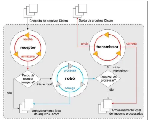
Based on a number of software pack-ages defined by the integration model, a client computer code denominated Client-CAD was developed. Such a code acts as an intermediate entity between the PACS and the CAD, with the objective of inte-grating the two systems by means of the DICOM communication protocol. The Cli-ent-CAD also comprises a software com-ponent known as i-CAD, especially uti-lized to change the serial number of medi-cal images in the digital format(19). Also, in a transparent manner, the Client-CAD pro-vides interfaces that allow the coupling with specific image processing modules for other acquisition modalities, as shown on Figure 1.
ex-presses a query in a databank based on the information comprised in the standard’s data dictionary, and may or may not com-prise filters to provide more specific re-sults. In the case of the herein described experiment, the client was configured to query only computed tomography (CT) studies, and the anatomic region of the CHEST. Therefore, as the PACS server receives a new chest HRCT study, the cli-ent requests to the PACS a copy of such study by sending image search and retrieval messages (query & retrieve). The retrieved images are then processed one at a time by the CAD system processing routine.
The CAD processing routine comprises five steps as follows: pre-processing, seg-mentation, characteristics extraction, clas-sification and post-processing. In the pre-processing, the image data (pixels) are ex-tracted from the DICOM file. In the next step, the lungs are segmented by means of an interactive algorithm for image thresh-olding(20)and the resulting image is divided into 20 × 20-pixel regions of interest (ROIs). During the classification step, each ROI is defined as belonging to one of the seven approached interstitial lung lesion classes as follows: normal, ground glass attenuation, reticular, nodular, honeycomb-ing, emphysema and consolidation. In the post-processing step, each ROI receives a color which identifies its characterization
class and a new DICOM file is generated with the results from the processing.
The CAD-PACS integration is com-pleted as the client, by means of the i-CAD software component, modifies the serial number of the new DICOM file and sends it back to the PACS server. Figure 2 depicts
the functioning principle of the Client-CAD code.
Based on the architecture of the client operation, the first process denominated re-ceptor represents an independent computer code which implements a DICOM service provider for images storage (storage service class provider – CStore SCP). It starts run-ning whenever the client requests a copy of a given imaging study stored at PACS. For each received image, the client invokes the robot process that, on its turn, initiates the CAD system image processing routine.
As the CAD routine is completed, the robot locally stores the image resulting from the processing and initiates the third and last process, the transmitter. In this stage, the i-CAD is utilized to change the serial number of the processed image. Completing the integration cycle, the trans-mitter sends DICOM messages for images storage request (storage service class user – CStore SCU) and sends the resulting im-age to the PACS server.
RESULTS
The experiment results are directly re-lated to the development of the
Client-Figure 2.Client-CAD functioning scheme. Functioning principle of the Client-CAD module created with basis on the CAD-PACS integration model. It illustrates the functioning of the receptor, robot and trans-mitter processes responsible for the processing and transmission of the CAD results to the PACS. Figure 1.Integration model architecture. Scheme presenting the integration model architecture and its
CAD software and to the success of the integration between the computer aided diagnosis system (CAD) for diffuse lung lesions and the medical images manage-ment system. The Client-CAD workflow comprises four vital tasks which contrib-uted to the integration success, as follows: – query & retrieval: the client periodi-cally queries the PACS server searching for new imaging studies by sending DICOM C-Find messages. The query is always per-formed by utilizing the data model for im-ages query and retrieval at the study level (study root query retrieve information modelFIND);
– CADprocessing: Once received and stored, the images are processed one at a time by the CAD routine initiated by the client. For each image, the processing re-sult consists of a JPEG image which com-prises relevant data that may be of assis-tance in the decision making by the radi-ologist;
– encapsulation: each resulting JPEG image is encapsulated by the client in a compatible DICOM format. Mandatory data in the dictionary, such as patients’ identification and name, for example, are copied from the original DICOM file. The difference between the original file and the
encapsulated version lies on the way the file is built. The encapsulated DICOM file is encoded from a JPEG image and follows the information model of the DICOM sec-ondary capture image storage SOP class;
– transmission: the resulting image transmission is performed by sending the DICOM C-Store SCU message imple-mented on the client. Thus, when the PACS server receives the encapsulated DICOM file, the file manager stores its contents in the same patient folder of the original file, but within a new image series according to the new serial number generated by the i-CAD code.
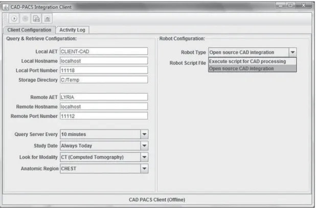
Figure 3 presents the interface of the cli-ent Clicli-ent-CAD code. It is possible to vi-sualize several of the utilized parameters. The fields on the left upper corner are uti-lized for the configuration of the DICOM communication between the client and the PACS server. Immediately below are the fields that identify the PACS to be queried. And below that, the last field establishes the scheduling configurations for the client’s queries and images retrieval.
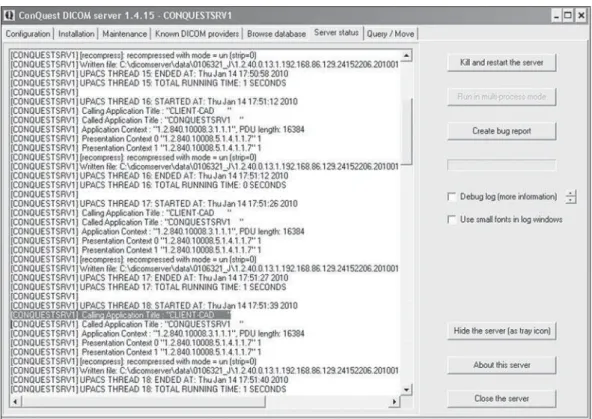
In order to illustrate the CAD-PACS communication, Figure 4 presents the in-teraction between the client and the PACS Conquest Server. In the highlighted area
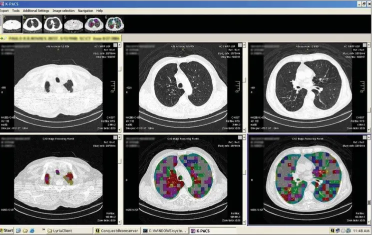
one visualizes an entity called “CLIENT-CAD” requesting the storage of an image resulting from the CAD processing. Once the resulting images are correctly stored in the PACS server, it is possible to visualize the new series created by the CAD-PACS integration on the display station. Figures 5 and 6 show the K-PACS viewer present-ing examples of original image series side beside the corresponding CAD system pro-cessed images.
DISCUSSION
The initial trials demonstrated a good potential for the proposed model for CAD-PACS integration, but an important limita-tion respects the types of files utilized dur-ing the integration workflow. Currently, DICOM and JPEG are the supported for-mats. Therefore, the integration model is compatible with CAD applications that provide JPEG images as a processing out-put. One should highlight that the DICOM encapsulated JPEG image does not provide the same diagnostic quality as the original image, because of the format’s inherent losses, and should therefore be utilized as a complementary information in the deci-sion making process based on the visual
Figure 5.Updated list of study series. K-PACS viewer query interface. On the highlighted area, three new processed images series after the CAD-PACS integration process completion.
Figure 4.DICOM Server interface. Conquest Server interface for the visualization of events and status. On the highlighted area, the interaction between the server and the Client-CAD can be seen.
inspection of the original images series. In case the CAD is not capable of providing the output in the JPEG format, a possible solution would be capturing and encapsu-lating the image on the computer screen as
the result is presented. Such a simple alter-native, however, may in a certain way com-promise the reading of results and impair the data interpretation by the physician, due to the various visual and text elements
distrib-uted on worksheets, charts, HTML docu-ments and even complete reports in PDF file format. Such data are relevant and, if integrated into the patient’s study in the PACS server, may provide a valuable source of information to assist the physi-cian in the decision making process. Such integration can be performed with methods available at DICOM structured reporting (SR), a new extension of the DICOM stan-dard which establishes stanstan-dards for encod-ing documents incorporatencod-ing references for medical images and related data, as well as services for data transmission and ex-change. An integration model based on the properties of the DICOM-SR would be more complete and would further allow interactive activities of data retrieval, for example, content based image retrieval (CBIR).
The actual contribution of a diagnosis aid tool depends on the synergy between the professional and the tool. Such a syn-ergic interaction does not generally occur naturally, demanding user training and
ap-propriateness of the computer solution. The experiment herein described presents a model of image processing tools integra-tion that is transparent within the workflow of a digital radiology environment. How-ever the assessment of the effective contri-bution of the implemented solution as a supporting tool for the decision making process, based on observer tests for the measurement of diagnostic accuracy was not comprised by the scope of the investi-gation in the present study.
CONCLUSION
With the massive dissemination of digi-tal apparatuses for the acquisition of medi-cal images, together with the adoption of data management solutions (PACS), the existence of imaging diagnosis centers working under the filmless radiology model has become increasingly common. In a filmless environment, the images are stored and distributed according to the DICOM standard functionalities. The
REFERENCES
1. Wiley G. The prophet motive: how PACS was developed and sold. Imaging Economics – May 2005. [acessado em 8 de agosto de 2011. Dis-ponível em: http://www.imagingeconomics.com/ issues/articles/2005-05_01.asp
2. Siegel EL, Kolodner RM. Filmless radiology: state of the art and future trends. In: Siegel EL, Kolodner RM, editors. Filmless radiology. New York, NY: Springer-Verlag; 1999. p. 3–20. 3. Azevedo-Marques PM, Salomão SC. PACS:
sis-temas de arquivamento e distribuição de imagens. Rev Bras Fís Méd. 2009;3:131–9.
4. Horii SC. A nontechnical introduction to DICOM. RSNA Informatics. [acessado em 8 de agosto de 2011]. Disponível em: http://www.rsna. org/technology/dicom/intro/index.cfm 5. Chen J, Bradshaw J, Nagy P. Has the picture
archiving and communication system (PACS) be-come a commodity? Journal of Digital Imaging. 2011;24:6–10.
6. van de Wetering R, Batenburg R. A PACS matu-rity model: a systematic meta-analytic review on maturation and evolvability of PACS in the hos-pital enterprise. Int J Med Inform. 2009;78:127– 40.
7. Vela JG, Bhaya A, Monteiro AMV, et al. Digitali-zação de filmes radiográficos com costura de imagens. Radiol Bras. 2011;44:233–7.
8. Parizoti A, Netto TG. Estudo de otimização de imagens em fluoroscopia intervencionista. Radiol Bras. 2009;42:375–8.
9. Ferreira DM, Cohrs FM, Lederman HM, et al. Comparação dos tempos de geração e digitação de laudos radiológicos entre um sistema eletrô-nico baseado em voz sobre IP (VoIP) e um sis-tema tradicional baseado em papel. Radiol Bras. 2010;43:7–12.
10. Nobre LF, von Wangenheim A. Software gratuito: uma opção para o radiologista? Radiol Bras. 2010;43(5):ix-x.
11. Barra FR, Barra RR, Barra Sobrinho A. Visuali-zadores de imagens médicas gratuitos: é possível trabalhar apenas com eles? Radiol Bras. 2010; 43:313–8.
12. Azevedo-Marques PM. Diagnóstico auxiliado por computador na radiologia. Radiol Bras. 2001;34: 285–93.
13. Doi K. Computer-aided diagnosis in medical im-aging: historical review, current status and future potential. Comput Med Imaging Graph. 2007;31: 198–211.
14. Azevedo CM, Alvarenga AV, Pereira WCA, et al. Análise computacional da textura de tumores de mama em imagens por ultrassom de pacientes submetidas a cirurgia conservadora. Radiol Bras. 2009;42:363–9.
15. Pádua RDS, Oliveira LF, Azevedo-Marques PM,
et al. Auxílio à detecção de anormalidade perfu-sional miocárdica utilizando atlas de SPECT e registro de imagens: resultados preliminares. Radiol Bras. 2008;41:397–402.
16. Caritá EC, Seraphim E, Honda MO, et al. Imple-mentação e avaliação de um sistema de gerencia-mento de imagens médicas com suporte à recu-peração baseada em conteúdo. Radiol Bras. 2008; 41:331–6.
17. Azevedo-Marques PM, Ponciano-Silva M, Salo-mão SC, et al. CAD-PACS integration: a frame-work for pattern recognition of diffuse lung dis-ease in HRCT. Int J Comput Assist Radiol Surg. 2009;4 Suppl 1:S177–83.
18. Le AH, Liu B, Huang HK. Integration of com-puter-aided diagnosis/detection (CAD) results in a PACS environment using CAD-PACS toolkit and DICOM SR. Int J Comput Assist Radiol Surg. 2009;4:317–29.