Evaluation of the p16 and Ki-67 Biomarkers as
Predictors of the Recurrence of Premalignant
Cervical Cancer Lesions after LEEP Conization
Avaliação dos biomarcadores p16 e Ki-67 como
preditores de recidivas de lesões pré-cancerígenas
do colo do útero após conização por cirurgia de alta
frequência
Paulo Macêdo de Oliveira Leite
1Luciene Tafuri
1Maria Zélia de Oliveira Costa
2Maria Inês de Miranda Lima
1Renata Toscano Simões
11Instituto de Ensino e Pesquisa, Santa Casa Belo Horizonte (IEP/SCBH), Belo Horizonte, Minas Gerais, Brazil
2Hospital São João de Deus, Fundação Geraldo Corrêa, Divinópolis, Minas Gerais, Brazil
Rev Bras Ginecol Obstet 2017;39:288–293.
Address for correspondence Paulo Macêdo de Oliveira Leite, MSc, Instituto de Ensino e Pesquisa, Santa Casa Belo Horizonte (IEP/SCBH), Rua Rio de Janeiro 324/704, 35500-009 Divinópolis, MG, Brazil (e-mail: [email protected]).
Keywords
►
biomarkers
►
conization
►
cervical intraepithelial
neoplasia
►
recurrence
Abstract
Objective
To evaluate the expressions of biomarkers p16 and K
i-67 in low-grade (LG)
or high-grade (HG) lesions, and to relate them to risk factors and the recurrence of these
lesions.
Methods
A retrospective case-control study of 86 patients with LG and HG lesions who
underwent a loop electrosurgical excision procedure (LEEP) between 1999 and 2004. The
control group was composed of 69 women with no recurrence, and the study group, of 17
patients with recurrence. All patients were followed-up over a two-year period after surgery,
and screened every six months, including cytology and colposcopy. Biopsy samples
collected from LEEP were submitted to immunohistochemical analysis for p16 and K
i-67.
The statistical analysis was performed using the Statistical Package for the Social Sciences
software (SPSS, IBM-SPSS, Inc., Chicago, IL, US), with a signi
fi
cant p
<
0.05.
Results
The biomarkers p16 and K
i-67, separately or combined, showed no relation to
recurrence on the total analysis. However, evaluating speci
fi
cally HG lesions, the positive
expression (2
þ
and 3
þ
) of p16/K
i-67 was associated with recurrence (0.010). In
addition, p16 isolated was also more expressive in HG lesions (2
þ
and 3
þ
,
p
¼
0.018),
but it was unrelated to recurrence.
Conclusion
Proteins p16 and K
i-67, both isolated and combined, are not reliable
primary markers for the recurrence of cervical lesions in the majority of LG lesions.
However, analyzing only the group with prior diagnosis of HG lesions, the expressions of
p16 and of p16/K
i-67 were associated with recurrence, and they may be useful in
monitoring these cases.
received June 12, 2016 accepted
December 15, 2016 published online February 23, 2017
DOI https://doi.org/ 10.1055/s-0037-1598643. ISSN 0100-7203.
Copyright © 2017 by Thieme Revinter Publicações Ltda, Rio de Janeiro, Brazil Original Article
Introduction
The search for markers to facilitate the diagnosis of diseases is a constant in scientific research to save resources, time and to prevent unnecessary treatments. Cervical cancer is the most common cancer among women in 45 countries of the world and, worldwide, 266 thousand women die of it each year;1it is preceded by cervical lesions that may or may not progress to invasion. They are associated with infection and with the persistence of the human papillomavirus (HPV) to progress to invasive carcinoma.2Through this process, the cells infected with high-risk oncogenic HPV alter the cell cycle, modifying the production of proteins p16 and Ki-67. The most common treatment for high grade (HG) lesions is cervical cone resec-tion using the loop electrosurgical excision procedure (LEEP). A major concern of the treatment is the recurrence of the lesion, as it may reappear without symptoms and more severely.
Proteins p16 and Ki-67 are, respectively, cell progression and proliferation markers. Protein p16 is a tumor suppressor from the Ink4a family that induces the hyperphosphorylation of the retinoblastoma protein (pRb), and has low expression in normal tissues.3Ki-67 is a nuclear protein present in cells during the active proliferation stage, but it is not expressed when cells are in the quiescent state.3The expression of both molecules simultaneously already denotes some problem in the cell cycle.4 The main objective of this study was to compare the expression of p16 and Ki-67, individually or
combined, with the recurrence of cervical cancer precursor lesions after a LEEP procedure and, also, to verify whether other factors contributed to this.
Methods
The study was approved by The Ethics and Research Commit-tee of Instituto de Ensino e Pesquisa, Santa Casa Belo Hori-zonte (no. 1.222.448), and only patients who agreed and signed the informed consent form (ICF) participated.
Sample Selection and Patient Monitoring
A total of 86 cases of cervical intraepithelial neoplasia (CIN) were evaluated, having been diagnosed by histopathology after LEEP surgery. The sample group was monitored from January 1999 to March of 2004 at a municipal healthcare center in the city of Belo Horizonte. All patients were re-evaluated every 6 months by oncotic cytology, colpos-copy and cervical biopsy, when indicated, and followed-up during 2 years to assess whether or not there was lesion relapse.
The total sample of 86 patients consisted of 17 patients with CIN1, 11 with CIN2, and 58 with CIN3. Of the total, 17 presented lesion recurrence. The study group was composed of the 17 recurrences, and the control group, of the other 69 patients. Apart from the biomarkers, both groups were evaluated considering sociodemographic data, previous health status and histological variables.
Palavras-chave
►
biomarcadores
►
conização
►
neoplasia
intraepitelial cervical
►
recidiva
Resumo
Objetivo
Avaliar as positividades dos biomarcadores p16 e K
i-67 em lesões de baixo
grau (BG) ou de alto grau (AG), e relacioná-las com os fatores de risco e com a recidiva
dessas lesões.
Métodos
Estudo retrospectivo caso-controle, com 86 pacientes com lesões de BG e
AG, submetidas à conização por cirurgia de alta frequência entre 1999 e 2004. O grupo
de controle foi constituído de 69 mulheres sem recidivas, e o grupo de estudo, de 17
pacientes que recidivaram. Todas as pacientes foram acompanhadas durante dois anos
após a cirurgia, com controle a cada seis meses, incluindo citologia e colposcopia. As
peças provenientes de cirurgia de alta frequência (CAF) foram submetidas a
imuno-histoquímica para p16 e K
i-67. A análise estatística foi realizada com o programa
Statistical Package for the Social Sciences (SPSS, IBM-SPSS, Inc., Chicago, IL, EUA), com
p
signi
fi
cante quando
<
0,05.
Resultados
Isoladamente ou em conjunto, p16 e K
i-67 não se relacionaram com as
recidivas quando analisados na totalidade dos casos. Entretanto, avaliando especi
fi
ca-mente as lesões de AG, a positividade (2
þ
e 3
þ
) do conjunto p16/K
i-67 foi relacionada
com recidiva (0,010). No mais, p16, isoladamente, foi também mais expresso nas lesões
de AG (2
þ
e 3
þ
,
p
¼
0,018), mas sem relação com recidiva.
Conclusão
Quando testadas na totalidade dos casos, as proteínas p16 e K
i-67,
separadas ou em conjunto, se mostraram ine
fi
cientes como marcadores primários de
recidiva de lesões precursoras. Entretanto, quando avaliadas somente no grupo
diagnóstico prévio de lesão de AG, as expressões das proteínas p16 e p16/K
i-67 têm
Immunohistochemical Markers
To evaluate p16 and Ki-67 expression levels, immunohis-tochemistry was performed using monoclonal antibody MIB 1 (Dako) for the Ki-67 in the dilution of 1:100, and G175–405 (Zeta) in the dilution of 1:100 for the p16. Both antigens were detected using HiDef Detection System, HRP Polymer System (Cell Marque, Rocklin, USA). All immunohis-tochemical studies were performed in the laboratory of Instituto Moacyr Junqueira, in Belo Horizonte, according to standard protocols.
The readings were done by two independent examiners who classified the slides according to the percentage of positive cells, as described by Zhong et al5(►Table 1).
Statistical Analysis
Numerical variables were tested for normality (Kolmogorov-Smirnov test) and the Student’s t-test was used in the calculations. At first, the analysis focused on the whole sample characteristics using tables of frequency for the categorical variables, and descriptive measures (mean, medi-an, 25th and 75th percentiles, minimum-value, maximum-value and standard deviation) for the quantitative variables. Sociodemographic, health and histopathologic variables and their relationship with recurrence, p16 and Ki-67 positivity, the margin compromise in LEEP etc. were analyzed by the Chi-Square Test. When necessary, Fisher’s Test was applied. In all tests, the significance level was of 5%. The statistical analyses were performed using the Statistical Package for the Social Sciences (SPSS, IBM-SPSS, Inc., Chicago, IL, US) software, version 20.0.
Results
The sociodemographic variables evaluated were age, parity, first intercourse, number of partners and smoking. They were equally distributed in the two groups, and no statistical significance was detected.
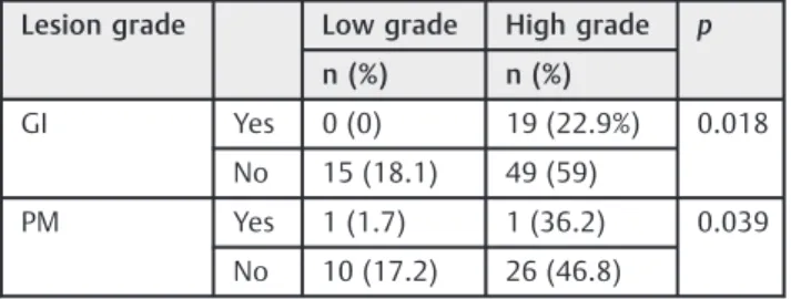
The histological factors for glandular involvement and compromised surgical margins had increased expression in HG lesions (p¼0.018 andp¼0.039 respectively,►Table 2).
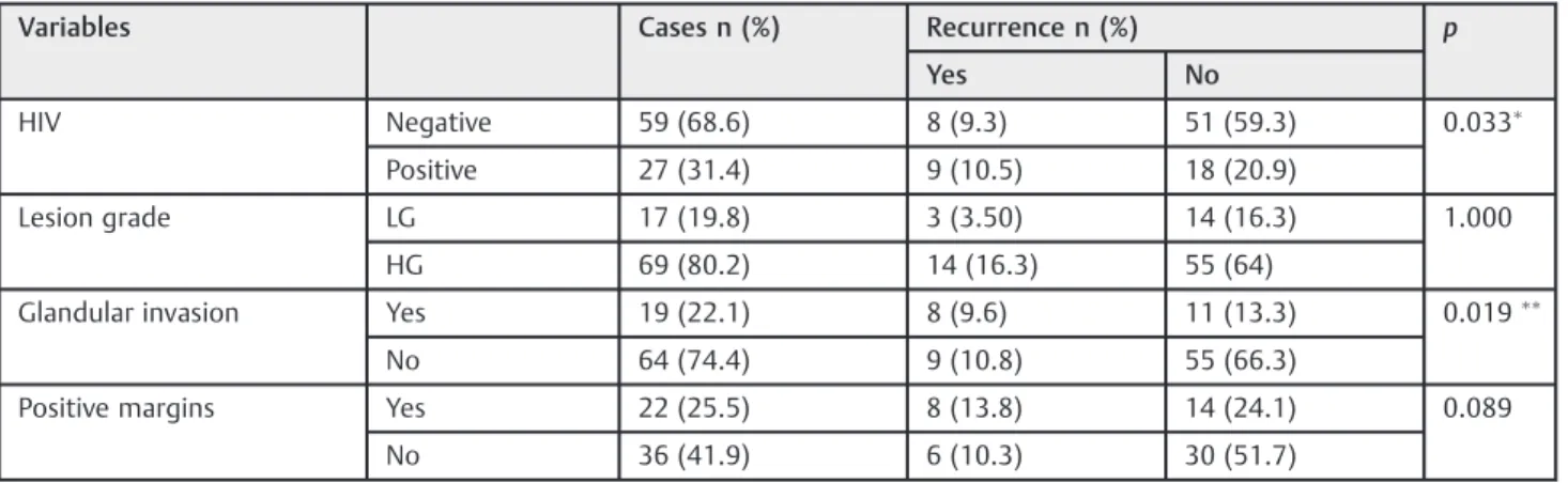
On the other hand, relevant associations with recurrence were found when evaluating the risk factors for human immunodeficiency virus (HIV) positivity (odds ratio [OR]: 0.31; 95% confidence interval [95%CI]: 0.135–0.937; p¼0.033) and glandular involvement in cervical lesions
(OR: 4.44; 95%CI: 1.40–14.06; p¼0.019). Data are listed
on►Table 3.
When the expressions of p16 and Ki-67, isolated or com-bined, were evaluated considering the same risk factors, disregarding the presence or absence of recurrence, a signifi -cant correlation was only found on p16 positive in HG lesions (OR: 4.713; 95%CI: 1.091–20.23;p¼0.018,►Table 4).
Specifically analyzing HG lesions that were p16/Ki-67 positive (2þand 3þ), comparing the presence/absence of
recurrence, a significant difference was found (OR: 0.19; 95% CI: 0.054–0.662; p¼0.010); however the percentage of
positive cells was higher in the HG group with no recurrence. The other variables were not significant (►Table 5).
Discussion
Most studies with biomarkers are limited to the correlation between percentage positivity and the presence and grading of pre-invasive lesions; nonetheless, few relate them to the recurrence of these lesions.6No literature was found with the same specific characteristics of this study. Therefore, the findings of this study were compared with each risk factor for which the markers were measured.
The group sample had a significant number of HIVþ
patients (31.4%), and it is known that this pathology is directly related to CIN recurrence, mainly when there is a decrease in CD4þ, indicating low immunity and a poor
control of the disease.7–9In the present study, it was observed that HIVþwomen had a higher recurrence of CIN than
HIV-women (52.9% and 26.1% respectively;p¼0.033). Thisfi
nd-ing is similar to that of Pantanowitz,10 who found 50% recurrence rates for high-grade squamous intraepithelial lesion (HSIL) and 75% for low-grade squamous intraepithelial lesion (LSIL) over a 6-month period evaluation. Russomano et al11reported similar results, suggesting that CIN recurrence is 42% higher in HIVþwomen. Tebeu et al8described the same
findings in a meta-analysis study that evaluated the number of CIN recurrences in HIVþ women undergoing LEEP with
clear surgical margins, in which the recurrence rate was of 20–75%. As in this study, they concluded that the presence of the HIV is a risk factor for CIN recurrence, even in the absence of any other important factors, such as compromised margins. There was no significant increase of p16 expression in HSIL in women who were HIVþcompared with those who were
Table 1 p16 and Ki-67 positivity according to the percentage of positivity
Marker Negative Low positive
Moderately positive
High positive
þ1 þ2 þ3
p16
<5% 5–25% 26–50% >50%
Ki-67
<5% 5–25% 26–50% >50%
Notes:Nuclear and cytoplasmic markers;nuclear marker. Source: Zhong et al.5
Table 2 Relationship between lesion grade and histological risk factors
Lesion grade Low grade High grade p
n (%) n (%)
GI Yes 0 (0) 19 (22.9%) 0.018
No 15 (18.1) 49 (59)
PM Yes 1 (1.7) 1 (36.2) 0.039
No 10 (17.2) 26 (46.8)
HIV-, and that corroborates thefindings of Nicol et al.,12who reported that the co-infection of HPV/HIV may result in alterations in the cervical cytokine profile, including factors such as interleukin-6, resulting in the decreased expression of p16. Although seropositivity for HIV has been proved to be a risk factor for recurrence,10,13–15 the cervical lesions that recurred have not expressed more biomarkers in HIVþ
wom-en than in HIV- womwom-en (p¼0.424), showing that the
markers cannot demonstrate if HIVþwomen are more prone
to recurrence.
Kodampur et al,16in a cohort study of 309 women with high-grade CIN who underwent LEEP, confirmed the increased need for further intervention when there was endocervical glandular involvement (p¼0.024), which is similar to our findings.
Glandular involvement is closely related to HSIL,17and it was
positively related to recurrence in the samples (p¼0.019),
which corroborates thefindings of Güdücü et al,18who observed that glandular extension is more present in HG lesions, thus demanding greater care when monitoring these cases.
A relationship was found between the histological risk factors, glandular involvement and compromised margins, to the high grade lesions (p¼0.018 and 0.039 respectively),
confirming the severity and greater care that these lesions require. Similar results were found by Kir et al (2012),17who suggested a greater attention to the treatment of HG lesions whenever such risk factors were observed.
Jin et al19compared groups with CIN recurrence after LEEP (348 cases and 1,608 controls), and found that glandular involvement and positive surgical margins increased the risk of relapse. The glandular extension has shown to be a Table 3 Relationship between risk factors and recurrence of CIN
Variables Cases n (%) Recurrence n (%) p
Yes No
HIV Negative 59 (68.6) 8 (9.3) 51 (59.3) 0.033
Positive 27 (31.4) 9 (10.5) 18 (20.9)
Lesion grade LG 17 (19.8) 3 (3.50) 14 (16.3) 1.000
HG 69 (80.2) 14 (16.3) 55 (64)
Glandular invasion Yes 19 (22.1) 8 (9.6) 11 (13.3) 0.019
No 64 (74.4) 9 (10.8) 55 (66.3)
Positive margins Yes 22 (25.5) 8 (13.8) 14 (24.1) 0.089
No 36 (41.9) 6 (10.3) 30 (51.7)
Abbreviations: CIN, cervical intraepithelial neoplasia; HIV, human immunodeficiency virus; LG, low-grade; HG, high-grade. Notes:
OR: 0.31; 95%CI: 0.135-0.937;
OR: 4.44; 95%CI: 1.40–14.06.
Table 4 Risk factors of CIN recurrence and relationship with biomarkers
p16 n (%) p Ki-67 n (%) p p16/Ki-67 n (%) p
Neg. Pos. Neg. Pos. Neg. Pos.
HIV Neg. 9
(10.5) 50 (58.1) 1.000 3 (3.5) 56 (65.1) 0.252 4 (4.7) 23 (26.5) 0.199 Pos. 4 (4.7) 23 (26.7) 5 (5.8) 22 (25.6) 3 (3.5) 56 (65.1)
PM Yes 2
(3.4) 20 (34.5) 0.697 2 (3.4) 20 (34.4) 0.697 1 (1.7) 31 (53.4) 0.235 No 6 (10.3) 30 (51.7) 6 (10.3) 30 (51.8) 6 (10.3) 20 (34.4)
GI Yes 1
(1.2) 18 (21.7) 0.280 1 (1.2) 18 (21.6) 0.675 1 (1.2) 18 (21.7) 0.314 No 12 (14.5) 52 (62.7) 7 (8.4) 57 (68.6) 6 (7.2) 58 (69.9) LG 6 (7.0) 11 (12.8)
0.018 3
(3.5) 14 (16.3) 0.189 3 (3.5) 14 (16.3) 0.189 HG 7 (8.1) 62 (72.1) 5 (5.8) 64 (74.5) 4 (4.7) 65 (75.6)
Abbreviations: CIN, cervical intraepithelial neoplasia; GI, glandular invasion; HG, high-grade; HIV, human immunodeficiency virus; LG, low-grade; Neg., negative; PM, positive margins; Pos., positive.
Note:
primary risk factor; however, compromised margins were not found to be a reliable predictor of recurrence, as opposed to several studies,20–22 and this may be because of the less expressive number of positive margin patients included in this study.
The main question of the current research was whether CIN recurrence could or could not be related to p16 and Ki-67 positive, data still unknown in literature. A study that re-sembles this was recently published by Fonseca et al.6They evaluated the markers p16 and p53 in 83 conization speci-mens, analyzing the recurrence predictors of high-grade CIN. They compared the grade of positive markers with relapse, and concluded that they could not foresee the disease’s recurrence after conization. Thefindings of that paper were supported by thefindings of this study, as the presence of p16 and Ki-67 could not be related to glandular involvement, positive margins or recurrence in the samples, suggesting that the dosage of p16/Ki-67 cannot be seen as effective in predicting the recurrence of these risk factors. A significant relation was found though, between p16 positive and HG lesions, leading to the conclusion that in HG lesions, changes in the cell cycle stand out, and that the increased expression of p16 reflects the subsequent inhibition of the pRb. This inhibition of the pRb induces cell immortalization and trans-formation, a main factor in the evolution of cancer lesions. The same was observed by Calil et al23 in a study of 174 biopsies of the cervix. A strong positive correlation between the expression of p16 and the severity of premalignant lesions was found. In contrast, p16 and Ki-67 (2þand 3þ), analyzed
together in HG lesions, were significantly associated with recurrence, suggesting that a strong positive HG lesion pro-tein expression would possibly have higher risks of recur-rence and, therefore, more attention should be given to these patients, as opposed to the negative or low expressions (1þ),
which would be less prone to recurrence.
Conclusion
The high positivity of p16/Ki-67 was a predictor of recurrence only in patients with HG lesions, suggesting that patients who fit the profile should be monitored closely. In addition, but independently, the research showed that HIV seropositivity and glandular invasion were recurrence risk factors, and also that compromised margins and glandular involvement are more common in severe lesions.
References
1 World Health Organization. Comprehensive cervical cancer con-trol: a guide to essential practice. 2nd ed. Geneva: WHO; 2014 2 Tornesello ML, Buonaguro L, Giorgi-Rossi P, Buonaguro FM. Viral
and cellular biomarkers in the diagnosis of cervical intraepithelial neoplasia and cancer. BioMed Res Int 2013;2013:519619 3 Brown CA, Bogers J, Sahebali S, Depuydt CE, De Prins F, Malinowski
DP. Role of protein biomarkers in the detection of high-grade disease in cervical cancer screening programs. J Oncol 2012; 2012:289315
4 Ikenberg H, Bergeron C, Schmidt D, et al; PALMS Study Group. Screening for cervical cancer precursors with p16/Ki-67 dual-stained cytology: results of the PALMS study. J Natl Cancer Inst 2013;105(20):1550–1557
5 Zhong P, Li J, Gu Y, et al. P16 and Ki-67 expression improves the diagnostic accuracy of cervical lesions but not predict persistent high risk human papillomavirus infection with CIN1. Int J Clin Exp Pathol 2015;8(03):2979–2986
6 Fonseca FV, Tomasich FD, Jung JE, Maestri CA, Carvalho NS. The role of P16ink4a and P53 immunostaining in predicting recur-rence of HG-CIN after conization treatment. Rev Col Bras Cir 2016; 43(01):35–41
7 Babkina N, Heller DS, Goldsmith LT, Houck KL. Cervical conization for cervical intraepithelial neoplasia (CIN) 2 and 3 in HIV-positive women: a case-control study. J Low Genit Tract Dis 2015;19(02): 110–114
8 Tebeu PM, Major AL, Mhawech P, Rapiti E. The recurrence of
cervical intraepithelial neoplasia in HIV-positive women: a review of the literature. Int J STD AIDS 2006;17(08):507–511
9 Clifford GM, Franceschi S, Keiser O, et al; Swiss HIV Cohort Study. Immunodeficiency and the risk of cervical intraepithelial neopla-sia 2/3 and cervical cancer: A nested case-control study in the Swiss HIV cohort study. Int J Cancer 2016;138(07):1732–1740 10 Pantanowitz L. Treatment failure and recurrence of cervical
in-traepithelial neoplasia in HIV-infected women. Womens Health (Lond) 2010;6(06):781–783
11 Russomano F, Paz BR, Camargo MJ, et al. Recurrence of cervical intraepithelial neoplasia in human immunodeficiency virus-in-fected women treated by means of electrosurgical excision of the transformation zone (LLETZ) in Rio de Janeiro, Brazil. Sao Paulo Med J 2013;131(06):405–410
12 Nicol AF, Golub JE, eSilva JR, et al. An evaluation of p16(INK4a) expression in cervical intraepithelial neoplasia specimens, includ-ing women with HIV-1. Mem Inst Oswaldo Cruz 2012;107(05): 571–577
Table 5 Relationship between recurrence and p16/Ki-67 expression (positive 2þand 3þ) and risk factors
Risk factor Recurrence n (%) p
Yes No
High-grade lesion
p16/Ki-67 positive
Yes 5 (7.2) 41 (59.4) 0.010
No 9 (13) 14 (20.3)
HIV presence
p16/Ki-67 positive
Yes 3 (11.1) 10 (37) 0.420
No 6 (22.2) 8 (29.6)
Positive margins
p16/Ki-67 positive
Yes 3 (13.6) 9 (40.9) 0.378
No 5 (22.7) 5 (22.7)
Glandular Involvement
P16/Ki-67 positive
Yes 5 (26.3) 9 (47.4) 0.603
No 3 (15.8) 2 (10.5)
Abbreviation: HIV, human immunodeficiency virus. Note:OR: 0.19; 95%CI: 0.054
13 Keller MJ, Burk RD, Xie X, et al. Risk of cervical precancer and cancer among HIV-infected women with normal cervical cytology and no evidence of oncogenic HPV infection. JAMA 2012;308(04): 362–369
14 Massad LS, Xie X, D’Souza G, et al. Incidence of cervical precancers among HIV-seropositive women. Am J Obstet Gynecol 2015; 212(05):606.e1–606.e8
15 Pantanowitz L, Michelow P. Review of human immunodeficiency virus (HIV) and squamous lesions of the uterine cervix. Diagn Cytopathol 2011;39(01):65–72
16 Kodampur M, Kopeika J, Mehra G, Pepera T, Menon P. Endocervical crypt involvement by high-grade cervical intraepithelial neoplasia after large loop excision of transformation zone: do we need a different follow-up strategy? J Obstet Gynaecol Res 2013;39(01): 280–286
17 Kır G, Karabulut MH, Topal CS, Yılmaz MS. Endocervical glandular
involvement, positive endocervical surgical margin and multi-centricity are more often associated with high-grade than low-grade squamous intraepithelial lesion. J Obstet Gynaecol Res 2012; 38(09):1206–1210
18 Güdücü N, Sidar G, Başsüllü N, Türkmen I, Dünder I. Endocervical
glandular involvement, multicentricity, and extent of the disease
are features of high-grade cervical intraepithelial neoplasia. Ann Diagn Pathol 2013;17(04):345–346
19 Jin J, Li L, Zhang F. Meta-analysis of high risk factors of residue or relapse of cervical intraepithelial neoplasia after conization. J Biol Regul Homeost Agents 2015;29(02):451–458
20 Kim TH, Han JH, Shin E, Noh JH, Kim HS, Song YS. Clinical
implication of p16, Ki-67, and proliferating cell nuclear antigen expression in cervical neoplasia: improvement of diagnostic ac-curacy for high-grade squamous intraepithelial lesion and predic-tion of resecpredic-tion margin involvement on conizapredic-tion specimen. J Cancer Prev 2015;20(01):70–77
21 Serati M, Siesto G, Carollo S, et al. Risk factors for cervical intraepithelial neoplasia recurrence after conization: a 10-year study. Eur J Obstet Gynecol Reprod Biol 2012;165(01):86–90 22 Lu HX, Chen YX, Ni J, Wan XY, Lü WG, Xie X. [Study on high risk
factors associated with positive margin of cervix conization in patient with cervical intraepithelial neoplasia]. Zhonghua Fu Chan Ke Za Zhi 2009;44(03):200–203