American Journal of Infectious Diseases 4 (4): 244-256, 2008 ISSN 1553-6203
© 2008 Science Publications
Corresponding Author: Michael P. Busch, Blood Systems Research Institute, 270 Masonic Ave, San Francisco, California 94118, USA Tel: 415-749-6615 Fax: 415-775-3859
Passive Transfer of HIV-1 Antibodies and Drug Resistant Virus during a Health Care
Worker Accident: Implications for HCW Post-Exposure Management
1,2
Carlos Fernando de Oliveira,
2Ricardo Sobhie Diaz,
3Abdallah Harmache,
3,4Lisa M. Frenkel,
5Phalguni Gupta,
3Gerald H. Learn,
3James I. Mullins and
1,6,7Michael P. Busch
1Blood Systems Research Institute (formerly part of Blood Centers of the Pacific),
San Francisco, CA, USA
2
Federal University of Sao Paulo, Sao Paulo, SP, Brazil
3University of Washington, Seattle, WA, USA.
4Children’s Hospital and Medical Center, Seattle, WA, USA
5University of Pittsburgh, Pittsburgh, PA, USA
6Blood Systems, Inc., Scottsdale, AZ, USA
7University of California, San Francisco, CA, USA
Abstract: Problem statement: We studied in detail a case in which a nurse caring for an HIV-infected child suffered a deep-laceration accident with contaminated blood. Approach: The patient had been treated with zidovudine (ZDV) and the nurse became infected despite prophylactic use of ZDV initiated 2 h after the accident. A reactive anti-HIV-1/2 EIA and an indeterminate western blot (gp120/160 reactivity) were obtained from the nurse on the day of the accident, suggesting pre-exposure infection. However, a negative western blot and positive DNA PCR were documented 10 days after the accident and seroconversion occurred an additional two weeks later. Results:
Phylogenetic analyses of HIV-1 tat and C2-C4-gp120 env regions confirmed that the nurse infected by two different HIV-1 strains present in the child. Strains present in both subjects revealed multi-nucleoside resistant HIV-1. Dilutional serological studies using 10 HIV-infected patients’ sera demonstrated that passive seroreactivity could occur with infusion of less than 1 uL of blood when highly sensitive assays are employed. Conclusion: This is the first well-documented case of passive HIV antibody detection after a percutaneous exposure. Reactive baseline serology should not be assumed to represent prior infection nor exclude prophylaxis. Transmission of drug-resistant HIV-1 corroborates the medical history and supports use of drug history and resistance testing to guide antiretroviral prophylaxis.
Key words: HIV-1, passive transfer of antibodies, health care work accident, bottleneck, transmission of resistant HIV-1
INTRODUCTION
The average risk of Health Care Worker (HCW) infection by the Human Immunodeficiency Virus (HIV) after a percutaneous exposure to contaminated blood is approximately 0.3%[1-3]. Factors linked to increased risk of infection after percutaneous exposure include whether the lesion is deep or caused by an instrument with visible blood and whether the source patient is at an advanced disease stage or has a high viral load[4,5.
Case-control studies have demonstrated the efficacy of antiretroviral prophylactic therapy in
In this study we report a case of occupational exposure that occurred in the United States in 1993. As will be demonstrated, despite following all the recommendations suggested at the time[13], blood exposure from a source patient infected this HCW with an antiretroviral drug-resistant HIV-1 strain. One of the factors that makes this case particularly important is that transient seroreactivity was documented by enzyme-linked immunoassay (third-generation HIV-1 and 2 EIA) and Western blot (indeterminate classification with gp120 +/- and gp160 1+bands) performed on the blood sample collected from the health professional 2 h after the exposure event. Thus, this case draws attention to the possibility of detecting passively transmitted antibodies if assays of extreme sensitivity are used. Such a finding could result in misdiagnosis of prior infection in the HCW and consequently, an inappropriate decision to discontinue post-exposure prophylactic therapy.
Case description: A nurse who was caring for an
HIV-infected child was covering a blood collection tube when it broke. The glass went through her glove and the skin of the palm of the hand, causing a cut one millimeter deep and three centimeters long that was grossly contaminated with the patient’s blood. The nurse promptly washed the lesion using iodated solution, water and soap. The contaminated blood had been collected from a three-year-old patient with documented HIV infection and clinical AIDS. The patient had been treated with ZDV 120 mg PO every 6 h during the 21 months preceding the accident. The viral load of the patient was not available, but the CD4 lymphocyte count measured three weeks earlier was 75 cells mm−3.
The written report of the accident indicates that during her first physical exam, the nurse did not show clinical abnormalities. She started prophylactic use of ZDV 2 h after the accident. A blood sample collected from the nurse on the same day of and subsequent to the accident was reactive for antibodies against HIV-1/2 tested using the Abbott® 3A77 EIA,a third-generation recombinant DNA antigen-sandwich EIA test. An indeterminate result (gp120 +/-, gp160 +) was obtained on the Western blot assay. Follow-up tests were requested and a Western blot of a sample collected 10 days after the accident was negative, but a DNA PCR from the same sample was positive. On the day these results were discussed with the nurse (13 days after the accident) she was reporting fatigue, malaise, loss of appetite, nausea and vomiting, symptoms which were
attributed to side effects of ZDV. It was decided to discontinue the use of ZDV at that time. Two weeks later the tests were compatible with complete seroconversion to HIV, with EIA reactivity and a positive Western blot.
The nurse was a white female in her thirties who had never had a blood transfusion. She denied use of injection drugs and reported that she had been celibate in the 6 years preceding the accident. Table 1 shows the progression of the diagnostic exams made on the nurse’s samples.
Further study detailed below was done to investigate the genetic linkage between the HIV-1 population present in the nurse and the putative source patient and to study the level of inoculum required for producing passive anti-HIV reactivity.
MATERIALS AND METHODS
Informed consent was obtained from the nurse and from the child’s parents prior to access and study of the stored samples.
Samples evaluated: Peripheral Blood Mononuclear
Cells (PBMC) collected from the nurse 20 days after the accident and serum from the child taken 10 days after the accident, were available for analysis. The samples were HLA typed through allele-specific PCR using radioactive markers[14] to verify that they belonged to different individuals. Aliquots of these samples were independently and simultaneously analyzed in three different institutes: Blood Centers of the Pacific (BCP), in San Francisco, California (specimens from the nurse and child), the University of Washington (UW) in Seattle (for specimens from the Nurse) and Children’s Hospital and Medical Center (CHMC) in Seattle (for specimens from the child).
Extraction of nucleic acids and PCR amplification of
HIV-1 sequences: For experiments done at BCP, DNA
from mononuclear cells and plasma RNA were extracted using the QIAamp Blood and QIAamp Viral RNA kits (QIAGEN Inc, Santa Clarita, CA), respectively. The extracted RNA was reverse transcribed to generate a complementary DNA that could be amplified by PCR[15]. Experiments performed at the UW and CHMC used in-house rapid lysis[16] to obtain cellular DNA.
base pair fragment (HXB2 coordinates 7001-7647; GenBank access number K03455). The env primer sequences and PCR conditions used are published elsewhere[17].
PCR products were amplified under conditions compatible with the end-point dilution technique described by[18,19] followed by DNA automated sequencing. At the BCP laboratories, Heteroduplex Mobility Analysis (HMA) was done using env (C2-V5) PCR products as described by[20] In a first experiment, env PCR products from the source patient were paired and analyzed by HMA. This procedure demonstrated the presence of two distinguishable virus populations among the patient's env amplicons. A representative PCR product from each of those two populations was used as a probe to trace similar sequences among all other env PCR products from both the child and the nurse using Heteroduplex Tracking Assay (HTA)[27]. After this initial screening, 13 env PCR products from the child and 17 from the nurse were selected for DNA sequencing. envPCR products independently generated at the UW and CHMC laboratories were DNA sequenced (13 from the child and 3 from the health care worker).
At the BCP laboratories, nested PCR of the same samples were performed to amplify the first exon of tat (HXB2 coordinates 5905-6133, 285 bp)[21] and the Reverse Transcriptase (RT) coding region of pol (HXB2 coordinates 2518-3320, 750 bp). The primers and PCR conditions used for RT amplification combined the procedure of Kozal et al.[22] for the first round and with that described by Frenkel et al.[23] for the second round.
DNA or cDNA sequencing: At BCP, sequencing was
done using the alfexpress automated sequencer for the separation of the sequencing products obtained with the Thermo Sequenase kit (Amersham Pharmacia Biotech, Piscataway, NJ). At the UW and CHMC, reactions were done with commercially available kits (Dye Terminator Sequencing kit, Applied Bio Systems, Foster City, CA) and separated on an automatic ABI model 377. In all sequencing reactions, products labeled with fluorescent molecules were present either in the primers or in the terminators included with the kit reagents. All the sequences produced for the env region (GeneBank accession numbers: pending) were compared with those already available in HIV-1 genome databases (local, GenBank and the Los Alamos HIV sequence database)
using BLAST[24,25]. Phylogenetic reconstructions were created using maximum likelihood estimation[26] under HKY models of evolution[27] (model details are available from the authors upon request) with the computer program PAUP* version 4.0b10[28]. Bootstrap support[29] of phylogenetic relationships were calculated using neighbor-joining trees. Only sequences generated at the BCP are presented in the alignment and phylogenetic analysis in this study, since the work generated at the UW and CHMC confirmed the overall results.
Serum dilution studies: Five-fold serial dilutions were prepared from the serum of 10 HIV seropositive patients (5 asymptomatic and 5 with clinical AIDS) at an interval of 1:53-1:512 using HIV-1 seronegative normal donor serum as diluent. Each dilution was tested using (1) EIAs containing HIV-1 viral lysate antigen (Abbott Diagnostic Systems, Abbott Park, IL and Genetic Systems, Redmond, WA); (2) third-generation antigen-sandwich EIAs for detection of HIV-1 and -2 (Abbott and Ortho Diagnostic Systems, Raritan, NJ); (3) HIV-1 viral lysate Western blot (Ortho) and (4) recombinant immunoblot assay (RIBA) for HIV-1 and -2 (Chiron Corporation, Emeryville, CA).
RESULTS
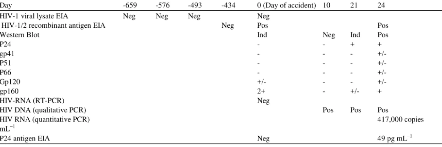
Table 1 shows the time course of samples evaluated from the nurse and child and summarizes results of the HIV serologic and viral nucleic acid amplification assays. Table 2 shows HLA typing that confirmed that the samples from the nurse (serum collected 10 days post exposure) and child (PBMC collected 20 days post accident) were genetically discordant and thus collected from different individuals.
Table 1: Laboratory studies of the nurse´s serial serum samples for direct (p24 antigen; RT-PCR; DNA PCR) or indirect (ELISA; Western blot) detection of HIV infection. The days are expressed relative to the date of the occupational accident
Day -659 -576 -493 -434 0 (Day of accident) 10 21 24
HIV-1 viral lysate EIA Neg Neg Neg Neg
HIV-1/2 recombinant antigen EIA Neg Pos Pos
Western Blot Ind Neg Ind Pos
P24 - - + +
gp41 - - - +/-
P51 - - - +/-
P66 - - - +/-
Gp120 +/- - - +/-
gp160 2+ - +/- +
HIV-RNA (RT-PCR) Neg
HIV DNA (qualitative PCR) Pos Pos Pos
HIV RNA (quantitative PCR) 417,000 copies
mL−1
P24 antigen EIA Neg 49 pg mL−1
Table 2: Allele-specific PCR for the determination of HLA type in the nurse and patient samples
DR 1 DR 2 DR 4 DR 7 DR 11 DQB 2 DQB 4 DQB 5 DQB 601 DQB 603 DQB 7 DQB 8
Nurse NEG NEG POS NEG NEG NEG NEG NEG NEG POS POS NEG
Patient POS NEG NEG POS NEG POS NEG POS NEG NEG NEG NEG
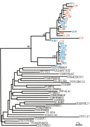
No sequence was sufficiently close so as to suggest a specimen contamination[30]. The closest sequence found was ~10% divergent (hiv509env; GenBank accession number L48063), a clade B virus from South Africa. This finding was consistent with the distance to an epidemiologically unlinked infection. In contrast, sequences from the nurse and child were more closely related (Fig. 2), with inter subject distances ranging from zero to 9% (4% average; Confidence Interval (CI) of 2-5%). In addition, two distinct populations of sequences were found within the child’s sample (mean distance between populations: 6.98%, range 6.47-7.53%).
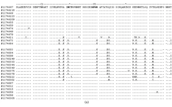
As may be seen in the alignment shown in Fig. 1, in addition to the remarkable nucleotide similarity, certain sequences from the nurse and child also shared a pattern of deletions. As is usually the case, deletions and insertions of nucleotides were not considered in our evolutionary distance studies. However, they can, as shown here, confer a specific signature to groups of sequences. The same sequences that show deletions also show a difference in amino acid sequence at the crown of the V3 loop (GPGR for GPGK) and loss of the highly conserved N-linked glycoslation site sequon spanning the N-terminus of the V3 loop (bold on the alignment, Fig. 1). The phylogenetic tree (Fig. 2) showing sequences from these two individuals in the context of sequences from epidemiologically unlinked HIV from the GenBank database, show that both groups of sequences from both individuals form a unique cluster. All of the foregoing data indicate a close
relationship between the viruses in the two infections, consistent with direct transmission from the child to the nurse.
Env region Heteroduplex Tracking Analysis (HTA):
(a)
(b)
Fig. 1a and b: Sequence alignment of amino acids encoded by amplicons obtained from the C2-V4 region of the child (identified by numbers starting with 11746) and the nurse (starting with 11760). Dots represent identities; dashes represent amino acids deletions. The GPGR motif at the crown of the V3 loop and the N-linked glycosylation site sequon are marked in bold (Panel a)
Tat region sequence and HTA analysis: The analysis
of the sequences of the tat viral gene region, although limited to a reduced number of amplicons, resulted in a similar pattern (Fig. 3). Although several sequences in the databases had more than 90% identity to the test sequences, comparisons of tat sequences from the nurse and child demonstrated a much closer relationship
Fig. 2: Maximum likelihood phylogram (-LnL = 4221.0130) representing the reconstruction of the phylogenetic relationships between the env (V3-V5) sequences obtained from the nurse (blue), the child (red) and 28 sequences chosen from GenBank. Ten random sequence addition iterations were used. Scale bar represents 1% genetic distance (0.01 substitutions/site). Bootstrap values are shown at nodes with greater than 70% support
tat PCR products 760026 and 746086 were used as probes in an HTA[21] with all the other tat PCR products available from both subjects. An additional 4 amplicons derived from the nurse’s sample were closely related to 746086 (only homoduplexes), but not to 760026 (only heteroduplexes). Conversely, two other tat amplicons available from the nurse were related to 760026. The child’s sample, however, had the reciprocal pattern of representation of the two forms, with 3 out of 50 related to 746086, while the other 47 formed homoduplexes with 760026 (data not shown).
Pol region sequence analysis: Finally, analysis of the sequences of the Reverse Transcriptase region of the pol gene from nurse and child samples was performed.
Fig. 3: Maximum likelihood phylogram (-LnL = 1229.0419) representing the reconstruction of the phylogenetic relationships between the tat sequences obtained from the nurse (blue), the child (red) and 18 sequences chosen from GenBank. Thirty-five random sequence addition iterations were used. Scale bar represents 1% genetic distance (0.01 substitutions/site). Bootstrap values are shown at nodes with greater than 70% support
There was, as expected for epidemiologically linked infections, near identity between the amplicons obtained from the two individuals (data not shown). They also demonstrated identical Nucleoside Associated Mutations[32], consistent with the known use of ZDV by the child (M41L, D67N, T69D, K70R, H208Y and L214F)[33-38].
0 2 4 6 8 10
-9 - 7
-5 - 3
-1
Log 10 (Dilution)
N
u
m
b
er
o
f
in
di
vi
du
al
s
w
it
h
p
o
si
ti
v
e
re
su
lt
s
(N
=
1
0
)
Fig. 4a: Five-fold serial dilutions were prepared with sera from 10 HIV-1 carriers (5 asymptomatic; 5 with clinical AIDS). The abscissa shows the dilution factor on a log10 scale. The ordinate
indicates the number of individuals with positive results on HIV-1 antibody tests. Open diamond: Abbott 3rd-generation HIV-1/2 combination EIA (3A77); open circle: Ortho 3rd-generation HIV-1/2 combination EIA; filled diamond: Abbott 2nd-gen HIV-1 EIA (3A11); filled triangle: Western blots; filled square: recombinant immunoblot assay (RIBA)-positive
-1.5 0.0 1.5
-9 -8 -7 -6 -5 -4 -3
Log10 (Dilution)
L
o
g
1
0
(
S
/C
o
)
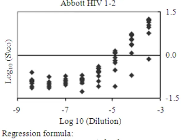
Fig. 4b: Optical density (OD) regression curve obtained from HIV-1 antibody detection of five-fold dilutions of 10 serum samples from HIV positive subjects (5 AIDS patients and 5 asymptomatic HIV-1 carriers) using 3rd-generation HIV-1/2 EIA (Abbott 3A77)
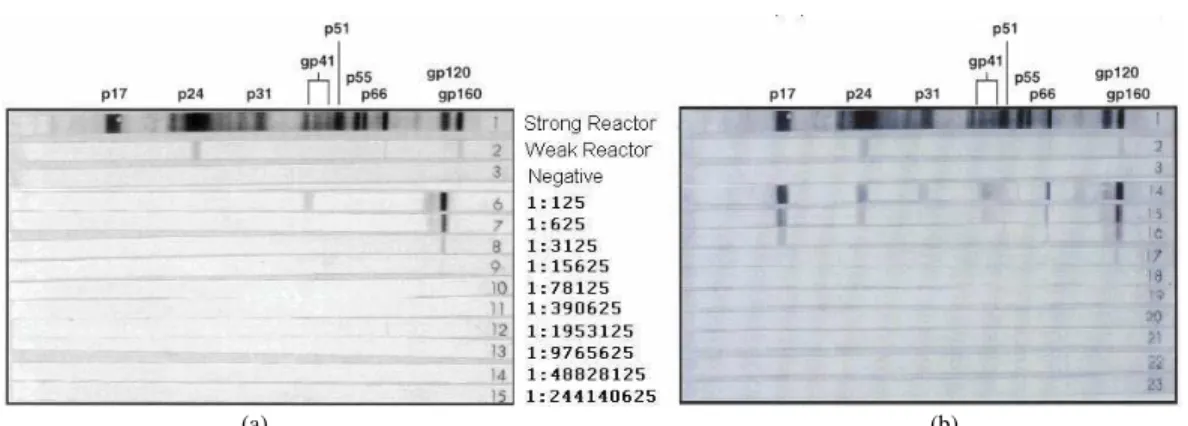
continued to show indeterminate results at dilutions up to 108 fold (Fig. 5) with an envelope-only band pattern similar to that in the nurse’s post-exposure sample (Table 1).
Fig. 4c: Optical Density (OD) regression curve obtained from HIV-1 antibody detection of five-fold dilutions of 10 serum samples from HIV positive subjects (5 AIDS patients and 5 asymptomatic HIV-1 carriers) using third generation EIA (Abbott 3A77HIV-1/2, or COMB)
(a) (b)
Fig. 5: Representative samples of Western blot HIV-1 positive patients’ results in serially diluted samples (five-fold). The figure legend between the Western blot images indicates the presented dilution. (a): Asymptomatic HIV carrier; (b): AIDS patient
As a final verification, we evaluated whether the Western blot reactivity seen on the baseline exam (i.e., gp120 +/-; gp160 +) could have been a false-positive result restricted to the detection of reactivity against a previously defined non-specific epitope of the HIV-1 envelope protein. Sayre et al.[41] have demonstrated that in some non-infected blood donors with false positive Western blot results, the reactivity is restricted to a 20-amino acid sequence of gp41 that is shared with non-HIV antigens; if the donor’s sample is pre-incubated with a solution containing this amino acid sequence, the Western blot reactivity is blocked. This procedure was done with the nurse’s baseline sample and the result remained the same, i.e., the peptide did not reduce the gp120 or 160 band reactivity (data not shown). Thus, all of our evidence is consistent with the hypothesis that the Western blot reactivity observed in the nurse was specific to HIV envelope and attributable to transient detection of passive HIV antibodies. Unfortunately, we did not have any left over sample from the source patient in this case to perform serial dilution studies.
DISCUSSION
The molecular genetic evidence shown in this case confirms that the child and the nurse had closely related viral populations, consistent with infections acquired from the same source or transmitted from one individual to the other. This, added to the evolving pattern of the serological and direct viral markers of HIV infection (PCR and p24 antigen) in the nurse’s samples, confirmed that she acquired the infection from the child as a result of the accident and rejected the possibility of a pre-existing infection resulting in the seroreactivity of
the baseline sample.
in the U.S., although most probably used first and second generation EIA, since testing was likely performed in hospital or commercial reference laboratories. This may explain the lack of documentation of detection of passively acquired antibodies in previous reports following HCW accidents[54-57].
Other studies have reported the detection of passively transferred antibodies in cases of post-transfusion hepatitis C[58,59] and HIV[60], which is not surprising given the large volume of plasma contained in blood components (20-250 mL). Our finding of baseline antibody reactivity against HIV in ultra-sensitive tests such as third generation EIA indicated that a moderate amount of contaminated blood entered the circulation of the exposed individual at the time of the accident, rather than indicating pre-existing infection. Whether such passive antibodies are indicative of high-level exposure and predictive of transmission warrants further study. In any event, these results indicate that special care must be taken in interpreting baseline tests at the time of making decisions involving initiation or continuation of prophylactic therapy following high-risk HCW exposures.
This case is also relevant in that it documents the transmission of a virus resistant to multiple nucleoside analogs from a patient to a health care worker after an occupational injury. Not surprisingly, the virus from the source patient had genotypic evidence for resistance not only to ZDV (M41L, D67N, K70R), but also to DDC, DDI (T69D) and with partial genotypic resistance to D4T and Abacavir (M41L, D67N, K70R) (http://hivdb.stanford.edu, www.ablnetworks.com). Although the patient had been treated for a long period of time with ZDV immunotherapy, it is currently known that the Nucleoside Analog Mutations selected by this drug can confer varying degrees of reduced susceptibility (cross resistance) to all nucleoside and nucleotide analogues reverse transcriptase inhibitors tested to-date[61]. These data and other showing that sources or potential source in occupational accidents involving health care works have antiretroviral resistant HIV[62], support guidelines recommending the use of HAART for post exposure prophylaxis in health care workers. The transmission of a virus containing genetic markers of resistance from an antiretroviral experienced patient also supports guidelines that take into consideration the medical history of the patient and anti-viral response profiles of the source patients’ virus
when deciding on antiretroviral prophylaxis in post-exposure settings[4]. Although the turnaround time for resistance testing (a few days to 2-3 weeks) makes it infeasible to guide initial post-exposure antiretroviral prophylaxis, it may be used to expeditiously adjust the drugs according to the resistance profile of the exposure source. Furthermore, prophylactic antiretroviral should preferentially choose a combination of reverse transcriptase inhibitors, since protease inhibitors will act after the cell is already infected.
CONCLUSION
This is the first well-documented case of passive HIV antibody detection after a percutaneous exposure. Reactive baseline serology should therefore not be assumed to represent prior infection nor exclude prophylaxis. Transmission of drug-resistant HIV-1 corroborates the medical history and supports use of drug history and resistance testing to guide antiretroviral prophylaxis.
ACKNOWLEDGEMENT
Dr. de Oliveira's stay in the US was made possible by financial support from the Fogarty International Foundation through the University of California, Berkeley, #D43-TW0003 and by the Brazilian Support Center for Education and Research (CAPES). Work at the University of Washington and the Children’s Hospital and Medican Center in Seattle was supported by grants from the US Public Health Service, including the UW Center for AIDS Research. We thank Maya Royz for the technical assistance and Barbara Johnson for assistance in manuscript preparation.
REFERENCES
1. Tokars, J.I., R. Marcus and D.H. Culver et al., 1993. Surveillance of HIV infection and zidovudine use among health care workers after occupational exposure to HIV-infected blood. The CDC Cooperative Needlestick Surveillance Group. Ann. Intern. Med., 118: 913-919.
http://www.annals.org/cgi/content/abstract/118/12/913 2. Gerberding, J.L., 1994. Incidence and prevalence
of human immunodeficiency virus, hepatitis B virus, hepatitis C virus and cytomegalovirus among health care personnel at risk for blood exposure: Final report from a longitudinal study. J. Infect. Dis., 170: 1410-1417.
3. Bell, D.M., 1997. Occupational risk of human immunodeficiency virus infection in healthcare workers: an overview. Am. J. Med., 102: 9-15. http://md1.csa.com/partners/viewrecord.php?reques ter=gs&collection=ENV&recid=4308919&q=Occu pational+risk+of+human+immunodeficiency+virus +infection+in+healthcare+workers%3A+an+overvi ew&uid=&setcookie=yes
4. Panlilio A.L., D.M. Cardo, L.A. Grohskopf, W. Heneine, C.S. Ross, 2005. Updated US public health service guidelines for the management of occupational exposures to HBV, HCV and HIV and recommendations for post-exposure prophylaxis.
MMWR Recomm. Rep., 54: 1-17.
http://www.ncbi.nlm.nih.gov/pubmed/16195697?dopt =Abstract
5. Gerberding, J.L., 2003. Clinical practice. Occupational exposure to HIV in health care settings. N. Engl. J. Med., 348: 826-833. http://www.ncbi.nlm.nih.gov/pubmed/12606738 6. Connor, E.M., R.S. Sperling and R. Gelber et al.,
1994. Reduction of maternal-infant transmission of human immunodeficiency virus type 1 with zidovudine treatment. Pediatric AIDS Clinical Trials Group Protocol 076 Study Group. N. Engl. J.
Med., 331: 1173-1180.
http://content.nejm.org/cgi/content/abstract/331/18/1173 7. Wade, N.A., G.S. Birkhead, B.L. Warren et al.,
1998. Abbreviated regimens of zidovudine prophylaxis and perinatal transmission of the human immunodeficiency virus. N. Engl. J. Med., 339: 1409-14.
http://www.ncbi.nlm.nih.gov/pubmed/9811915 8. Mandelbrot, L., J. Le Chenadec and A. Berrebi et al.,
1998. Perinatal HIV-1 transmission: Interaction between zidovudine prophylaxis and mode of delivery in the French Perinatal Cohort. Jama, 280: 55-60.
http://cat.inist.fr/?aModele=afficheN&cpsidt=2306298 9. Guay, L.A., P. Musoke and T. Fleming et al., 1999.
Intrapartum and neonatal single-dose nevirapine compared with zidovudine for prevention of mother-to-child transmission of HIV-1 in Kampala, Uganda: HIVNET 012 randomised trial. Lancet,
354: 795-802.
http://www.ncbi.nlm.nih.gov/pubmed/10485720 10. Cohen, O.J. and A.S. Fauci, 1999. Transmission of
drug-resistant strains of HIV-1: Unfortunate, but inevitable. Lancet, 354: 697-698. DOI: 10.1016/S0140-6736(99)90106-X
11. Salomon, H., M.A. Wainberg and B. Brenner et al., 2000. Prevalence of HIV-1 resistant to antiretroviral drugs in 81 individuals newly infected by sexual contact or injecting drug use. Investigators of the Quebec Primary Infection Study. Aids, 14: F17-F23.
http://cat.inist.fr/?aModele=afficheN&cpsidt=1330035 12. Little, S.J., S. Holte and J.P. Routy et al., 2002.
Antiretroviral-drug resistance among patients recently infected with HIV. N. Engl. J. Med., 347: 385-94.
http://www.ncbi.nlm.nih.gov/pubmed/12167680 13. CDC, 1990. Public health service statement on
management of occupational exposure to human immunodeficiency virus, including considerations regarding zidovudine postexposure use. MMWR
Recomm Rep., 39: 1-14.
http://www.cdc.gov/mmwr/preview/mmwrhtml/000 01556.htm
14. Lee, T.H., N.S. Sakahara, E.W. Fiebig, D.F. Hirschkorn, D.K. Johnson and M.P. Busch, 1998. Quantitation of white cell subpopulations by polymerase chain reaction using frozen whole-blood samples. Viral activation transfusion study. Transfusion, 38: 262-270.
http://cat.inist.fr/?aModele=afficheN&cpsidt=2214 543
15. Diaz, R.S., L. Zhang, M.P. Busch, J.W. Mosley and A. Mayer, 1997. Divergence of HIV-1 quasispecies in an epidemiologic cluster. Aids, 11: 415-22. http://www.ncbi.nlm.nih.gov/pubmed/9084787 16. Kalish, M.L., A. Baldwin and S. Raktham et al.,
1995. The evolving molecular epidemiology of HIV-1 envelope subtypes in injecting drug users in Bangkok, Thailand: Implications for HIV vaccine trials. Aids, 9: 851-7.
http://www.ncbi.nlm.nih.gov/pubmed/7576318 17. Delwart, E.L., M.P. Busch, M.L. Kalish, J.W. Mosley
and J.I. Mullins, 1995. Rapid molecular epidemiology of human immunodeficiency virus transmission. AIDS Res. Hum. Retroviruses, 11: 1081-93. http://www.ncbi.nlm.nih.gov/pubmed/8554905 18. Simmonds, P., P. Balfe, C.A. Ludlam, J.O. Bishop
19. Rodrigo, A.G., P.C. Goracke, K. Rowhanian and J.I. Mullins, 1997. Quantitation of target molecules from polymerase chain reaction-based limiting dilution assays. AIDS Res. Hum. Retroviruses, 13: 737-42.
http://www.ncbi.nlm.nih.gov/pubmed/9171217 20. Delwart, E.L., E.G. Shpaer and J. Louwagie et al.,
1993. Genetic relationships determined by a DNA heteroduplex mobility assay: Analysis of HIV-1 env genes. Science, 262: 1257-61. DOI: 10.1126/science.8235655
21. Diaz, R.S., C.F. De Oliveira, R. Pardini, E. Operskalski, A.J. Mayer and M.P. Busch, 1999. HIV type 1 tat gene heteroduplex mobility assay as a tool to establish epidemiologic relationships among HIV type 1-infected individuals. AIDS Res. Hum. Retroviruses, 15: 1151-1156. http://www.ncbi.nlm.nih.gov/pubmed/10480628 22. Kozal, M.J. and N. Shah, et al., 1996. Extensive
polymorphisms observed in HIV-1 clade B protease gene using high-density oligonucleotide arrays. Nat. Med., 2: 753-759. http://www.ncbi.nlm.nih.gov/pubmed/8673920 23. Frenkel, L.M. and L.E. Wagner, S.M. Atwood,
T.J. Cummins and S. Dewhurst, 1995. Specific, sensitive and rapid assay for human immunodeficiency virus type 1 pol mutations associated with resistance to zidovudine and didanosine. J. Clin. Microbiol., 33: 342-347. http://jcm.asm.org/cgi/content/abstract/33/2/342 24. Karlin, S. and S.F. Altschul, 1990. Methods for
assessing the statistical significance of molecular sequence features by using general scoring schemes. Proc. Natl. Acad. Sci., 87: 2264-2268. http://www.ncbi.nlm.nih.gov/pubmed/2315319 25. Altschul, S.F., T.L. Madden and A.A. Schaffer et al.,
1997. Gapped BLAST and PSI-BLAST: A new generation of protein database search programs. Nucleic Acids Res., 25: 3389-3402. http://www.ncbi.nlm.nih.gov/pubmed/9254694 26. Felsenstein, J., 1981. Evolutionary trees from DNA
sequences: a maximum likelihood approach. J. Mol. Evol., 17: 368-76. DOI: 10.1007/BF01734359 27. Hasegawa, M., H. Kishino and T. Yano, 1985.
Dating of the human-ape splitting by a molecular clock of mitochondrial DNA. J. Mol. Evol., 22: 160-174. http://www.ncbi.nlm.nih.gov/pubmed/3934395 28. Swofford, D.L., 1999. PAUP* 4.0: Phylogenetic
Analysis Using Parsimony (*and Other Methods). 4.0b2a Edn., Sinauer Associates, Inc., Sunderland, Massachusetts, Sunderland, MA., USA.
29. Felsenstein, J., 1985. Confidence limits on phylogenies: an approach using the bootstrap. Evolution, 39: 783-791.
http://iphylo.org/~rpage/challenge/www/uri/75417d 0075a983340bd5088134155fb9
30. Learn, G.H., Jr., B.T. Korber, B. Foley, B.H. Hahn, S.M. Wolinsky and J.I. Mullins, 1996. Maintaining the integrity of human immunodeficiency virus sequence databases. J. Virol., 70: 5720-5730. http://jvi.asm.org/cgi/content/abstract/70/8/5720 31. Lole, K.S., R.C. Bollinger and R.S. Paranjape et al.,
1999. Full-length human immunodeficiency virus type 1 genomes from subtype C-infected seroconverters in India, with evidence of intersubtype recombination. J. Virol., 73: 152-160. http://www.ncbi.nlm.nih.gov/pubmed/9847317 32. Zhu, T., H. Mo and N. Wang et al., 1993.
Genotypic and phenotypic characterization of HIV-1 patients with primary infection. Science, 26HIV-1: 1179-1181. DOI: 10.1126/science.8356453 33. Larder, B.A., K.E. Coates and S.D. Kemp, 1991.
Zidovudine-resistant human immunodeficiency virus selected by passage in cell culture. J. Virol., 65: 5232-5236.
http://www.pubmedcentral.nih.gov/articlerender.fc gi?artid=249001
34. Larder, B.A. and S.D. Kemp, 1989. Multiple mutations in HIV-1 reverse transcriptase confer high-level resistance to zidovudine (AZT). Science, 246: 1155-1158. DOI: 10.1126/science.2479983 35. Kellam, P., C.A. Boucher and B.A. Larder, 1992.
Fifth mutation in human immunodeficiency virus type 1 reverse transcriptase contributes to the development of high-level resistance to zidovudine. Proc. Natl. Acad. Sci., 89: 1934-1938. http://www.pubmedcentral.nih.gov/articlerender.fc gi?artid=48568
36. Lawrence, J., J. Schapiro and M. Winters et al., 1999. Clinical resistance patterns and responses to two sequential protease inhibitor regimens in saquinavir and reverse transcriptase inhibitor-experienced persons. J. Infect. Dis., 179: 1356-1364. http://www.ncbi.nlm.nih.gov/pubmed/10228055 37. Fitzgibbon, J.E., R.M. Howell, C.A. Haberzettl,
S.J. Sperber, D.J. Gocke and D.T. Dubin, 1992. Human immunodeficiency virus type 1 pol gene mutations which cause decreased susceptibility to 2',3'-dideoxycytidine. Antimicrob Agents Chemother, 36: 153-7.
38. Kemp, S.D., C. Shi, S. Bloor, P.R. Harrigan, J.W. Mellors and B.A. Larder, 1998. A novel polymorphism at codon 333 of human immunodeficiency virus type 1 reverse transcriptase can facilitate dual resistance to zidovudine and L-2',3'-dideoxy-3'-thiacytidine. J. Virol., 72: 5093-5098.
http://jvi.asm.org/cgi/content/abstract/72/6/5093 39. Lenes, B., 1995. Formulas for Clinical Volumes,
Calculations of Leukocyte Reduction and Mathematics of Washout Curves. American Association of Blood Banks Press, Bethesda, Maryland, USA.
40. Mast, S.T., J.D. Woolwine and J.L. Gerberding, 1993. Efficacy of gloves in reducing blood volumes transferred during simulated needlestick injury. J. Infect. Dis., 168: 1589-15892. http://www.ncbi.nlm.nih.gov/pubmed/8245553 41. Sayre, K.R., R.Y. Dodd, G. Tegtmeier, L. Layug,
S.S. Alexander and M.P. Busch, 1996. False-positive human immunodeficiency virus type 1 western blot tests in noninfected blood donors. Transfusion, 36: 45-52.
http://cat.inist.fr/?aModele=afficheN&cpsidt=2988400 42. Wolfs, T.F., G. Zwart, M. Bakker and J. Goudsmit,
1992. HIV-1 genomic RNA diversification following sexual and parenteral virus transmission. Virology, 189: 103-10.
http://www.ncbi.nlm.nih.gov/pubmed/1376536 43. McNearney, T., Z. Hornickova and R. Markham et al.,
1992. Relationship of human immunodeficiency virus type 1 sequence heterogeneity to stage of disease. Proc. Natl. Acad. Sci., 89: 10247-10251. http://www.pubmedcentral.nih.gov/articlerender.fc gi?artid=50315
44. Wolinsky, S.M., C.M. Wike and B.T. Korber et al., 1992. Selective transmission of human immunodeficiency virus type-1 variants from mothers to infants. Science, 255: 1134-1137. DOI: 10.1126/science.1546316
45. Ait-Khaled, M. and V.C. Emery, 1993. Sequence variation within the human immunodeficiency virus V3 loop at seroconversion. J. Med. Virol., 41: 270-274.
http://cat.inist.fr/?aModele=afficheN&cpsidt=3878824 46. Zhang, L.Q., P. MacKenzie, A. Cleland, E.C. Holmes,
A.J. Brown and P. Simmonds, 1993. Selection for specific sequences in the external envelope protein of human immunodeficiency virus type 1 upon primary infection. J. Virol., 67: 3345-56. http://www.pubmedcentral.nih.gov/articlerender.fc gi?artid=237678
47. Cornelissen, M., G. Mulder-Kampinga and J. Veenstra et al., 1995. Syncytium-inducing (SI) phenotype suppression at seroconversion after intramuscular inoculation of a non-syncytium-inducing/SI phenotypically mixed human immunodeficiency virus population. J. Virol., 69: 1810-1818.
http://jvi.asm.org/cgi/content/abstract/69/3/1810 48. Diaz, R.S., E.C. Sabino, A. Mayer, C.F. deOliveira,
J.W. Mosley and M.P. Busch, 1996. Lack of dual HIV infection in a transfusion recipient exposed to two seropositive blood components. AIDS Res. Hum. Retroviruses, 12: 1291-1295.
http://www.ncbi.nlm.nih.gov/pubmed/8870851 49. Long, E.M., H.L. Martin, Jr. and J.K. Kreiss et al.,
2000. Gender differences in HIV-1 diversity at time of infection. Nat. Med., 6: 71-5. DOI: 0.1038/71563 50. Ritola, K., C.D. Pilcher and S.A. Fiscus et al.,
2004. Multiple V1/V2 env variants are frequently present during primary infection with human immunodeficiency virus type 1. J. Virol., 78: 11208-11218. DOI: 10.1128/JVI.78.20.11208-11218.2004
51. Zaaijer, H.L., P.V. Exel-Oehlers, T. Kraaijeveld, E. Altena and P.N. Lelie, 1992. Early detection of antibodies to HIV-1 by third-generation assays. Lancet, 340: 770-772.
http://www.ncbi.nlm.nih.gov/pubmed/1356186 52. Barrera, J.M., B. Francis and G. Ercilla et al.,
1995. Improved detection of anti-HCV in post-transfusion hepatitis by a third-generation ELISA. Vox Sang, 68: 15-18.
http://www.ncbi.nlm.nih.gov/pubmed/7536987 53. Busch, M.P., L.L. Lee and G.A. Satten et al., 1995.
Time course of detection of viral and serologic markers preceding human immunodeficiency virus type 1 seroconversion: Implications for screening of blood and tissue donors. Transfusion, 35: 91-97. http://www.ncbi.nlm.nih.gov/pubmed/7825218 54. Pratt, R.D., J.F. Shapiro, N. McKinney, S. Kwok
and S.A. Spector, 1995. Virologic characterization of primary human immunodeficiency virus type 1 infection in a health care worker following needlestick injury. J. Infect. Dis., 172: 851-854. http://www.ncbi.nlm.nih.gov/pubmed/7658081 55. Jochimsen, E.M., 1997. Failures of zidovudine
postexposure prophylaxis. Am. J. Med., 102: 52-57. http://www.ncbi.nlm.nih.gov/pubmed/9845497 56. Cardo, D.M., D.H. Culver and C.A. Ciesielski et al.,
1997. A case-control study of HIV seroconversion in health care workers after percutaneous exposure. Centers for Disease Control and Prevention Needlestick Surveillance Group. N. Engl. J. Med., 337: 1485-1490.
57. Ippolito, G., V. Puro, J. Heptonstall, J. Jagger, G. De Carli and N. Petrosillo, 1999. Occupational human immunodeficiency virus infection in health care workers: Worldwide cases through September 1997. Clin. Infect. Dis., 28: 365-83. http://cat.inist.fr/?aModele=afficheN&cpsidt=1692655 58. Vallari, D.S., B.W. Jett, H.J. Alter, L.T. Mimms, R.
Holzman and J.W. Shih, 1992. Serological markers of posttransfusion hepatitis C viral infection. J. Clin. Microbiol., 30: 552-556.
http://www.pubmedcentral.nih.gov/articlerender.fc gi?artid=265107
59. Barcena, R., A. Gonzalez, C. Martin-de-Argila, C. Ulibarrena, J. Graus and L.A. Grande, 1994. Prevalence of antibodies to hepatitis C virus after blood transfusion in heart surgery. Postgrad. Med. J., 70: 572-575.
http://www.pubmedcentral.nih.gov/articlerender.fc gi?artid=2397685
60. Salomao, R., J.S. Oliveira, C.F. Oliveira, R.S. Diaz, H. Minelli and F. Valdetaro, 2000. Passive transfer of HIV-1 antibodies and absence of HIV infection after the transfusion of HIV-1-seropositive red cells. Transfusion, 40: 252-253.
DOI: 10.1046/j.1537-2995.2000.40020252.x
61. Whitcomb, J.M., N.T. Parkin, C. Chappey, N.S. Hellmann and C.J. Petropoulos, 2003. Broad nucleoside reverse-transcriptase inhibitor cross-resistance in human immunodeficiency virus type 1 clinical isolates. J. Infect. Dis., 188: 992-1000. http://www.ncbi.nlm.nih.gov/pubmed/14513419 62. El-Far, F., E.A. Medeiros, C.T. Gasparoto and
R.S. Diaz, 2005. Antiretroviral drug resistance among patients with human immunodeficiency virus who act as sources or potential sources in occupational accidents involving healthcare workers. Infect. Control Hosp. Epidemiol., 26: 782-788.