Relative frequency and estimated minimal frequency of Lysosomal Storage
Diseases in Brazil: Report from a Reference Laboratory
Roberto Giugliani
1, 2, 3, 4, 5, Andressa Federhen
4, Kristiane Michelin-Tirelli
1, Mariluce Riegel
1,3and
Maira Burin
11
Medical Genetics Service, Hospital das Clínicas de Porto Alegre (HCPA), Porto Alegre, RS, Brazil.
2Department of Genetics, Universidade Federal de Rio Grande do Sul (UFRGS), Porto Alegre, RS, Brazil.
3Post-Graduate Program in Genetics and Molecular Biology, Universidade Federal de Rio Grande do Sul
(UFRGS), Porto Alegre, RS, Brazil.
4
Post-Graduate Program in Child and Adolescent Health, Universidade Federal de Rio Grande do Sul
(UFRGS), Porto Alegre, RS, Brazil.
5
Clinical Research Group on Medical Genetics, Hospital das Clínicas de Porto Alegre (HCPA), Porto
Alegre, RS, Brazil.
Abstract
Lysosomal storage diseases (LSDs) comprise a heterogeneous group of more than 50 genetic conditions of inborn errors of metabolism (IEM) caused by a defect in lysosomal function. Although there are screening tests for some of these conditions, diagnosis usually depends on specific enzyme assays, which are only available in a few laborato-ries around the world. A pioneer facility for the diagnosis of IEM and LSDs was established in the South of Brazil in 1982 and has served as a reference service since then. Over the past 34 years, samples from 72,797 patients were referred for investigation of IEM, and 3,211 were confirmed as having an LSD (4.41%, or 1 in 22), with 3,099 of these patients originating from Brazil. The rate of diagnosis has increased over time, in part due to the creation of diagnos-tic networks involving a large number of Brazilian services. These cases, referred from Brazilian regions, provide in-sight about the relative frequency of LSDs in the country. The large amount of data available allows for the estimation of the minimal frequency of specific LSDs in Brazil. The reported data could help to plan health care policies, as there are specific therapies available for most of the cases diagnosed.
Keywords: Lysosomal storage diseases, epidemiology, reference center, biochemical genetics, Brazil.
Received: October 15, 2016; Accepted: November 10, 2016.
Introduction
Lysosomal storage diseases (LSDs) comprise a heter-ogeneous group of more than 50 genetic progressive condi-tions caused by a defect in lysosomal function (Giugliani, 2012a). LSDs have a wide range of disease manifestations, including hydrops fetalis, neurocognitive decline, dysmor-phia, hepatosplenomegaly and musculoskeletal abnormali-ties (Kingmaet al., 2015).
LSDs usually result from a deficiency in an enzyme involved in the degradation of macromolecules, or some-times, from a problem in the transport of molecules across the lysosomal membrane (Futerman and van Meer, 2004). The diseases are typically classified according to the type of material that accumulates (Vellodi, 2005). Clinical
fea-tures vary from mild to severe, and these conditions are not evident at birth in most cases, with features becoming ap-parent usually in childhood. Most cases have severe mani-festations, high morbidity and shortened life spans (Giu-gliani, 2012a). It is clear that most LSDs are heterogeneous and have a broad continuum of clinical severity and age at presentation, making their early identification difficult and causing a significant delay between disease onset and diag-nosis (Wilcox, 2004).
Although LSDs are classified as rare diseases, the fre-quency is significant when the group is considered as a whole, varying from one case in every 4,000 to 9,000 births across different studies (Fulleret al., 2006). In countries where consanguinity rates are high, an increased incidence of inherited disorders is observed and can be as high as 1 in 2,200 in Saudi Arabia (Moammaret al., 2010).
In a retrospective study in Australia, the incidence of LSDs as a group was calculated to be 1 in 7,700, ranging from 1 in 57,000 for Gaucher disease to as low as 1 in 4.2
DOI: http://dx.doi.org/10.1590/1678-4685-GMB-2016-0268
Send correspondence to Andressa Federhen. Medical Genetics Service, Hospital de Clinicas de Porto Alegre, Rua Ramiro Barcelos 2350, 90035-903 Porto Alegre, RS, Brazil. E-mail: [email protected]
million for sialidosis (Meikleet al., 1999). Similar rates were found in a study conducted by Poorthuiset al.(1999) in the Netherlands, where the combined LSD frequency was 1 in 7,100 live births, with Pompe disease being the most prevalent at 1 in 50,000 (Poorthuiset al., 1999).
Specific protocols for selective screening of inborn errors of metabolism (IEM) in high-risk patients were introduced by the middle of the last century in several coun-tries. Improvements in analytical equipment and tech-niques for assaying metabolites have allowed the diagnosis of an increasing number of disorders (Hoffmann, 1994).
Based on the experience of developed countries, a reference laboratory for the detection of IEM was estab-lished in 1982 in Southern Brazil at the Medical Genetics Service (MGS) of Hospital de Clínicas de Porto Alegre (HCPA). Currently, this facility is one of the most compre-hensive laboratories for the diagnosis of lysosomal storage diseases in Latin America. This laboratory is a well-known reference center in Brazil and has been receiving samples since 1982, not only from Brazil but also from many other countries. The aim of this study was to report the experi-ence from this referexperi-ence laboratory for LSD diagnosis, to estimate the relative frequency and minimal frequency of these diseases in Brazil, and to compare this information to the reported frequencies from other countries.
Methods
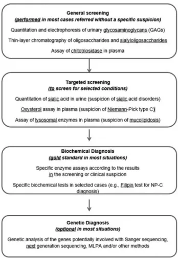
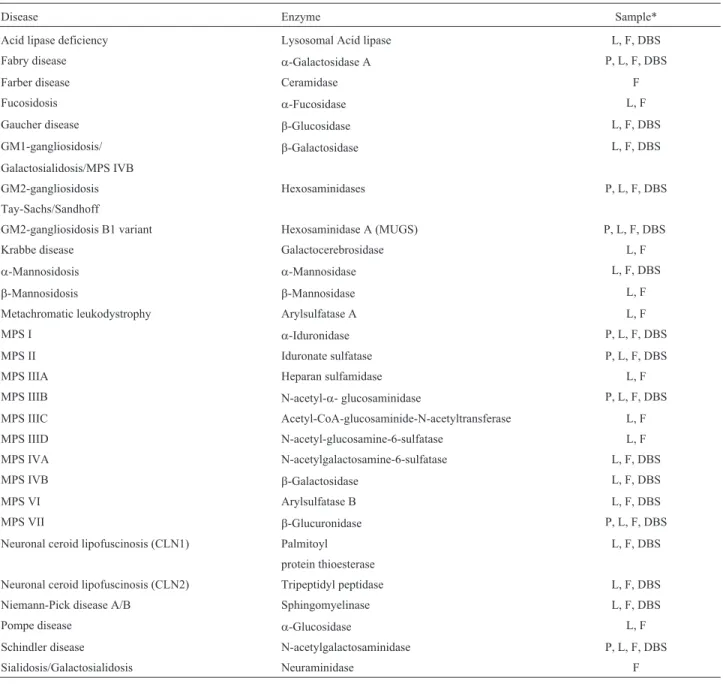
Patient and laboratory records from individuals who had been diagnosed with an LSD at MGS/HCPA from 1982 to 2015 were analyzed. For some cases, the LSD investiga-tion was initiated by urine screening, with quantitainvestiga-tion and electrophoresis of urinary glycosaminoglycans (GAGs). Thin-layer chromatography of oligosaccharides and sialy-loligosaccharides, chitotriosidase assays in plasma, and other selected procedures were performed according to clinical suspicion. The diagnoses were usually confirmed by specific fluorimetric, colorimetric or radioisotopic en-zyme assays and/or by identification of pathogenic muta-tions, typically from blood samples (dried blood spots-DBS, plasma, leukocytes), and, when necessary, us-ing fibroblasts cultivated from skin biopsies. When only DBS were available, enzyme assays were considered diag-nostic when performed at least twice (in two independent samples). When necessary (mainly during the earlier years of the study period), the samples were sent to reference lab-oratories in other countries for complementary analyses. A laboratory workflow chart is shown in Figure 1, and the en-zyme assays performed in the laboratory are listed in Table 1.
Since 1988, prenatal diagnosis has been offered for those families for which a previous LSD diagnosis was well established in an index case, or for previously identified heterozygous couples (or for women carrying mutations for an X-linked disorder).
Results
From 1982 to 2015, 72,797 high-risk patients were investigated for IEM, as referred by several services from different regions of Brazil, other countries in Latin Amer-ica, and occasionally Africa or Asia. During this period, an IEM diagnosis was confirmed in 4,489 (6.44% of all pa-tients investigated) cases, and of these papa-tients, 3,211 had LSDs (71.6% of the IEM cases and 4.41% of all patients in-vestigated). From these 3,211 cases for which an LSD diag-nosis was confirmed, 3,099 were from Brazil. The number of cases diagnosed according to the Brazilian state of origin is shown in Table 2, and the distribution of these diagnoses, including the percentage of the Brazilian population living in each region, is shown in Figure 2. The origins of samples sent from foreign countries are shown in Table 3.
Considering only the 3,099 confirmed Brazilian cases, the most common LSDs diagnosed (over 50 cases each) were Gaucher disease (725 cases), MPS II (343 cases), MPS VI (238 cases), MPS I (225 cases), acid sphin-gomyelinase deficiency/ASMD (199 cases), MPS IVA (153 cases), GM1 gangliosidosis (176 cases), Niemann-Pick C (150 cases), Metachromatic Leukodystrophy (150 cases), Tay-Sachs disease (122 cases), Fabry disease (104
cases), MPS IIIB (88 cases), Krabbe disease (96 cases), MPS IIIA (53 cases), and MPS IIIC (52 cases). It is interest-ing to note that of all patients diagnosed with Tay-Sachs disease, 44% were of the B1 variant. The number of diag-nosed cases for each LSD from the 1982-2015 period is shown in Table 4.
One-hundred twenty cases were evaluated by prena-tal diagnosis since 1988, and a positive result was found in 34 pregnancies (28.3%). The majority of prenatal diagno-ses were in pregnancies at risk for GM1 gangliosidosis (37 cases), Mucopolysaccharidosis type I (16 cases), Tay-Sachs diseases (14 cases), Mucopolysaccharidosis type II (12 cases) or Metachromatic Leukodystrophy (10 cases).
Figure 3 shows the number of diagnosed patients by time period, divided as follows: 1982/1991 - implementa-tion of diagnostic methods, few enzyme assays performed; 1992/1999 - growing number of enzyme assays performed in the laboratory; 2000/2007 - introduction of enzyme as-says using dried blood spots, establishment of the MPS Brazil Network to facilitate diagnosis; and 2008/2015 -new screening protocols for LSDs in high-risk patients, im-plementation of the LSD Brazil network. The number of di-agnoses increased significantly when we compared the first ten years (196 cases) with the next 24 years (3,019 cases) of the study period. In the last 16 years, approximately 160 new cases were identified per year.
Table 1- Specific enzyme assays performed for the diagnosis of LSDs in MGS/HCPA.
Disease Enzyme Sample*
Acid lipase deficiency Lysosomal Acid lipase L, F, DBS
Fabry disease a-Galactosidase A P, L, F, DBS
Farber disease Ceramidase F
Fucosidosis a-Fucosidase L, F
Gaucher disease b-Glucosidase L, F, DBS
GM1-gangliosidosis/ b-Galactosidase L, F, DBS
Galactosialidosis/MPS IVB
GM2-gangliosidosis Hexosaminidases P, L, F, DBS
Tay-Sachs/Sandhoff
GM2-gangliosidosis B1 variant Hexosaminidase A (MUGS) P, L, F, DBS
Krabbe disease Galactocerebrosidase L, F
a-Mannosidosis a-Mannosidase L, F, DBS
b-Mannosidosis b-Mannosidase L, F
Metachromatic leukodystrophy Arylsulfatase A L, F
MPS I a-Iduronidase P, L, F, DBS
MPS II Iduronate sulfatase P, L, F, DBS
MPS IIIA Heparan sulfamidase L, F
MPS IIIB N-acetyl-a- glucosaminidase P, L, F, DBS
MPS IIIC Acetyl-CoA-glucosaminide-N-acetyltransferase L, F
MPS IIID N-acetyl-glucosamine-6-sulfatase L, F
MPS IVA N-acetylgalactosamine-6-sulfatase L, F, DBS
MPS IVB b-Galactosidase L, F, DBS
MPS VI Arylsulfatase B L, F, DBS
MPS VII b-Glucuronidase P, L, F, DBS
Neuronal ceroid lipofuscinosis (CLN1) Palmitoyl L, F, DBS
protein thioesterase
Neuronal ceroid lipofuscinosis (CLN2) Tripeptidyl peptidase L, F, DBS
Niemann-Pick disease A/B Sphingomyelinase L, F, DBS
Pompe disease a-Glucosidase L, F
Schindler disease N-acetylgalactosaminidase P, L, F, DBS
Sialidosis/Galactosialidosis Neuraminidase F
Considering the period from 2000 to 2013 (for which the number of live births is available in the Brazilian Health System Database), 2,092 patients were diagnosed with an LSD. During this period, 41,719,041 live births occurred with 1 case of LSD per 19,942 live births. The minimal fre-quency estimated for each LSD in Brazil is presented in Ta-ble 5.
Discussion
As there are only few other laboratories investigating selected LSDs in Brazil, it is not possible to say that the re-sults presented in this report represent the overall data for LSDs in Brazil. However, as shown in Table 2 and Figure 2, it is clear that the reference laboratory covers the whole
Figure 2- Percentage of LSDs diagnoses from different Brazilian regions (percentage of the Brazilian population living the region indicated be-tween parenthesis).
Table 2- Number of cases diagnosed from each Brazilian state, consider-ing the 3,038 patients for whom this information was available.
Region/States Number of patients diagnosed with LSD
South Region
Paraná 172
Santa Catarina 82
Rio Grande do Sul 539
Total 793
Southeast Region
Minas Gerais 248
São Paulo 934
Rio de Janeiro 246
Espirito Santo 52
Total 1480
Centerwest Region
Mato Grosso 2
Goias 50
Distrito Federal 76
Mato Grosso do Sul 1
Total 129
Northeast Region
Maranhão 29
Ceará 105
Piauí 17
Rio Grande do Norte 31
Paraíba 55
Pernambuco 157
Alagoas 39
Sergipe 0
Bahia 139
Total 572
North Region
Acre 3
Rondônia 0
Amazonas 25
Pará 36
Roraima 0
Amapá 0
Total 64
TOTAL 3038
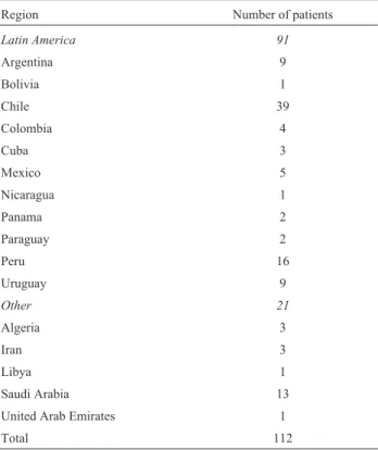
Table 3- Number of LSD diagnoses in patients from foreign countries*.
Region Number of patients
Latin America 91
Argentina 9
Bolivia 1
Chile 39
Colombia 4
Cuba 3
Mexico 5
Nicaragua 1
Panama 2
Paraguay 2
Peru 16
Uruguay 9
Other 21
Algeria 3
Iran 3
Libya 1
Saudi Arabia 13
United Arab Emirates 1
Total 112
country, as cases were identified in all Brazilian regions. The cases were nearly evenly distributed among the Brazil-ian regions, with a relative overrepresentation of the South and Southeast (possibly due to the location of the reference
laboratory and the increased availability of better health system facilities in these regions) and a relative underre-presentation of the Northeast, Center-West and North (pos-sibly due to health system deficiencies and/or logistics difficulties in sending samples to the reference laboratory).
The continuous introduction of diagnostic methods during the study period, such as specific enzyme assays, may explain the increased diagnostic rates during the later years. Additionally, the incorporation of enzyme assays performed using DBS in the first years of this century sim-plified the collection and shipment of samples and may have also played a role in increasing LSD diagnoses, even in services located in distant regions and/or in foreign coun-tries. Additionally, some enzyme assays were introduced only more recently, such as for instance the lysosomal acid lipase assay, which has only been available since 2012.
In a previous study published by our group, Gaucher disease and GM1 gangliosidosis were the conditions with the highest incidence in our population (Coelho et al., 2001). At the time that study was published, no specific treatment for MPS was available. In the present investiga-tion, we can see that MPS I, MPS II and MPS VI are also among the most frequent LSDs diagnosed. Similar results have also been observed in Colombia, where this group of diseases, mainly represented by MPS, is more frequently reported (Barrera and Uribe, 1994). This finding may re-flect a higher awareness of referring health professionals on diseases that have specific treatment available.
LSDs were detected in 4.41% of all samples referred for IEM investigation, representing the most frequent IEM group in our service (71.6% of IEM diagnosis). It is impor-tant to emphasize that our laboratory was the first to offer specific diagnosis of LSD in Brazil, and is also the most complete LSD laboratory in the country. It soon became recognized as a specialized center for these conditions, which may explain the high proportion of this group of dis-orders among the cases of diagnosed IEM. We have to
Figure 3- Number of lysosomal storage diagnosis by period (1982 to 2015).
Table 4- Lysosomal storage diseases diagnosed from 1982 to 2015 in Brazilian patients*.
Lysosomal storage disease Number of con-firmed diagnoses
Additional prob-able diagnosis**
Mucopolysaccharidoses
Mucopolysaccharidosis type I 225 11 Mucopolysaccharidosis type II 343 4 Mucopolysaccharidosis type IIIA 52
-Mucopolysaccharidosis type IIIB 88 -Mucopolysaccharidosis type IIIC 52
-Mucopolysaccharidosis type IVA 153 -Mucopolysaccharidosis type IVB 13 -Mucopolysaccharidosis type VI 238 3
Mucopolysaccharidosis type VII 20 -Multiple sulphatase deficiency 6 -Glycoproteinoses
Aspartylglucosaminuria 1
-Fucosidosis 4
-Galactosialidosis 19
-a-Mannosidosis 7 1
Mucolipidosis II/III 41 8
Sialidosis 14
-Sphingolipidoses
Fabry disease 104 3
Gaucher disease 725 2
GM1 gangliosidosis 175
-GM2 Tay-Sachs disease (44% B1) 121 3
GM2 Sandhoff disease 28
-Krabbe disease 96
-Metachromatic Leukodystrophy 150
-Niemann-Pick type A/B disease 199 5
Other LSDs
Lysosomal acid lipase deficiency 10 7 Neuronal Ceroid lipofuscinosis 1
(CLN1)
3
-Neuronal Ceroid lipofuscinosis 2 (CLN2)
14 3
Niemann-Pick type C 150
-Pompe disease 47 9
Salla disease 1
-Total 3099 59
* Classified as proposed by Kingmaet al., 2015).
Giugliani
et
al.
Brazil1 Australia2 The
Netherlands3
British Columbia3
Portugal3 Czech
Republic3
Eastern Province of Saudi Arabia2
United Arab Emirates3
Disease Clinical phenotype Present study Meikle
et al., 1999
Poorthuis et al., 1999
Applegarth et al., 2000
Pinto et al., 2004
Poupetova et al., 2010
Moammar et al., 2010
Al-Jasmi et al., 2013
a-N-Acetylgalactosaminidase deficiency Schindler disease; Kanzaki dis-ease
0.20 0 0
Acid lipase deficiency Cholesterol ester storage
dis-ease; Wolman disease
0.011 0.19 0.58 0.27 1.0 0
Aspartylglucosaminuria 0 0.05 0.13 1.72 0
Cystinosis 0.52 0.68 1.0 0.25
Danon disease
Fabry disease Fabry disease 0.22 0.85 0.21 0.29 0.12 0.52 5.0 0.25
Farber lipogranulomatosis Farber disease 0.96
Fucosidosis 0.004 0.05 0 0 2.02
Galactosialidosis types I/II 0.02 0.04 0.39 0.77 0 1.0 0
Gaucher disease Gaucher disease 1.43 1.75 1.16 0.39 1.35 1.13 0.25
Globoid cell leukodystrophy Krabbe disease 0.14 0.71 1.35 0.29 1.21 0.4 0
Glycogen storage disease II Pompe disease 0.10 0.68 2.00 0.87 0.7 0.37 2.66
GM1-gangliosidosis types I/II/III 0.18 0.26 0.41 0.19 0.62 0.26 2.0 4.66
GM2-gangliosidosis type AB
GM2-gangliosidosis type I (B variant) Tay-Sachs disease 0.21 0.50 0.41 0.29 3.13 0.3 0.74
GM2-gangliosidosis type II (O variant) Sandhoff disease 0.04 0.26 0.34 0.19 1.49 0.19 5.0 1.21
a-Mannosidosis 0.016 0.09 0.09 0.12 0.38 1.0 1.51
b-Mannosidosis 0.13 0.12 0.16 0
Metachromatic leukodystrophy 0.21 1.09 1.42 0.58 1.85 0.69 1.5
Mucolipidosis type I Sialidosis types I/II
Mucolipidosis types II/III I-cell disease; pseudo-Hurler
polydystrophy
0.06 0.31 0.24 0.29 2.7 0.22 1.35
Mucolipidosis type IIIC pseudo-Hurler polydystrophy
Mucolipidosis type IV
MPS I Hurler; Hurler-Scheie; Scheie
syndrome
0.31 1.14 1.19 0.58 1.33 0.72 4.0 0.25
MPS II Hunter Syndrome 0.71 0.74 0.65 0.10 1.09 0.43 0
MPS IIIA Sanfilippo syndrome 0.08 0.88 1.16 0.29 0 0.47 0
of
lysosomal
diseases
in
Brazil
37
Brazil1 Australia2 The
Netherlands3
British Columbia3
Portugal3 Czech
Republic3
Eastern Province of Saudi Arabia2
United Arab Emirates3
Disease Clinical phenotype Present study Meikle
et al., 1999
Poorthuis et al., 1999
Applegarth et al., 2000
Pinto et al., 2004
Poupetova et al., 2010
Moammar et al., 2010
Al-Jasmi et al., 2013
MPS IIIC Sanfilippo syndrome 0.09 0.07 0.21 0.12 0.42
MPS IIID Sanfilippo syndrome 0 0.09 0.10 0 0
MPS III (all types) Sanfilippo syndrome 0.29 2.0
MPS IVA Morquio syndrome 0.21 0.59 0.22 0.48 0.6 0.71 1.41
MPS IVB Morquio syndrome 0.016 0.14 0.02
MPS IV (both types) Morquio syndrome 0.22 4.0
MPS VI Maroteaux-Lamy syndrome 0.37 0.43 0.15 0.48 0.42 0.05 8.0 2.51
MPS VII Sly syndrome 0.026 0.05 0.24 0.29 0 0.02
MPS IX 0
Multiple sulphatase deficiency 0.011 0.07 0.05 0.10 0.48 0.26 0
Neuronal Ceroid Lipofuscinosis 1 (CLN1) Santavuori disease 0.0024
Neuronal Ceroid Lipofuscinosis 2 (CLN2) Jansky-Bielschowsky disease 0.02
Neuronal Ceroid Lipofuscinosis 3 (CLN3) Batten disease 5.0
Neuronal Ceroid Lipofuscinosis 5 (CLN5) Finnish variant late-infantile neuronal ceroid lipofuscinosis
Neuronal Ceroid Lipofuscinosis 6 (CLN6) Variant late-infantile neuronal ceroid lipofuscinosis
Neuronal Ceroid Lipofuscinosis 8 (CLN8) Northern epilepsy
Niemann-Pick type A/B Niemann-Pick disease 0.33 0.40 0.53 0.6 0.33 5.0 0.25
Niemann-Pick type C Niemann-Pick disease 0.304 0.47 0.35 2.2 0.91 1.0 0.25
Prosaposin deficiency Atypical Gaucher disease
Pycnodysostosis
Sialic acid storage disease Infantile free sialic acid storage disease; Salla disease
0.0024 0.19 0.07 0.19 0.02 0
Sialidosis 0.02 0.02 0.05 0 0.07 0
Sialuria
1
Total number of cases diagnosed from 2000 to 2013 (14 years) divided by the total number of births in the same period.
2Total number of diagnosed cases within a certain period of time divided by the total number of births in the same period.
3
highlight that LSDs are clinically more evident to physi-cians due to their phenotypic appearance, and are, thus, more promptly suspected than other IEM.
Due to the rarity of the LSDs and the relatively so-phisticated methods required for their diagnosis, the identi-fication of these conditions represents a challenge to clinicians, especially in developing countries. Aiming to improve the access of families and health professionals to information and diagnosis of LSDs, an innovative project was set up in Brazil, initially for MPS and later for other LSDs. In 2004, a partnership among Brazilian medical ser-vices that addresses MPS patients was created, the MPS Brazil Network, with headquarters at MGS/HCPA. Since then, the network provides a wide range of information about MPS, performs the laboratory tests necessary for the diagnosis, and organizes regular meetings in order to keep families updated with the most recent advances in the field. The network’s objective is not only to provide the tests for diagnosis, but also to support research, courses, workshops and training for other services interested in MPS to support earlier diagnosis of these conditions. This initiative is sup-ported with public and private grants, which enable it to provide the services free of charge to the requesting physi-cian, making information and diagnostic tests available even for families that usually do not have access to sophisti-cated healthcare facilities (Giugliani, 2012b). Since the MPS Brazil Network initiated its activities, the average number of patients diagnosed with MPS by year has dou-bled. The success of this template in MPS diagnosis has stimulated the creation of a similar network for Niemann-Pick type C (the NPC Brazil Network) and for other lyso-somal diseases (the LSD Brazil Network). This model was probably a leading factor for the significant increase in the number of cases diagnosed in the 2000-2007 and 2008-2015 periods compared to the diagnoses made in the 1982-1991 and 1982-1991-1999 periods, as shown in Figure 3.
These results indicate that LSDs, although individu-ally rare, may be frequent when the investigation is concen-trated in reference laboratories, with 1 out of 22 patients identified with an LSD among the cases referred for suspi-cion of an inherited metabolic disease. The relative fre-quency of LSDs, as shown in Table 3, provides a useful guideline for health authorities to plan the care of these pa-tients because there are specific therapies available for many of the most frequent conditions. Although the mini-mal frequency for each LSD displayed in Table 5 may represent an underestimate (as many cases are still undiagnosed and include data from only one of the diagnos-tic centers), this is the first attempt to make this estimate for Brazil. A large number of cases enables centers to obtain experience in managing these conditions, to perform natu-ral history studies, and to participate in clinical trials. It is important to mention that the majority of patients that were identified could benefit from the therapeutic alternatives
that are already available for these conditions, or from those which are being developed.
Acknowledgments
The authors would like to thank the biologists, bio-chemists, biomedical, undergraduate and graduate students who contributed along 34 years to the work developed at our center, as well as all physicians who referred the pa-tients to be included in this sample. We are also grateful to the foreign laboratories that kindly performed confirmatory analyses in selected cases. The laboratory acknowledges the support received from CNPq, CAPES, FAPERGS, FIPE/HCPA, FAURGS, Fundação Médica do RS, and sev-eral non-governmental and private entities that contributed to the diagnostic activities that were developed.
References
Al-Jasmi FA, Tawfig N, Berniah A, Ali BR, Taleb M, Hertecant JL, Bastaki F and Souid AK (2013) Prevalence and novel mutations of lysosomal storage disorders in United Arab Emirates: LSD in UAE. JIMD Rep 10:1-9.
Applegarth DA, Toone JR and Lowry RB (2000) Incidence of in-born errors of metabolism in British Columbia, 1969-1996. Pediatrics 105:e10.
Barrera LA and Uribe A (1994) Errores innatos del metabolismo (EIM) Ocho años de investigacion em Colombia. Proceed-ings of the 11th Latin American Congress of Genetics. Puerto Vailarta, Mexico.
Coelho JC, Burin MG, Wajner M, Vargas C, Souza FTS and Giugliani R (2001) Selective screning of 18,000 high-risk Brazilian patients for the detection of inborn erros of metab-olism. Rev HCPA 21:286-292.
Fuller M, Meikle PJ and Hopwood JJ (2006) Epidemiology of lysosomal storage disease: An overview. In: Mehta A, Beck M and Sunder-Plassman G (eds) Fabry Disease: Perspec-tives from 5 Years of FOS. Oxford PharmaGenetics, Ox-ford, pp 9-20.
Futerman AH and van Meer G (2004) The cell biology of lysosomal storage disorders. Nat Rev Mol Cell Biol 5:554-565.
Giugliani R (2012a) Newborn screening for lysosomal diseases: Current status and potential interface with population medi-cal genetics in Latin America. J Inherit Metab Dis 35:871-877.
Giugliani R (2012b) Mucopolysacccharidoses: From understand-ing to treatment, a century of discoveries. Genet Mol Biol 35(4 Suppl):924-931.
Hoffmann GF (1994) Selective screening for inborn errors of me-tabolism-past, present and future. Eur J Pediatr 153(Suppl 1):S2-S8.
Kingma SD, Bodamer OA and Wijburb FA (2015) Epidemiology and diagnosis of lysosomal storage disorders; challenges of screening. Best Pract Res Clin Endocrinol Metab 29:145-157.
Meikle PJ, Hopwood JJ, Clague AE and Carey WF (1999) Preva-lence of lysosomal storage disorders. JAMA 281:249-254. Moammar H, Cheriyan G, Mathew R and Al-Sannaa N (2010)
Eastern Province of Saudi Arabia, 1983-2008. Ann Saudi Med 30:271.
Pinto R, Caseiro C, Lemos M, Lopes L, Fontes A, Ribeiro H, Pinto E, Silva E, Rocha S, Marcão A,et al.(2004) Preva-lence of lysosomal storage diseases in Portugal. Eur J Hum Genet 12:87-92.
Poorthuis BJ, Wevers RA, Kleijer WJ, Groener JE, de Jong JG, van Weely S, Niezen-Koning KE and van Diggelen OP (1999) The frequency of lysosomal storage diseases in The Netherlands. Hum Genet 105:151-156.
Poupetová H, Ledvinová J, Berná L, Dvoráková L, Kozich V and Elleder M (2010) The birth prevalence of lysosomal storage
disorders in the Czech Republic: Comparison with data in different populations. J Inherit Metab Dis 33:387-396. Vellodi A (2005) Lysosomal storage disorders. Br J Haematol
128:413-431.
Wilcox WR (2004) Lysosomal storage disorders: The need for better pediatric recognition and comprehensive care. J Pediatr 144:S3-S14.
Associate Editor: Carlos F. M. Menck