Serotonergic neurons in the caudal
raphe nuclei discharge in association
with activity of masticatory muscles
1Departamento de Fisiologia e Biofísica, Instituto de Ciências Biomédicas,
Universidade de São Paulo, 05508-900 São Paulo, SP, Brasil L.E. Ribeiro-do-Valle1
Abstract
There is a dense serotonergic projection from nucleus raphe pallidus and nucleus raphe obscurus to the trigeminal motor nucleus and serotonin exerts a strong facilitatory action on the trigeminal motoneu-rons. Some serotonergic neurons in these caudal raphe nuclei increase their discharge during feeding. The objective of the present study was to investigate the possibility that the activity of these serotonergic neurons is related to activity of masticatory muscles. Cats were implanted with microelectrodes and gross electrodes. Caudal raphe single neuron activity, electrocorticographic activity, and splenius, digastric and masseter electromyographic activities were recorded during active behaviors (feeding and grooming), during quiet waking and during sleep. Seven presumed serotonergic neurons were identi-fied. These neurons showed a long duration action potential (>2.0 msec), and discharged slowly (2-7 Hz) and very regularly (interspike interval coefficient of variation <0.3) during quiet waking. The activ-ity of these neurons decreased remarkably during fast wave sleep (78-100%). Six of these neurons showed tonic changes in their activity positively related to digastric and/or masseter muscles activity but not to splenius muscle activity during waking. These data are consistent with the hypothesis that serotonergic neurons in the caudal raphe nuclei play an important role in the control of jaw movements.
Correspondence
L.E. Ribeiro-do-Valle Departamento de Fisiologia e Biofísica
Instituto de Ciências Biomédicas Universidade de São Paulo 05508-900 São Paulo, SP Brasil
E-mail: ribeiro@bmb.icb1.usp.br
Research supported by FAPESP and CNPq.
Received June 4, 1996 Accepted November 11, 1996
Key words
•Masticatory muscles
•Serotonergic neurons
•Caudal raphe nuclei
•Feeding behavior
•Grooming behavior
The trigeminal motor nucleus receives a dense serotonergic projection (1,2) which originates from nucleus raphe pallidus, nucleus raphe obscurus and, to a lesser ex-tent, from nucleus raphe dorsalis (3,4). Sero-tonergic fibers terminate in close proximity to the cell body and proximal processes of trigeminal motoneurons (1,5). Serotonin was shown to strongly facilitate trigeminal moto-neurons (6,7). These findings suggest an im-portant role for the serotonergic system in
the control of jaw muscles activity.
It is well known that the activity of the serotonergic neurons in the raphe nuclei is related to the level of arousal of the organism (for a review, see Ref. 8). Thus, the tonic serotonergic influence on the trigeminal motoneurons during waking may contribute to maintaining jaw position and facilitating jaw movements during feeding, grooming, and defense/aggression behaviors.
neurons in nucleus raphe dorsalis selectively increase their activity during feeding and grooming behaviors (9,10). Many serotoner-gic neurons in nucleus raphe pallidus and nucleus raphe obscurus behave similarly (11). These results raise the interesting possibility that there may be additional changes in the serotonergic influence on the trigeminal mo-toneurons associated with the activation of these motoneurons by central mechanisms during behaviors that involve oral move-ments. In the present study we investigated this hypothesis.
Adult cats of either sex were used. Hous-ing conditions of the animals and the exper-imental protocol were reviewed and approved by the Comissão de Fiscalização de Pesquisa com Animais (COFIPA) of São Paulo City. For single neuron recording the animals were stereotaxically implanted with a microdrive consisting of two inner cannulas that could be moved along outer guide cannulas by turning a small screw. Two bundles of 6 microelectrodes (32- and 64-µm diameter Formvar-insulated nichrome wires) were lowered through the inner cannulas to the coordinates AP -11.6 and -12.6, LM ±0.0 and DV -0.2 of the Snider and Niemer (12) atlas of the cat brain and then glued to the top of these cannulas. Gross electrodes were implanted into the digastric, masseter and splenius muscles (the digastric and masseter muscles are the major opening and jaw-closing muscles, respectively) and threaded into the parietal, frontal, temporal and retroorbital bones to record muscular activ-ity (EMG), cortical activactiv-ity (EEG) and eye movements (EOG). The microdrive and elec-trode implantation procedures are described in detail by Heym et al. (13).
Testing was initiated after a recovery period of 2 weeks following surgery. All microelectrodes were screened 2 to 3 times a day for the presence of stable, isolated single-unit activity (signal to noise ratio >3). In the absence of this activity the microelectrodes were advanced in small steps (about 80 µm)
by moving the microdrive screw 1/4 turn. When a recordable spike was found the ani-mals were further tested. Neuronal activity as well as the EEG and 3 EMG were re-corded during quiet waking, slow wave sleep and fast wave sleep, and while the subject was exhibiting spontaneous behaviors such as feeding and grooming. All signals were recorded on paper and on videotape. Sponta-neous behaviors of the subjects and neuronal activity were recorded together on another videotape.
After advancing the microelectrodes about 5 mm into the brainstem, the animals were sacrificed and their hindbrain was pro-cessed histologically to locate the recorded neurons (for details see Ref. 13).
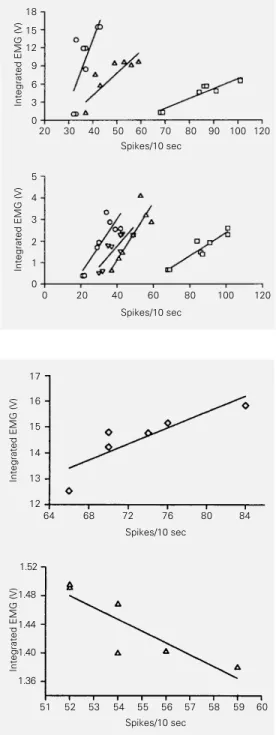
over a period of 1 msec. b) A possible gross tonic relationship between neuronal activity and muscular activity was determined by correlating the number of spikes per two 10-sec epochs of quiet waking, drinking milk, licking lips and licking forelimbs/washing face with the corresponding integrated mus-cular voltages. c) A possible fine tonic rela-tionship between neuronal activity and muscular activity was examined by correlat-ing the number of spikes per six 10-sec epochs of drinking milk with the corresponding in-tegrated muscular voltages. (For both b) and c) the Spearman rank correlation test was used.) Seven neurons that showed a long-dura-tion aclong-dura-tion potential (>2.0 msec) and slow (2 to 7 Hz) and very regular (interspike interval coefficient of variation <0.3) discharge dur-ing quiet wakdur-ing were recorded in two ani-mals. All of them systematically changed their activity across the wake-sleep cycle. The firing rate decreased from quiet waking to slow wave sleep (the respective means ± SEM for six of these neurons were: 3.86 ± 0.73 and 3.15 ± 0.76 Hz; P = 0.02) and from slow wave sleep to fast wave sleep (the respective means ± SEM for the same six neurons were: 3.15 ± 0.76 and 0.24 ± 0.17 Hz; P = 0.02). One of these neurons was located in the dorsal third, 2 in the middle third and the remaining 4 in the ventral third of the medulla, in a stripe extending 0.5 mm to each side of the midline between the caudal and the rostral poles of the inferior olive. These landmarks delimit reasonably well the rostrocaudal extension of nucleus raphe pallidus and nucleus raphe obscurus in the cat (14). These neurons were classi-fied as serotonergic on the basis of these functional characteristics and anatomical location (for criteria for identification of serotonergic neurons see Ref. 10).
Six neurons could be evaluated during a 60-sec period of drinking milk. None of them exhibited phasic changes in its activity related to muscle activity.
All seven neurons were evaluated during
quiet waking, drinking milk, licking lips and licking forelimbs/washing face. The dis-charge of these neurons tended to increase from quiet waking to licking lips to licking forelimbs/washing face and drinking milk. Muscle activity changed considerably from one behavior to another. Digastric muscle activity tended to increase from quiet wak-ing to lickwak-ing forelimbs/washwak-ing face to lick-ing lips and drinklick-ing milk; masseter muscle activity tended to increase from quiet wak-ing to lickwak-ing lips to drinkwak-ing milk and lick-ing forelimbs/washlick-ing face; and splenius muscle activity tended to increase from quiet waking to drinking milk to licking lips and licking forelimbs/washing face. Five of the neurons changed their firing rate in relation to muscle activity. For 1 neuron the firing rate was related to digastric muscle activity
(rS = 0.77; P = 0.04). For 2 other neurons the
firing rate was related to masseter muscle
activity (rS = 0.79 and rS = 0.79; P = 0.04).
For the last 2 neurons the firing rate was
related to digastric (rS = 0.90 and rS = 0.90; P
= 0.02) and masseter (rS = 0.83 and rS = 0.90;
P = 0.03 and P = 0.02, respectively) muscles activity (Figure 1).
Of the 6 neurons that could be evaluated during a 60-sec period of drinking milk only 2 discharged in association with muscle activity. One fired in relation to digastric muscle
activ-ity (rS = 0.90; P = 0.04) and the other in relation
to splenius muscle activity (rS = -0.88; P =
0.04) (Figure 2). The former neuron did not show any relationship between its activity and digastric muscle (or masseter muscle) activity when several behaviors were considered.
by decreasing resting membrane conductance to potassium ions. We propose that the sero-tonergic input, possibly by modulating the depolarization level of the trigeminal moto-neurons in a tonic and graded way, actively contributes to the determination of the gen-eral intensity of masticatory muscles con-traction appropriate for each behavior.
We observed only one case in six of fine tonic coupling between neuronal firing and masticatory muscles activity but five cases in seven of gross tonic coupling. This obser-vation suggests that the presumed active se-rotonergic modulation of trigeminal moto-neurons excitability may be more important for causing coarse general changes in mus-cular activity than for causing fine ones. It is possible that serotonin specifically facili-tates the recruitment of additional, higher threshold, trigeminal motoneurons by the central pattern generators that control jaw movements.
Also highly significant was the fact that in a sample as small as 7 neurons we could identify 6 neurons whose activity was re-lated to jaw muscles activity. This fact corre-lates well with the anatomical observation of a very high density of serotonergic projec-tions from nucleus raphe pallidus and nucleus raphe obscurus to the trigeminal motor nucleus (1,2). Veasey et al. (11), also using cats, recorded 29 serotonergic neurons in nucleus raphe pallidus and nucleus raphe obscurus. Twelve of these neurons increased their discharge during feeding (licking pu-reed meat and water). The much smaller percentage of serotonergic cells presumably related to the control of jaw movements found in their study could be explained by the fact that the subjects exhibited only one behavior with these movements and prob-ably developed the same general intensity of masticatory muscle contraction.
It is not known whether the serotonergic neurons that supposedly influence mastica-tory muscles activity also participate in the control of other body muscles. Since five of
Figure 1 - Gross relationship be-tween the electrical activity of putative serotonergic neurons and the integrated electrical ac-tivity of two masticatory muscles (digastric muscle, top panel; masseter muscle, bottom panel). Two 10-sec samples were taken during each of the following be-havioral activities: quiet waking, drinking milk, licking lips and lick-ing forelimbs/washlick-ing face. !, neuron #14041; #, neuron #24151;
@
, neuron #14021; , neuron #14173; P, neuron #14194. Correlations were sig-nificant at levels from 0.02 to 0.04.Integrated EMG (V)
18 15 12 9 6 3 0
20 30 40 50 60 70 80 90 100 120 Spikes/10 sec
Integrated EMG (V)
5 4 3 2 1 0
0 20 40 60 80 100 120 Spikes/10 sec
Figure 2 - Fine relationship be-tween the electrical activity of putative serotonergic neurons and the integrated electrical ac-tivity of the digastric muscle (top panel) and the splenius muscle (bottom panel). Six 10-sec samples were taken during feed-ing behavior (drinkfeed-ing milk). p, neuron #14171; #, neuron #24151. In both cases the corre-lation was significant at the 0.04 level.
Integrated EMG (V)
17
16
15
14
13
12
64 68 72 76 80 84 Spikes/10 sec
Integrated EMG (V)
1.52
1.48
1.44
1.40
1.36
51 52 54 57 58 59 Spikes/10 sec
53 55 56 60
Serotonin by itself does not activate lum-bar (15) and facial (16) motoneurons but renders them more responsive to glutamater-gic excitation. Katakura and Chandler (6) reported the same facilitatory action of sero-tonin on digastric motoneurons. According to Vandermaelen and Aghajanian (16), sero-tonin may cause membrane depolarization
the neurons related to the masticatory muscles were not related to the splenius muscle, some degree of specificity is suggested. Only one of 10 feeding-responsive serotonergic cells tested by Veasey et al. (11) was not activated during a treadmill-induced locomotion task. However, their finding is not conclusive be-cause during locomotion jaw position must be actively held against destabilizing inertial and gravitational forces. Serotonergic activ-ity may have been increased by higher ner-vous centers to augment the gain of the stretch reflex of the jaw-closing muscles and, consequently, stiffness of muscles (17). No change in jaw muscles activity phasically related to serotonergic neurons activity was found in the present study. This is not surprising since the action of serotonin on motoneurons has been described to be of
slow onset and to have a prolonged time course (6,15). Related phasic changes in serotonergic neurons activity and physiologi-cal functions were observed only rarely in other studies (11,13).
The results of the present study are in agreement with the views of Jacobs and Azmitia (8) that the serotonergic system has a tonic modulatory role in the nervous sys-tem. Jacobs and Fornal (18) proposed that one of the primary functions of the seroto-nergic system is to facilitate motor output. Our main finding supports this hypothesis.
Acknowledgments
We thank Mr. Roberto Vieira and Mr. Ademar Petri Filho for their valuable techni-cal assistance.
References
1. Steinbusch HWM (1981). Distribution of serotonin-immunoreactivity in the central nervous system of the rat - cell bodies and terminals. Neuroscience, 6: 557-618. 2. Cropper EC, Eisenman JS & Azmitia EC (1984). 5-HT-immunoreactive fibers in the trigeminal nuclear complex of the rat.
Ex-perimental Brain Research, 55: 515-522.
3. Fritschy J-M, Lyons WE, Molliver ME & Grzanna R (1988). Neurotoxic effects of p-chloroamphetamine on the serotoniner-gic innervation of the trigeminal motor nucleus: retrograde transport study. Brain
Research, 473: 261-270.
4. Fort P, Luppi P-H, Sakai K, Salvert D & Jouvet M (1990). Nuclei of origin of monoaminergic, peptidergic, and cholin-ergic afferents to the cat trigeminal motor nucleus: a double-labeling study with chol-era-toxin as a retrograde tracer. Journal of
Comparative Neurology, 301: 262-275.
5. Kolta A, Dubuc R & Lund JP (1993). An immunocytochemical and autoradiograph-ic investigation of the serotoninergautoradiograph-ic in-nervation of trigeminal mesencephalic and motor nuclei in the rabbit. Neurosci-ence, 53: 1113-1126.
6. Katakura N & Chandler SH (1990). An ion-tophoretic analysis of the pharmacologi-cal mechanisms responsible for trigemi-nal motoneurotrigemi-nal discharge during masti-catory like activity in the guinea pig.
Jour-nal of Neurophysiology, 63: 356-369.
7. Ribeiro-do-Valle LE, Metzler CW & Jacobs BL (1991). Facilitation of masseter EMG and masseteric (jaw-closure) reflex by se-rotonin in behaving cats. Brain Research, 550: 197-204.
8. Jacobs BL & Azmitia EC (1992). Structure and function of the brain serotonin sys-tem. Physiological Reviews, 72: 165-229. 9. Ribeiro-do-Valle LE, Fornal CA, Litto WJ & Jacobs BL (1989). Serotonergic dorsal raphe unit activity related to feeding/ grooming behaviors in cats. Society for
Neuroscience Abstracts, 15: 1283.
10. Fornal CA, Metzler CW, Marrosu F, Ribeiro-do-Valle LE & Jacobs BL (1996). A subgroup of dorsal raphe serotonergic neurons in the cat are strongly activated during oral-buccal movements. Brain
Re-search, 716: 123-133.
11. Veasey SC, Fornal CA, Metzler CW & Jacobs BL (1995). Response of seroton-ergic caudal raphe neurons in relation to specific motor activities in freely moving cats. Journal of Neuroscience, 15: 5346-5360.
12. Snider RS & Niemer WT (1970). A
Stereo-taxic Atlas of the Cat Brain. University of
Chicago, Chicago.
13. Heym J, Steinfels GF & Jacobs BL (1982). Activity of serotonin-containing neurons in the nucleus raphe pallidus of freely moving cats. Brain Research, 251: 259-276.
14. Jacobs BL, Gannon PJ & Azmitia EC (1984). Atlas of serotonergic cell bodies in the cat brainstem: an immunocytochemi-cal analysis. Brain Research Bulletin, 13: 1-31.
15. White SR & Neuman RS (1980). Facilita-tion of spinal motoneurone excitability by 5-hydroxytryptamine and noradrenaline.
Brain Research, 188: 119-127.
16. Vandermaelen CP & Aghajanian GK (1982). Serotonin-induced depolarization of rat facial motoneurons in vivo: compari-son with amino acid transmitters. Brain
Research, 239: 139-152.
17. Lund JP, Drew T & Rossignol S (1984). A study of jaw reflexes of the awake cat during mastication and locomotion. Brain
Behavior and Evolution, 25: 146-156.
18. Jacobs BL & Fornal CA (1993). 5-HT and motor control: a hypothesis. Trends in