5º CONGRESSO NACIONAL EM BIOMECÂNICA
5
thPortuguese Congress on Biomechanics
Fórum de Cultura e Arte de Espinho
–
FACE
Espinho
–
8 e 9 de Fevereiro de 2013
~ PROGRAMA ~
4
2
Organização do CNB2013 |
CNB2013 Organization2.1
Comité Organizador |
Organizing Committee Renato M. Natal Jorge, FEUP João Manuel R.S. Tavares, FEUP
Jorge Américo de Oliveira Pinto Belinha, IDMEC-FEUP Marco Paulo Lages Parente, IDMEC-FEUP
Pedro Alexandre Lopes de Sousa Martins, IDMEC-FEUP
2.2
Comité Cientifico |
Scientific Committee Adélia Sequeira, IST-UTL
António Completo, Universidade de
Aveiro
António Silva, UTAD
António Torres Marques, FEUP
António Veloso, FMH-UTL
Arcelina Marques, ISEP-IPP
Aurélio Faria, UBI
Cristina Mateo Martinez, IDMEC
Eduardo Borges Pires, IST-UTL
Elza Fonseca, IPB
Fernanda Gentil, IDMEC, ESTSP-IPP
Fernando Simões, IST-UTL
Filipa Manuel Machado Sousa
Gilberto Costa, FMUP
Helena Moreira
João Folgado, IST-UTL
João Levy Melancia, FML, U. Lisboa
João MCS Abrantes, U. Lusófona
João Paço, Hospital CUF, FML-U. Lisboa
João Paulo Flores Fernandes, U. Minho
João Paulo Vilas-Boas, FADEUP
João Santos Baptista , FEUP
Joaquim Silva Gomes, FEUP
Jorge Ambrósio, IST-UTL
José Alberto Ramos Duarte, FADEUP
José Carlos Reis Campos, FMDUP
José Manuel Casanova, FM, U.Coimbra
José Oliveira Simões, U. Aveiro
Kostas Gianikellis, U.Extremadura-España
Leandro Machado, FADEUP
Lidia Carvalho, UA
Luís Roseiro, ISEC-IPC
Luisa Sousa, FEUP
Manuel Gutierres, FMUP
Mário Augusto Vaz, INEGI, FEUP
Mario Forjaz Secca, FCT-UNL
Miguel Tavares da Silva, IST-UTL
Miguel Velhote Correia, FEUP
Paulo Piloto, IPB
Paulo Rui Fernandes, IST-UTL
Ronaldo Eugénio Calçada Dias Gabriel
Rui Barreiros Ruben, IPL
Rui Lima, IPB
Rui Miranda Guedes, FEUP
Santos Rubim, ESTSP-IPP
2.3
Organização Institucional |
Institutional Party Laboratório de Biomecânica do Porto
Faculdade de Engenharia da Universidade do Porto
Instituto de Engenharia Mecânica - Pólo FEUP
Instituto de Engenharia Mecânica e Gestão Industrial
6
Programa Detalhado |
Detailed Program6.1
Geral |
GeneralSecretariado
Secretariat
Registo no CNB2013
CNB2013 RegistrationSexta-feira, 08 de Fevereiro | Friday, February 8th
08:00
Abertura da área de Registo.
Entrega da documentação aos delegados.
Registration area opening.
Individual documentation delivery.
Auditório
Auditorium
Cerimónia de Abertura
Opening ceremonySexta-feira, 08 de Fevereiro | Friday, February 8th
09:00
Boas vindas aos delegados. Sessão de Abertura presidida por:
Presidente da Sociedade Portuguesa da Biomecânica: Professor Mário Vaz
Com a presença do Presidente da Câmara Municipal de Espinho: Dr. Pinto Moreira
Welcome to all delegates Opening session by:
President of the Portuguese Society of Biomechanics: Professor Mário Vaz With the presence of the President of the Municipal of Espinho: Dr. Pinto Moreira
Auditório
Auditorium
Assembleia Geral da Sociedade Portuguesa de Biomecânica
General Assembly of the Portuguese Society of BiomechanicsSexta-feira, 08 de Fevereiro | Friday, February 8th
16
Sala 2 Room 2
Sessão
Session 2.3
Biomecânica Cardiovascular, Biofluidos e
Hemodinâmica
Cardiovascular and Hemodynamic Bio-fluids Biomechanics Chairs: Henrique Almeida, Luisa Sousa
11:30 3223
Cell-free layer measurements in a bifurcation microchannel comparison between a manual and automatic methods
D. Bento, D. Pinho, E. Pinto, T. Yaginuma, T. Correia, J. Lima, A.I. Pereira, R. Lima
11:45 3232
Enhanced velocity of red blood cells in highly branched vessels: influence of vessels Diameter
B. Pires, L. Pimparel, D. Pinho, R. Lima, R. Dias
12:00 3237
Desenvolvimento de um microdispositivo biomédico para a separação de células sanguíneas
S. Novais, D. Pinho, A.I. Pereira, M. Mujika, S. Arana, R. Lima
12:15 3247
Image filtering, contrast enhancement and deformation analysis of complex anatomical conduits and microcirculation experiments
A.J. Joao, A.M. Gambaturo, A. Sequeira
12:30 3255 Optimização de forma multi-objectivo de um stent coronário N. Ribeiro, J. Folgado, H. Rodrigues
12:45 3306
Numerical study of the flow of a blood analog fluid in a bifurcation with a stenosis: Pulsatile flow and elasticity of the fluid
S.I.S Pinto, E.D. Costa, J.B.L.M. Campos, J.M.R. Miranda.
Sala 3 Room 3
Sessão
Session 2.4
Biomecânica do Sistema Músculo-esquelético
Musculoskeletal System BiomechanicsChairs: António Ramos, Aurélio Faria
11:30 3089
Utilização de técnica termográfica, para determinação de desequilíbrios musculares, durante o transporte de laptop em mochila suportada por um único ombro
J. Rocha, L. Queijo, J. Santos
11:45 3330
Simulação numérica dos danos nos músculos do pavimento pélvico durante um parto vaginal
P. Maia, M.P.L. Parente, R.M. Natal Jorge, A.A. Fernandes
12:00 3041 Análise cinemática da marcha no 3º trimestre de gravidez e pós-parto M. Branco, R. Santos-Rocha, L. Aguiar, F. Vieira, A.P. Veloso
12:15 3059
Relação das forças reativas do apoio durante o caminhar e a atividade física em mulheres pós-menopáusicas
J. Fonseca, R. Gabriel, J. Tavares, F. Aragao, A. Leite, J. Faria, H. Moreira
12:30 3064
Forças reactivas do apoio durante o caminhar e densidade mineral óssea do calcâneo em mulheres pós-menopáusicas
M. Pereira, R. Gabriel, F. Aragão, J. Fonseca, A. Leite, A. Faria, H. Moreira
12:45 3099
Contribuição dos momentos de força do membro inferior no salto unilateral (hopping)
: h P• •I'll 1·11 :I ( Cl'l .j I''S',. 1·1\l\ II'C 11' .11
:-R :,I :- . ll.djnr~· .. l \ 1.1 {.~ T.P .Irt,, I holll ,h .• \ 11.1 .. P.ilclllt.
r·.
\. 1.~. \ 1.111111. , J·.cl·I J"'l' .H•. l'tttl \l . ..!.d. ~ c. ~. t1l I l . rlHIJ .:«•t.:
CELL-FREE LAYER MEASUREMENTS IN A BIFURCATION
MICROCHANNEL: COMPARISON BETWEEN A MANUAL AND AUTOMATIC METHODS
David Bento1, Dimw Pinlto1'1, Elmano Pinto1, Tomoko l'agimmw1, Teresa Correia1•3, Jose Lima1•4,
Ana I. Pereira1'5, Cal'la S. Feruandes1, Ricardo Dia/·1, Rui Lima1'1 1 ESTiG, Po(vtechnic Institute of Braganr;a. IPB, Braganr;a, Portugal;
/ c/rll'it/hl!lll 0.1 tllllflk t1. ( 'I 'l!i!! a / 11 i {)h. /.11 .' ,•/ IIW IIIltJ illf t) 11 /11 )/ lll ((i /. c'O III
~ CEFT. Faculdade de E11ge11haria da Universidade do Porto (FEUP) , Poria, Portugal; ,'dimui.ruim.·c.ricar,/o, /: 11 iph.p/
3CIMOIESA, Po~ v t e c/mi c Institute of Braganr;a. Portugal; Icurreio alioh./ll
4
lNESC. Faculdade de E11genharia da Universidade do Porta (FEUP). Porta, Portugal; j//imo ,, il!h.nt
5
Aigoritmi, U11iversidade do Mi1!110; a(>.:reira•a•iph.(lt
KEYWORDS: Cell-free layer, Bifurcation, Automatic Method.
ABSTRACT: In the present work, in vitro blood flowing through a bifurcation microchannel was studied. The aim was to develop different automatic methods capable to measure the cell-free layer (CFL) thickness in the input and output of the microchannel. The results were
compared with the trajeclories of the CFL obtained manually. The method with the besl results was the me/hod that uses the binarization.
1 INTRODUCTION
Blood flow in microcirculation shows
several hemodynamic phenomenons, in
vivo and in vitro . Hence, over the years in vitro blood studies in microchannels have been extensively performed in order to obtain an understanding of blood rheology and its flow dynamics
11,
2]. The Fahraeus-Lindqvist effect is one of the physical reasons for the tendency of RBCs to migrate toward the centre of the microtube resulting in a marginal cell-free layer (CFL) at regions adjacent to the wallf3
j. Recently several studies showed strong evidence that the formation of the CFL is affected by the geometry of the microchannel [4-7] and the physiological conditions of the working fluid, such as the haematocrit (Het) 18, 91. Until now, most of the experimental data resulted from the studies have been analysed manually. The manual methods can be highly reliable but they are relatively time consuming and can also introduce usererrors into the data. So, as an outcome it is essential to develop image analysis methods able to process the data automatically. In
this point, image analysis plays an
extremely important role to obtain
information about the blood rheology
r
10]. In this study, the experimental phase was performed using a confocal microscopy system. It was used the image analysis techniques to measure several trajectories of the CFL in a microchannel with a diverging and converging bifurcation. In this study it is compare one manual method and two automatic methods developed in Mat LabJ
5 and 6, Conclusion and Future Work respectively.
2
EXPERIMENTAL SET-UPThe confocal micro-PlY system used in this study consists of an inverted microscope
(IX? I; Olympus) combined with a hi oh-o
speed camera (i-SPEED LT). The
microchannel was placed on the stage of the inverted microscope and a pressure-driven flow was kept constant based on a syringe pump (PHD ULTRA). The confocal imaoes 0 have the resolution of 800x600 pixel at a frame rate of lOO frames/s.
The working fluid used in this study was Dextran 40 (Dx-40; Otsuka Medicine) containing 5 and 10% (± 2%) i.e., Het= 5 and 10 by volume of RBCs. The blood was collected from a healthy sheep and was added heparin to prevent clotting. Further, the cells were separated from blood by centrifugation.
3
IMAGEANALYSIS3.1
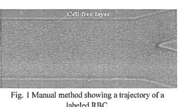
MANUAL METHODA manual tracking plugin (MTrackJ) [6] of an image analysis software (Image J, NIH)
[7] was used to track individual RBC flowing around the boundary of the RBCs core. By using MTrackJ plugin, the
centroid of the selected RBC was
automatically computed.
Fig. l Manual method showing a trajectory of a labeled RBC.
After obtaining
x
and y positions, the data were exported for the determination of each individual RBC trajectory.3.2
AUTOMATI C METHODSAll image sequences were processed using image processing techniques provide by MatLab Ill].
Firstly, a median filter was applied to each frame to remove the noise from the imaoes 0 , by using a 3x3 mask. Then the intensity of each pixel in the frame sequences was evaluated to obtain an image with the maximum intensity. In the Method A, we used the previous image, and found the edges of the channels by using the Canny algorithm ( ll] (Fig. 2 a)). After selected the region of interest, upper and lower CFL trajectories were automaticall y measured. In the case of the Method B, the image with the maximum intensity was converted into a binary image (Fig. 2 b)) , and the upper
and lower CFL trajectories were
automatically measured.
n)
b)
Fi g. 2: Automatic Methods: a) Method A; b) Method B.
4
RESULTS AND DISCUSSIONThe results were taken in the inflow and outflow of the microchannels, Fig. 3. The flow rate used was a 500nllmin and two fluids were studied, respectively with 5 and
10% of Het.
Inflow
I
Flow dUuct1 on
_....
In Table I, the results from the three methods are presented.
Table I - Comparison between the different methods.
Het Manual Automatic Automatic
(/tm) A tJim)
n
(/tm)5% 16.0249 11.6508 22.58 15
Inflow
10 % 11.7096 8.2707 12.2182
5% 14.4679 6.7014 21.321
Outflow
10% 10.7721 3.7087 15.2006
The data obtained from both automatic methods presents some different results from the manual data. However, the results obtained from the Method B, as shown in the Fig. 4, present best results because they have a similar behavior between both.
a ~t am•tl • Autrn!L1tic B • :\lll'""·" ic A
75
70
0
lnOo\\ 5'• lnOow to•, OutOo\\' ~ ·. OutOowiO' o
Fig. 4 Comparison between the manual and the two automatic methods.
The Method A, by applying the Canny filter
along the channel, have a constant
decreases of the CFL thickness and this behavior is not observed in the case of the manual method. Though, in the case of the Method B that use the binarization, presents the same comportment of the manual method. Other result analysis that is possible to observe for the three methods is the CFL decreasi ng from the inflow to the outflow and that the hematocrit has a
considerable influence in the CFL
thickness, increasing the hematocrit the CFL decreases.
5 CONCLUSIONS
The present study indicates that the proposed automatic Method B have the best agreement to data obtained from the manual
From the inflow to the outflow is possible to observe a decreasing of the CFL thickness and the CFL tend to decrease as the Het increases. Hence, the Method B may be a promising way to carry on this study.
6
FUTURE WORKIn this type of study the quality of the image sequence plays a crucial rule and as result we plan to obtain sequence of images with more quality and to use an objective lens with better resolution. Additionally, we also plan to improve the current automatic method to obtain similar results to those obtained manually.
ACKNOWLEDGMENTS
The authors acknowledge the financial support provided by: PTDC/ SAU-BEB/108728/2008, PT DC/SA U-BEB/ I 05650/2008,
PTDC/EMEMFE/0991 09/2008 and PTDC/SA U ENB/116929/2010 from the FCT (Science and Technology Foundation) and COMPETE, Portugal.
REFERENCES
Ill A. A. Pries, D. Ncuhaus, P. Gaclllgcns, ''Blood viscosity in tube flow: dependence on diamete r and hc mutoc rit". Am J Physiol 263, H 1770-H 1778, 1992.
12] H. Goldsmith, G. Cokelet, P. Gaehtgens, " Robin Fahraeus: evolution of hi s concepts in cardiovascular phys iology" Am. J. Physiol., 257, I-11005-Hl00 15, 1989.
13 1 C. Caro, T. Pcdlcy, R. Schro ter, W. Seed, Tir e mechanics oftlte circulation , Oxford University Press, 1978.
141 D. Pinho, Determination ami clwracterbllion of red
Mood cells trajectories: a semi-automatic metltod, Master in
Biomedical Technology, Polytechnic Institute of Brag an~a.
Portugal, 2011 (in portugucsc ).
[51 R. Lima, S. Wada, S. T anaka, M. Takcda, et al , " In vitro
blood flow in a rectangular PDMS microchannel:
experimental observations usi ng a confocal m icro-PlY system", Biomedical Microdcviccs, 10.2. 153 -167, 2008.
16 1 R. Lima, T. Ishikawa, Y. lmai, et al, " Radial dispersion of red blood cells in blood flowing through glass capillaries: role of hematocrit and geometry", J ournal of Biomechanics 41 , 2188-2197, 2008.
171 V. Leble. R. Lima, R. P. Dias, C . S. Fcrnandes, T.
lshikawa, Y. Imai, T . Yamaguchi, "Asymmetry of red blood cell motions in a microchannel with a diverging and converging bifurcation" Biomicrofiuidics 5. 044 120, 2011.
181 H. Fujiwara. T. Ishika wa, R. Lima, et al., " Red blood
r .b · . ~t1 b c.t d •l I Jt.•n l'tnl 1,, l ~ h11. 1 tt• I t:ll' '· l ••ll ••·l'' '. ;,~ 1 1l o~l
191 V. Garcia, R. Dias. R. Lima, /11 \litro BlooJ Flow
Belzal'irmr ill Mir·roclwmr<!l.l with Simple am/ Complex Gt'ometrit!s. Applied Biological Engineering - Principles and Praclice, Ganesh R. Naik (ed.), lnTcch, 17. 394-4 16. 2012.
1101 D. Pinho, R. Lima, A. I. Pereira, F. Gayubo,
"Aulo malic !racking of labelcd red blood cells in
microchnnncls" , 1nl. 1. Numer. Mclh. Biomcd. Engng., DOl :
I 0. 1002/cnm. . 20 12.
1111 S. L. Eddins. R. C. Gonzalez, R. E. Woods, Di!!illll