UNIVERSIDADE DE LISBOA FACULDADE DE CIÊNCIAS
DEPARTAMENTO DE BIOLOGIA VEGETAL
Contribution to unveiling the roles played by small non-coding
RNAs in the biology and pathogenesis of Burkholderia cepacia
complex bacteria
Soraia Isabel da Silva Guerreiro
Mestrado em Microbiologia Aplicada
Dissertação orientada por:
Professor Doutor Jorge Humberto Gomes Leitão (IST) Professor Doutor Rogério Paulo de Andrade Tenreiro (FCUL)
ACKNOWLEDGMENTS
First, I would like to especially thank to Professor Jorge Leitão for his availability, optimism and exchange of scientific knowledge during this thesis, as well as for all the time spent correcting this manuscript. Thank you!
I also thank to Professor Isabel Sá-Correia for kindly receiving me in the BSRG group at Instituto Superior Técnico.
To Professor Rogério Tenreiro, I am thankful for accepting to be my internal supervisor.
I also would like to express my deepest gratitude and admiration to Dr. Sílvia Sousa, who was always there to help me. I recognize the amount of work I was and I thank you for all that you have done as well as for the critical thinking in fundamental scientific issues. Thank you very much, Sílvia. I will be eternally grateful to you.
To Joana Feliciano who was always available to answer my questions, even the dummiest ones, and to help me through this thesis. I am so thankful for all the talks we had, especially those to lift my spirit. Someday I want to be as strong as you are. No words can acknowledge you enough. I wish you all the best.
To Diana and Marília, I thank their incredible support and patience when I was a little grumpy. We made it, girls!
I would also like to thank to all the people of the 6th floor for the many laughs shared at lunch time and suggestions.
Last but not least, I have a very special thanks to my family. Mom and dad, thank you for your unconditional support, even though you are far away, and for helping me pursue my education. Big sis, thank you for all the support, encouragement, optimism and ears pulling throughout this year. Despite we were both in the same situation, when I was ready to throw the towel you were there to not let me to do it and I recognize it was also hard for you. Thank you for being the best sister and for giving me the best brother I could ask for. I also want to thank Sérgio for the patience, understanding and caring.
ABSTRACT
The development of high-throughput sequencing techniques and continued decrease of associated costs, together with advances in bioinformatics and increasing availability of powerful software for nucleotide and amino acid sequence analyses contributed to the exponential increase of available microbial genomes. These developments have unveiled several novel sequences presenting a size within the range of 50-500 nucleotides (nt) and encoding the now called small non-coding regulatory RNAs (sRNAs). These sRNAs are frequently encoded in intergenic regions, often partially overlapping with the 5’ or 3’ untranslated regions of the vicinal genes or annotated-opening reading frames (ORFs). The current known function for some sRNAs comprises the gene expression regulation through interference RNA mechanisms, mainly at the posttranscriptional level.
Bacterial sRNAs may regulate their targets (messenger RNAs) through positive or negative regulation mechanisms. To carry out a negative regulation, the sRNA base pairs with its target in the region containing the initiation codon and/or the ribosome-binding site (RBS), precluding the ribosome binding and consequently preventing the mRNA translation.
The Burkholderia cepacia (Bcc) complex comprises nowadays 20 validated and phylogenetically-related species of Gram-negative bacteria. These bacteria are phenotypically similar and genotypically distinct, being widely distributed in different ecological niches such as soil, water, plants, animals and in humans. Some Bcc bacteria have potential application as bioremediation and biocontrol/biopesticides agents due to their unusual metabolic abilities, which include xenobiotics metabolism, production of antifungal compounds and promotion of plant growth.
However, Burkholderia species have emerged in 1980s as important opportunistic pathogens, especially to cystic fibrosis (CF) patients. Bcc bacteria have gained the attention of medical and scientific community because they can cause in CF patients a rapid evolving clinical state known as the cepacia syndrome, which can cause a fast and necrotizing pneumonia, septicemia and ultimately results in the patient early death. Bcc bacteria produce a wide variety of virulence factors, possessing intrinsic resistance mechanisms to different antibiotics which lead to a difficult eradication. Over the last years, some Bcc species have also been recognized as emerging nosocomial pathogenic agents in hospitalized non-CF patients, particularly in cancer patients.
To identify potential genes involved in the Bcc bacteria virulence, researchers from iBB (Instituto de Bioengenharia e Biociências) prepared mutant libraries derived from B. contaminans IST408 and B.
cenocepacia J2315 by random plasposon mutagenesis. This work aims to characterize a mutant derived
from B. cenocepacia J2315, identified in an attenuated virulence screening using as model of infection the nematode Caenorhabditis elegans. This mutant, B. cenocepacia SJ2, contains the plasposon inserted in the intergenic region of B. cenocepacia J2315 chromosome 1, located between the coding gene of DNA gyrase subunit A (GyrA) enzyme and the coding gene of an outer membrane protein of the OmpA family.
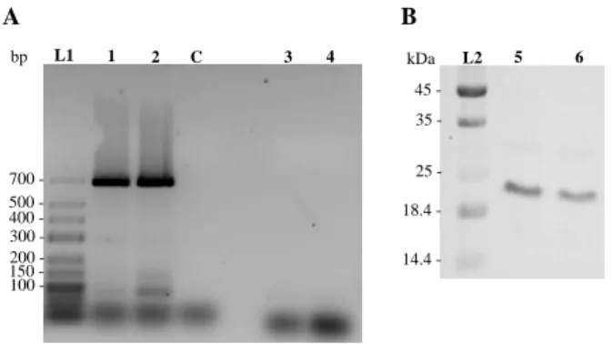
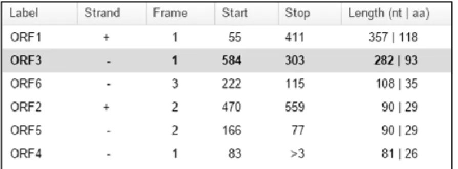
Bioinformatic and Southern blot analyses allowed the identification of the plasposon insertion in the mutant B. cenocepacia SJ2. The plasposon is located 8 nucleotides (nt) after the predicted 5’ UTR beginning of the B. cenocepacia J2315 OmpA-like protein. Therefore, the presence and functionality of the transcript ompA were evaluated by RT-PCR and Western blot, respectively. The results obtained indicated that the plasposon insertion did not affect ompA mRNA transcription or functionality. Bioinformatic analyses also led to the identification of a possible opening reading frame (ORF) in the complementary strand (named ORF3) and encoding a 93 amino acids protein. The presence of this ORF was supported by Northern blot assays previously performed. These assays allowed the identification of
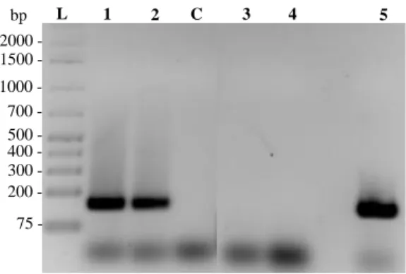
a transcript from the complementary strand. However, the amplification of complementary DNA ends (5’ and 3’ RACE) using specific primers for ORF3 region demonstrated that the transcript in study has a 5’ end with a lower size than the expected transcript of the potential protein (ORF3). A transcript containing approximately 179 base pairs and non-interrupted by the plasposon was identified. In the presence of these results, primers for the potential DNA sequence of ORF3 were designed and the results showed that this is the region interrupted by the plasposon.
Due to the absence of initiation and termination codons in the region closer to the 179 base pairs transcript, it was hypothesized that the transcript corresponds to a sRNA (MavA). In this work, the presence of two genetic elements, a sRNA and a protein, in the intergenic region of B. cenocepacia J2315 chromosome 1 is described. The coding sequences of both the sRNA and protein are overlapped in, at least, 100 nucleotides.
Bioinformatic analysis of the genetic elements showed that they are conserved among the Burkholderia genus. Moreover, the sequence of ORF3 protein also showed to be identical to the sequence of one protein encoded by an annotated ORF in the B. multivorans genome and to five proteins of B.
pseudomallei.
The assays performed with the strain containing the ORF3 interrupted by the plasposon revealed the involvement of this protein in the resistance of B. cenocepacia J2315 strain to heat-shock stress (50 ºC), susceptibility to the detergent sodium dodecyl sulphate (SDS), biofilms formation, cellular hydrophobicity and antibiotics resistance (imipenem, ceftazidime and tetracycline). In general, these results suggest that this protein is likely involved in the maintenance of the outer membrane integrity and virulence of B. cenocepacia J2315 towards the nematode C. elegans.
MavA sRNA identified in the work herein presented was predicted to be a functional homologue of IstR-2 sRNA of Escherichia coli K-12 MG1655. Preliminary results of the MavA sRNA overexpression characterization indicate a possible direct or indirect role in the overexpression of ribosomal protein S12. Phenotypic analysis also showed the involvement in the swimming motility of B. cenocepacia J2315 strain and in resistance to thermal (50 ºC) stress. In addition, this sRNA is not involved in the virulence of B. cenocepacia J2315 towards the C. elegans nematode.
Overall, the results presented in this study contribute to a better knowledge of the intergenic region under study and to the identification of a putative ORF and a sRNA, contributing to a better understanding of the biology of Bcc bacteria and the role of sRNAs in the regulation of the expression of putative virulence factors. The identification of MavA sRNA and ORF3 protein highlight the importance of these studies in identifying genetic elements that might be exploited as targets for the development of effective treatments to the B. cenocepacia J2315 strain infections.
Keywords
RESUMO
O recente desenvolvimento de técnicas de elevado rendimento para sequenciar genomas e a diminuição contínua do custo associado, conjuntamente com os avanços na bioinformática e a disponibilidade de número crescente de ferramentas bioinformáticas para análise de sequências nucleotídicas e aminoacídicas, tornou possível o aumento exponencial dos genomas microbianos disponíveis. Consequentemente, têm vindo a ser descobertos diversos pequenos transcritos com ação regulatória denominados de pequenos RNAs reguladores não codificantes (sRNAs), com tamanhos compreendidos entre 50-500 nucleótidos. Estes sRNAs têm sido identificados essencialmente em regiões intergénicas, junto a genes ou grelhas de leitura (ORFs) anotadas. A função conhecida de um grupo importante desses sRNAs consiste na regulação da expressão génica, principalmente ao nível pós-transcricional, por mecanismos de interferência de RNA.
Os sRNAs bacterianos podem exercer nos seus alvos (RNAs mensageiros) uma regulação positiva ou negativa. No caso de ser exercida uma regulação negativa o sRNA estabelece com o seu alvo um emparelhamento na região que contém o codão de iniciação e/ou a região de ligação do ribossoma (RBS), tornando este local inacessível para a ligação do ribossoma e impedindo, assim, a tradução do mRNA.
O complexo Burkholderia cepacia (Bcc) é atualmente constituído por 20 espécies de bactérias Gram-negativas validadas e filogeneticamente próximas. As bactérias que compõem este grupo são fenotipicamente semelhantes e genotipicamente distintas, podendo ser isoladas de várias fontes. São exemplo de fontes o solo, a água, a rizosfera de plantas, animais e Humanos. Devido às suas capacidades metabólicas invulgares, algumas estirpes do Bcc apresentam um elevado potencial de aplicação ao nível do biocontrolo, biorremediação e agricultura, pois são capazes de metabolizar xenobióticos, produzir compostos com atividade antifúngica e promover o crescimento de plantas.
Contudo, na década de 80 as bactérias incluídas no género Burkholderia, incluindo as englobadas no complexo Bcc, emergiram como agentes patogénicos oportunistas em indivíduos com fibrose quística (FQ).
O facto de as bactérias deste complexo produzirem vários fatores de virulência, apresentarem mecanismos de resistência a um largo espetro de antibióticos sendo difíceis de erradicar e de causarem em doentes com FQ infeções por vezes acompanhadas por um estado clínico de evolução rápida que inclui pneumonia necrotizante e septicémia, levando à morte do doente – síndrome cepacia -, faz com que tenham merecido uma elevada atenção da comunidade médica e científica. Acresce que algumas espécies do Bcc têm vindo a ser reconhecidas nos últimos anos como sendo agentes patogénicos nosocomiais emergentes em doentes hospitalizados, principalmente em doentes oncológicos.
Com o objetivo de identificar possíveis genes envolvidos na virulência de bactérias do Bcc, investigadores do iBB (Instituto de Bioengenharia e Biociências) construíram bibliotecas de mutantes de B. contaminans IST408 e B. cenocepacia J2315 utilizando plasposões. As bibliotecas de mutantes foram rastreadas tendo como objetivo a identificação de mutantes que exibiam virulência atenuada, utilizando como modelo de infeção o nemátodo Caenorhabditis elegans. O trabalho aqui apresentado teve como objetivo a caracterização de um mutante identificado no rastreio acima mencionado, contendo um plasposão inserido na região intergénica do cromossoma 1 de B. cenocepacia J2315, localizada entre o gene codificante da subunidade A da enzima ADN girase (GyrA) e o gene codificante de uma proteína da membrana externa do tipo A (OmpA).
Através de análises bioinformáticas e experimentais, foi possível localizar no mutante B. cenocepacia SJ2 a inserção do plasposão 8 nucleótidos (nt) após o início da região 5’ não traduzida (5’ UTR) do gene
que codifica a proteína do tipo OmpA. Neste sentido, foram avaliados a presença e a funcionalidade do transcrito ompA por ensaios de RT-PCR e Western blot, respetivamente. Os resultados obtidos confirmaram que a inserção do plasposão não afetou a transcrição nem a funcionalidade do transcrito correspondente ao gene ompA.
As análises bioinformáticas realizadas permitiram ainda a identificação de uma possível grelha de leitura aberta (ORF) na cadeia complementar da região intergénica (designada de ORF3), codificando para uma proteína de aproximadamente 93 aminoácidos. A confirmação da presença desta ORF foi suportada por ensaios de Northern blot realizados anteriormente e que permitiram a identificação de um transcrito a partir da cadeia complementar. No entanto, análises de amplificação em cadeia pela polimerase (PCR) das extremidades do ADN complementar (5’ e 3’ RACE) com sequências oligonucleotídicas iniciadoras específicas para a região da ORF3 demonstraram que o transcrito em causa possui uma extremidade 5’ menor que o esperado para o transcrito da possível proteína (ORF3), permitindo assim a identificação de um transcrito com cerca de 179 pares de bases que se verificou não ser interrompido pelo plasposão. Deste modo, tendo em conta os resultados aqui apresentados foram desenhadas sequências oligonucleotídicas iniciadoras específicas para a sequência de ADN da possível ORF3, tendo-se verificado que esta era a região interrompida pelo plasposão.
Devido à ausência de codões de iniciação e de terminação na região próxima do transcrito de 179 pares de base, colocou-se a hipótese deste transcrito corresponder a outro elemento genético, nomeadamente um sRNA, que se denominou de MavA. Neste trabalho é descrita a existência de dois elementos genéticos na região intergénica do cromossoma 1 de B. cenocepacia J2315, um sRNA e um gene que codifica uma proteína, sendo que as sequências codificantes de ambos encontram-se parcialmente sobrepostas em, pelo menos, 100 nucleótidos.
As análises bioinformáticas realizadas dos elementos genéticos permitiram a sua identificação como sendo ambos conservados no género Burkholderia. Além disso, demonstraram também que a proteína ORF3 é idêntica à sequência de uma proteína codificada por uma ORF anotada no genoma de B.
multivorans e à de cinco proteínas de B. pseudomallei.
Ensaios realizados com a estirpe contendo o plasposão a interromper a ORF3 demonstraram que esta proteína está envolvida na resistência da estirpe B. cenocepacia J2315 a choque térmico (50 ºC), suscetibilidade à presença do detergente dodecil sulfato de sódio (SDS), capacidade para formação de biofilmes e hidrofobicidade das células, bem como na resistência a antibióticos (imipeneme, ceftazidima e tetraciclina). De um modo geral, estes resultados apontam para que esta proteína esteja envolvida na manutenção da integridade da membrana externa e virulência da estirpe B. cenocepacia J2315 para o nemátodo C. elegans.
O sRNA MavA identificado neste trabalho foi bioinformaticamente previsto como tendo como seu homólogo funcional o sRNA IstR-2 de Escherichia coli K-12 MG1655. Os resultados preliminares da caracterização do sRNA MavA através da sua sobrexpressão demonstram um possível envolvimento direto ou indireto na sobrexpressão da proteína ribossomal S12. As análises fenotípicas mostraram ainda o seu envolvimento na mobilidade da estirpe B. cenocepacia J2315 por swimming e na resistência ao choque térmico (50 ºC). Os resultados obtidos, baseados em ensaios de morte lenta do nemátodo C.
elegans sugerem que este sRNA não está envolvido na virulência de B. cenocepacia J2315 neste modelo
animal de infeção.
Os resultados obtidos no presente trabalho contribuem para um conhecimento mais aprofundado da região intergénica estudada, tendo permitido identificar 2 elementos genéticos parcialmente sobrepostos que codificam uma proteína e um sRNA. Este trabalho constitui assim um contributo para o melhor
conhecimento da biologia das bactérias do complexo Bcc e do papel dos sRNAs na regulação da expressão de fatores de virulência. A identificação do sRNA MavA e da proteína ORF3 realça a importância deste tipo de estudos na identificação de elementos genéticos que possam ser explorados como alvos para o desenvolvimento de estratégias terapêuticas mais eficazes com vista ao tratamento das infeções causadas pela estirpe B. cenocepacia J2315.
Palavras-chave
Complexo Burkholderia cepacia; região intergénica; pequenos RNAs regulatórios não codificantes; regulação pós-transcricional; grelha de leitura aberta
TABLE OF CONTENTS
Acknowledgments I
Abstract III
Resumo V
Table of contents IX
List of figures XIII
List of tables XIV
Abbreviations XV
1. Introduction 1
1.1 Cells harbour different classes of RNAs 1
1.2 Bacterial small non-coding RNAs (sRNAs) 1
1.3 General regulatory RNAs classes 2
1.3.1 Regulatory 5’ UTR elements 2
1.3.2 Cis-encoded antisense sRNAs 3
1.3.3 Trans-encoded sRNAs 4
1.3.3.1 mRNAs 3’ regions are rich reservoirs for sRNAs 5
1.3.4 Regulatory sRNAs of protein activity 6
1.4 Cell advantages of regulating with sRNAs 7
1.5 Burkholderia cepacia complex (Bcc) – overview 7
1.5.1 Bcc species as opportunistic pathogens in Humans 9
1.5.2 Major virulence factors of the Bcc 10
1.5.3 Identification of novel Bcc virulence factors 11
2. MATERIALS AND METHODS 13
2.1 Bacterial strains, plasmids, nematode and culture conditions 13
2.2 Molecular biology techniques 14
2.2.1 Extraction and purification of genomic and plasmid DNA 14
2.2.2 Construction of a plasmid expressing the MavA sRNA 14
2.2.3 Insertion of foreign DNA in bacterial cells 14
2.2.3.1 Heat-shock transformation of E. coli cells 14
2.2.3.2 Triparental mating of B. cenocepacia cells 15
2.2.5 Extraction and purification of total RNA 15 2.2.6 Reverse transcription-polymerase chain reaction (RT-PCR) 16
2.2.7 Rapid amplification of cDNA ends (RACE) 16
2.2.8 Analysis of total soluble proteins by Tricine-SDS-PAGE 17
2.2.9 Western blot experiments 17
2.3 Nematode killing assays 18
2.3.1 Maintenance of C. elegans and egg preparation 18
2.3.2 C. elegans killing assays 18
2.4 Biofilm formation and cell surface hydrophobicity assays 18
2.4.1 Biofilm formation ability 18
2.4.2 Bacterial adhesion to hexadecane 19
2.5 Motility assays 19
2.5.1 Swimming assays 19
2.5.2 Twitching motility assays 19
2.6 Stress susceptibility experiments 19
2.6.1 Heat stress susceptibility 19
2.6.2 Anionic detergent shock experiments 20
2.7 Antibiotic susceptibility testing 20
2.8 Statistical analysis 20
2.9 Bioinformatics analysis 21
2.9.1 MavA sRNA bioinformatics analysis 21
2.9.2 Hypothetic ORF bioinformatics analysis 21
3. RESULTS AND DISCUSSION 23
3.1 Characterization of the B. cenocepacia J2315 chromosome 1 intergenic region flanked by
genes gyrA and ompA 23
3.2 Biocomputational analysis and phenotypic characterization of the hypothetical protein
encoded by ORF3 26
3.2.1 The hypothetical protein ORF3 is conserved among Burkholderia and has high homology
with annotated ORFs 26
3.2.2 B. cenocepacia J2315 ORF3 does not impair growth ability 28 3.2.3 B. cenocepacia SJ2 mutant strain forms thicker biofilms and has an increased cell surface
hydrophobicity 29
3.2.4 The twitching motility of B. cenocepacia SJ2 strain is apparently affected 30 3.2.5 B. cenocepacia SJ2 strain shows increased mortality at 50 ºC and cell lysis rate in the
presence of SDS 30
3.2.6 The B. cenocepacia SJ2 strain is more susceptible to ceftazidime, imipenem and tetracycline 32 3.2.7 B. cenocepacia SJ2 mutant strain exhibits decreased ability to kill the nematode C. elegans
3.3 Biocomputational analysis and phenotypic characterization of MavA sRNA 33 3.3.1 MavA sRNA is specific of the Burkholderia genus and has one functional homologue 33
3.3.2 MavA putative targets in B. cenocepacia J2315 genome 36
3.3.3 MavA overexpression leads to S12 protein overexpression 38 3.3.4 MavA overexpression does not affect B. cenocepacia J2315 ability to grow in minimal
medium 40
3.3.5 MavA overexpression affects B. cenocepacia J2315 resistance to heat-shock 40 3.3.6 MavA overexpression does not affect biofilm formation 41 3.3.7 MavA sRNA overexpression does not affect the ability of B. cenocepacia J2315 to kill the
nematode C. elegans significantly 42
4. CONCLUDING REMARKS AND FUTURE PERSPECTIVES 44
References 46
LIST OF FIGURES
Figure 1.1 Schematic representation of the three levels of gene expression regulation involving
regulatory RNAs. 2
Figure 1.2 Regulatory mechanisms employed by cis-encoded antisense sRNAs are based on two
configurations. 3
Figure 1.3 Regulatory mechanisms employed by trans-encoded sRNAs. 4
Figure 1.4 Major mechanisms of action of the RNA-binding protein Hfq. 5
Figure 1.5 sRNAs from bacterial 3’ UTRs. 6
Figure 3.1 Detection by RT-PCR of the transcript from the ORF putatively encoding an OmpA-like
protein, located in B. cenocepacia J2315 chromosome 1 (A) and assessment of the ompA mRNA
functionality by Western blot (B). 23
Figure 3.2 ORF prediction. 24
Figure 3.3 Analysis of 5’ and 3’ Rapid Amplification of cDNA Ends (RACE) in 2% (w/v) agarose gel
electrophoresis. 25
Figure 3.4 Detection by RT-PCR of the transcript obtained by 5’ and 3’ RACE from B. cenocepacia
J2315 chromosome 1. 25
Figure 3.5 Detection of the transcript from the non-annotated ORF3 from B. cenocepacia J2315 by
RT-PCR. 26
Figure 3.6 Genetic organization of the IGR locus interrupted by the plasposon. 26
Figure 3.7 Prediction of the consensus secondary structure of B. cenocepacia J2315 ORF3 protein, by
JPred4 web server. 27
Figure 3.8 Growth curves of B. cenocepacia strains J2315 and SJ2. 28
Figure 3.9 Tricine-SDS-PAGE analysis of the total soluble proteins obtained from B. cenocepacia
J2315 or B. cenocepacia SJ2 strains. 29
Figure 3.10 Assessment of biofilm formation by B. cenocepacia J2315 or B. cenocepacia SJ2 strains
in polystyrene microtiter plates. 29
Figure 3.11 Assessment of B. cenocepacia J2315 and B. cenocepacia SJ2 cells hydrophobicity. 30
Figure 3.12 Comparison of B. cenocepacia J2315 and B. cenocepacia SJ2 swimming and twitching
motility. 30
Figure 3.13 B. cenocepacia SJ2 mutant strain is more susceptible to heat-shock stress than the WT
strain. 31
Figure 3.14 B. cenocepacia SJ2 mutant strain exhibits an increased cell lysis rate upon exposure to
SDS, when compared to B. cenocepacia J2315. 31
Figure 3.15 Comparison of E. coli OP50, B. cenocepacia J2315 and B. cenocepacia SJ2 ability to kill
the nematode C. elegans. 33
Figure 3.16 Prediction of putative promoters and Rho-independent transcription terminators of MavA.
34
Figure 3.17 Prediction of the secondary structure of MavA sRNA. 35
Figure 3.18 Results of the predicted homologue retrieved from the analysis with sRNAMap tool. 36 Figure 3.19 Tricine-SDS-PAGE analysis of the total soluble proteins obtained from Bcc strains under
study. 38
Figure 3.20 Growth curve comparison. 40
Figure 3.21 MavA overexpression in B. cenocepacia J2315 cells increases its resistance to heat-shock
stress. 41
Figure 3.22 Biofilm formation by B. cenocepacia J2315, B. cenocepacia J2315 (pMLS7) and B.
Figure 3.23 MavA sRNA influences swimming and twitching motility 42
Figure 3.24 Comparison of E. coli OP50 (purple bars), B. cenocepacia J2315, B. cenocepacia J2315
(pMLS7) and B. cenocepacia J2315 (pMya2) ability to kill the nematode C. elegans. 43
LIST OF TABLES
Table 1.1 Burkholderia cepacia complex (Bcc) species, their main sources and biotechnological
applications according to the List of Prokaryotic Names with Standing in Nomenclature (LPSN) 8
Table 2.1 Bacterial strains, plasmids and nematode used in this study. 13
Table 3.1 Antibiotic susceptibility of B. cenocepacia J2315 and B. cenocepacia J2315 derived mutant
SJ2. 32
Table 3.2 Predicted mRNA targets of MavA in B. cenocepacia J2315 genome with TargetRNA2 tool.
ABBREVIATIONS
aa Amino acids
ANI Average nucleotide identity
BATH Bacterial adhesion to hydrocarbon
Bcc Burkholderia cepacia complex
BLAST Basic local alignment tool
bp Base pair
cAMP 3’,5’-cyclic adenosine monophosphate
cDNA Complementary DNA
CF Cystic fibrosis
CFTR Cystic Fibrosis Transmembrane Conductance Regulator
CFU Colony forming units
CGD Chronic granulomatous disease Csr Carbon storage regulator protein
EPS Exopolysaccharides
iBB Institute for Bioengineering and Biosciences
IGR Intergenic region
kDa Kilodalton
LB Lennox Broth
LBB Luria-Bertani Broth
LPS Lipopolysaccharide
LPSN List of Prokaryotic Names with Standing in Nomenclature Lrp Leucine-responsive regulatory protein
MH Mueller Hinton
MIC Minimal inhibitory concentration MLST Multilocus sequence typing
MMC Mytomicin C
mRNA Messenger RNA
NADPH Nicotinamide adenine dinucleotide phosphate NCBI National center for biotechnology information
nt Nucleotides
OD Optical density
Omp Outer membrane protein
ORF Open reading frame
PCR Polymerase chain reaction
PIA Pseudomonas Isolation Agar
QS Quorum-sensing
RBS Ribosome-binding site
RNase Ribonuclease
RNAseq RNA sequencing
ROS Reactive oxygen species
RPM Rotations per min
rRNA Ribosomal RNA
SD Shine-Dalgarno
SD Standard deviation
SDS Sodium Dodecyl Sulphate
sRNA Small non-coding RNA
StrR Streptomycin resistant
tRNA Transfer RNA
TSS Transcription start sites
UTRs Untranslated regions
WT Wild-type
1. INTRODUCTION
1.1 Cells harbour different classes of RNAs
The work developed by Hoagland et al. [1] has played a major role in helping us to understand the DNA translation mechanism to proteins. Transfer RNAs (tRNAs) were first described by this research group as “soluble RNAs” that when bound to amino acids (aa) caused their activation. However, an important piece of the puzzle was still missing and it was only in 1961 that the concept of messenger RNA (mRNA) based on the Jacob-Monod Model [2] was biochemically demonstrated by Brenner et al. [3] and Gros
et al. [4].
Cells harbor several types of RNA molecules with different functions, which can be divided into two major classes i) protein coding RNAs and ii) non-coding RNAs [5]. These two classes are discriminated based on the presence or absence of an open reading frame (ORF), respectively [5–7]. mRNA contains the genetic information copied from DNA coded in triplets of “letters” and is the only RNA belonging to the class (i) [8]. Non-coding RNAs (ii) are a much more diverse group and due to their function as regulators in different steps of gene expression, they are recognized also as regulatory RNAs. Therefore, non-coding RNA class includes, among others, a) tRNAs which are responsible for the growth of the polypeptide chains due to the presence of a three base sequence that base pairs with its complementary triplet present in mRNA [1,8]; b) ribosomal RNAs (rRNAs) that are associated with proteins consisting of essential structures of the translation process - ribosomes, and c) small non-coding RNAs (sRNAs), a relatively new class of non-coding RNAs that can have housekeeping or exclusively posttranscriptional regulatory roles [9,10].
1.2 Bacterial small non-coding RNAs (sRNAs)
Bacteria are present in different ecological niches where they are often exposed to changing and hostile environments such as those encountered during host infection processes. The changes that occur in these environments can be fast and enclose fluctuations in pH, nutrient availability, temperature and presence of reactive oxygen and nitrogen species (reviewed in [11,12]).
However, bacteria can adapt to these conditions, i.e. they are able to maintain cellular homeostasis and thrive, due to modifications in gene and protein expression [7,13]. These alterations are caused by a regulation that can occur at different stages of gene expression (such as transcription and translation) and can affect mRNA stability, as well as DNA maintenance or silencing. The mechanisms involved in these gene regulatory networks are based on changes in RNA conformation, protein-DNA, RNA-RNA and RNA-DNA interactions (Fig. 1.1) (reviewed in [14]).
Technical advances in nucleic acid sequencing methodologies have significantly contributed to the amount of known microbial genomes presently available. Consequently, it is possible to produce biocomputational genome-wide searches. Allied to these techniques, approaches relying on direct detection are important to define transcription start sites (TSS) and 5’ untranslated regions (5’ UTRs), such as tilling arrays and high-throughput RNA sequencing (RNAseq). These techniques allowed the discovery of sRNAs loci not only in several pathogenic and non-pathogenic bacteria but also in the other two domains of life (Archaea and Eukarya) [9,12,15,16].
sRNAs are an heterogeneous group of molecules [14]. They are primarily encoded in chromosomes, being most of them located in intergenic regions (IGRs), between annotated protein-coding genes and in the complementary strand of ORFs [17,18], although they have also been found in plasmids [19]. There are two relevant features in sRNAs namely a) their size and b) structure. Despite the absence of a
consensus among the authors about the size of these molecules, it is in general within the range of 50-500 nucleotides (nt) [12,15,20]. Regarding its structure, sRNAs enclose an initial stable stem-loop and they are often associated with Rho-independent terminators of transcription which consist in a GC-rich stem-loop followed by a set of U’s. These stem-loops may be the main reason for the high stability observed in these molecules [21,22].
Figure 1.1 Schematic representation of the three levels of gene expression regulation involving regulatory RNAs.
Transcription regulation is a process mainly mediated by protein-DNA interactions which occurs at the gene promoter region (i). Posttranscriptional regulation. The majority of the regulatory RNAs known so far act at this level of regulation causing conformational changes in the binding molecules or enzymes (ii). Posttranscriptional modification is relevant due to the fact that some proteins are produced in an inactive form and have to undergo covalent or conformational changes to display their activity (iii). Adapted from [23].
The majority of the sRNAs identified so far belong to Escherichia coli. Although their functions are not fully described, it has been shown that they mainly act as negative regulators of gene expression [21]. In fact, the first chromosomally encoded regulator identified was the E. coli MicF RNA, which binds imperfectly to the 5’ region of the ompF mRNA near the start codon. In view of this, translation of the mRNA encoding the major outer membrane porin F type (OmpF) is inhibited [24].
1.3 General regulatory RNAs classes
In a first approach, two classes of regulatory non-coding RNAs can be distinguished based on the location of their target: cis-encoded and trans-encoded molecules. The cis-encoded molecules are RNA regulators encoded in the same genetic location of the target RNA (directly on the same - sense- or on the opposite strand - antisense), whereas trans-encoded refers to regulators that modulate the expression of a mRNA transcribed from a distinct genetic locus [12,14].
1.3.1 Regulatory 5’ UTR elements
Riboswitches are a group of non-coding RNAs with a regulatory function. However, they are not a standard sRNA since they are transcribed from the 5’ UTR of the mRNA that they regulate, being cis-encoded and cis-acting elements [25]. In response to several environmental signals, both chemical and physical, these regulatory elements change their conformation leading to the modulation of expression of downstream genes [25]. The riboswitch conformation is altered upon binding of the leader sequence to small molecules, repressing transcription or translation through the disruption of transcriptional terminators/antiterminators or preventing binding to the ribosome-binding site (RBS) region [14].
DNA mRNA Protein Transcription (Transcriptional regulation) Translation (Posttranscriptional regulation) Posttranslational modification i) ii) iii) Stress
e
RNA thermometers are a group of these molecules which specifically respond to fluctuations in temperature, being particularly important in pathogenic bacteria since the host body temperature induces expression of specific virulence genes [26]. Their regulation is achieved by an interchange between two conformations: a) “closed” structure, present at low temperatures where the Shine-Dalgarno (SD) sequence and/or AUG start codon are inaccessible for translation initiation and b) “open” structure, present at high temperatures when the secondary inhibitory structure around the RBS melts [12]. These RNA elements are composed by the “aptamer region” domain which functions as ligand-binding domain and the “expression platform” domain, being the effector domain on the downstream genes [27].
1.3.2 Cis-encoded antisense sRNAs
In addition to the environmental sensors aforementioned there is another class of cis-encoded RNA regulators, being the majority of them constitutively expressed [14]. sRNAs residing on plasmids were the first to be described as members of this class [28].
Their mechanism of action consists on base pairing with their target mRNAs. Since these sRNAs are mainly transcribed from the DNA strand opposite of their target, generally overlapping the 5’ end, they are named antisense sRNAs [13]. Consequently, they display extended regions of full complementarity with their target, around 75 or more nucleotides, and can exert a downregulation on the target genes. This downregulation can occur at the level of mRNA stability, transcription or translation (Fig. 1.2) [14].
Figure 1.2 Regulatory mechanisms employed by cis-encoded antisense sRNAs are based on two configurations. Base
pairing between a sRNA (encoded opposite to the 5’ UTR of its target mRNA) and its target, prevents the ribosome binding to the RBS. This event inhibits the translation process and promotes sRNA-mRNA complex degradation (A). A few sRNAs are encoded in regions complementary to the sequence separating ORF’s (B) and base pairing of these sRNAs can target RNases causing mRNA cleavage (1) or transcriptional termination through the cessation of RNA polymerase activity due to a putative loop formation (2). In the last case, a reduction in the expression levels of downstream genes is induced. The grey arrows represent the promoter. Adapted from [14].
A few years ago, a new subset of these sRNAs was identified as negative regulators of the expression of mRNAs encoding potentially toxic proteins, when present in high intracellular concentrations [29]– toxin-antitoxins (reviewed in [30]). In this case, the antisense sRNA acts as an antitoxin and it is
Inhibition of translation RNAs degradation 30S 50S mRNA Ribosome sRNA RBS A) RNase sRNA mRNA sRNA ORF 2 mRNA cleavage sRNA mRNA Transcription termination B) 1 2 ORF 1 ORF sRNA
responsible for maintaining a low basal level of toxin expression. Although this mechanism is not fully understood, it is thought that it occurs substantially by a translation block (reviewed in [19,29]). In addition to the already mentioned role as antitoxin, the group of cis-encoded antisense sRNAs can also be involved in direct processing of mRNAs encoded in its opposite strands (Fig. 1.2. B1) [19]. The
E. coli GadY is a cis-encoded RNA that it is encoded in the opposite strand of the operon gadXW,
overlapping the 3’ end of the gadX mRNA [31]. Opdyke et al. [31] and Tramonti et al. [32] have shown that in cells in the stationary phase, the GadY antisense sRNA levels increase, promoting the generation of base pairing with the 3’ UTR of gadX mRNA. This binding promotes the cleavage of the duplex
gadX-gadW, which is involved in the response to acidic conditions, increasing the intracellular levels of gadX transcript. Therefore, GadY acts as a positive posttranscriptional regulator of gadX.
1.3.3 Trans-encoded sRNAs
Unlike cis-encoded sRNAs, trans-encoded sRNAs are only expressed under stress conditions. These sRNAs have been encountered in specific conditions such as i) iron limitation [33], ii) oxidative stress [34], iii) low temperature [35] and iv) accumulation of non-metabolizable phosphoglucose molecules such as glucose-phosphate [36].
As already mentioned, trans-encoded sRNAs are encoded in a different genetic location relatively to their mRNA target [12,14]. In view of this, a limited complementarity is shared between the sRNA and its target, usually in discontinuous patches. In this case, the region where the sRNA overlaps the target mRNA is known as the “seed sequence” [19]. Seed regions of only 7 contiguous bases have been found for some sRNAs as being required for regulation [37,38].
The base pairing between sRNA and mRNA can result in positive or negative regulations. In both cases, mRNA translation and/or stability can be affected. However, the majority of trans-encoded sRNAs known so far, display a negative regulation through translational inhibition, mRNA degradation or both (Fig. 1.3).
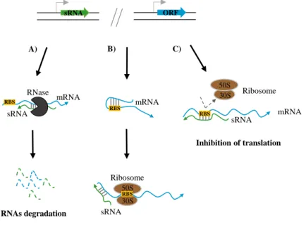
Figure 1.3 Regulatory mechanisms employed by trans-encoded sRNAs.
The imperfect binding between the sRNA and its target is often linked with enzymes involved in the degradation of the duplex sRNA-mRNA, such as RNase E or RNase III (A). Trans-encoded sRNAs can have positive effects in some targets through an anti-antisense mechanism, which causes remodeling of the intrinsic inhibitory secondary structure covering the RBS and consequently allowing translation initiation (B). Besides the negative regulation through RNAs degradation, sRNAs can base pair with the SD sequence or the start codon of its target and sequester the RBS (C). The grey arrows represent the promoter. Adapted from [12].
Almost 50 years ago, Franze de Fernandz et al. [39] identified Hfq protein in E. coli as an important host factor to the replication of RNA bacteriophage Qβ. However, it was only in the last decade that this global metabolism regulator, acting as a RNA chaperone, was found to be required by several
Gram-sRNA ORF sRNA mRNA A) RNase RNAs degradation RBS mRNA Induction of Translation B) 30S 50S mRNA Ribosome sRNA RBS C) Inhibition of translation sRNA Ribosome 30S 50S RBS RBS
positive and Gram-negative bacteria at the posttranscriptional level of regulation. This Sm-like protein appears to act as a mediator of the short and imperfect interaction established between trans-encoded sRNAs and their target mRNAs, due to their limited complementarity. In this case, it stimulates pairing between sRNA-mRNA by increasing the annealing rates (reviewed in [40]). The protein has the ability to bind not only sRNAs, but also mRNAs and in both cases it binds A-/U- rich sequences near hairpins [41]. Furthermore, Hfq is also involved in regulation through the modulation of the sRNA levels (Fig. 1.4).
Unlike what would be expected, the Hfq protein is not required in all cases of limited pairing but the reason behind this observation is still poorly understood.
Figure 1.4 Major mechanisms of action of the RNA-binding protein Hfq. Hfq associates with a sRNA, creating a complex
that interacts with the RBS of the target mRNA and inhibits translation (A). The complex sRNA-Hfq-mRNA disrupts the secondary structure present in the 5’ UTR region of the mRNA and enables the translation process (B). The turnover rate of sRNA and mRNA is modulated by Hfq through protection against degradation or by the recruitment of RNase E, respectively (C and D). Adapted from [40,42].
1.3.3.1 mRNAs 3’ regions are rich reservoirs for sRNAs
The majority of sRNAs that have been investigated and characterized are encoded by free-standing genes located in IGRs and having a single output function associated. However, some protein-coding genes have been observed to possess dual-function since they produce from its 5’ region a mRNA template for translation and from its 3’ region a regulatory RNA, which will act as an antisense regulator on other mRNAs, through a shared promoter and/or terminator [43–46]. In the last years, a few examples of sRNAs overlapping mRNAs 3’ region were reported [47,48]. Recent studies based on co-immunoprecipitation of Hfq and associated RNAs, allied to RNA-seq techniques with higher resolution, confirmed that mRNAs 3’ regions are large reservoirs for new sRNAs [49,50].
Regarding their biogenesis, sRNAs that are originated from 3’ regions of known mRNAs can be distinguished into two different types: sRNAs transcribed from an insider promoter of the overlapping mRNA gene or 3’ UTR (type I) (Fig. 1.5 A) and sRNAs resulting from an internal processing of the initial mRNA (type II) (Fig.1.5 B). However, in both types there is a shared use of the Rho- independent transcription terminator present in the mRNA. The Salmonella dapB mRNA represents the first example of dual output via the 3’ UTR of a conserved gene being included in type I sRNAs, since DapZ sRNA overlaps the 3’ UTR of the dapB mRNA and is transcribed from an mRNA-internal promoter that exists upstream of the stop codon of the conserved dapB gene (Fig.1.5 A). This sRNA is completely independent from the transcription of dapB and has a different outcome associated, since the dapB gene is essential for the biosynthesis of lysine and DapZ causes a translational repression of genes that encode major ABC transporters [50]. The cpxP mRNA belonging to the Cpx regulon of Salmonella is a perfect
example of type II sRNAs, due to its associated dual output. CpxQ sRNA is the first processed 3’ UTR multi-target regulator to be known. It overlaps the 3’ UTR of the cpxP mRNA and both share the same promoter, which makes CpxQ sRNA dependent of cpxP transcription (Fig. 1.5. B). After transcription of the parental mRNA, CpxQ is generated through processing in 3’ region of the coding sequence by RNase E cleavage, a few nucleotides downstream of cpxP stop codon (Fig. 1.5. B) [48]. Although CpxP protein and CpxQ noncoding RNA act in the same pathway, they perform different but complementary functions since CpxP is a periplasmic protein involved in envelope stress [51] and CpxQ sRNA is a cytoplasmic effector that repress mRNAs from several envelope proteins [48].
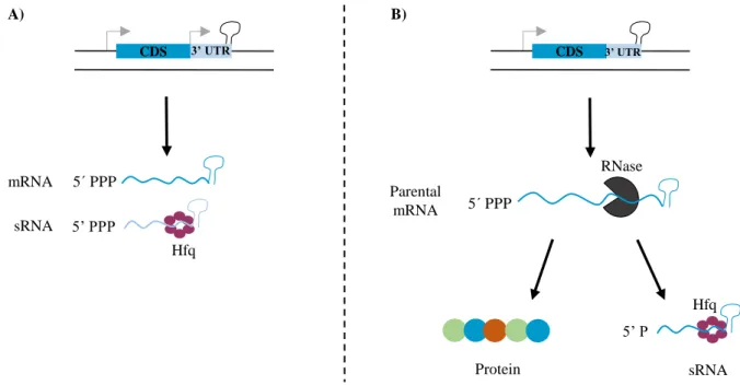
Figure 1.5 sRNAs from bacterial 3’ UTRs. Transcription of sRNAS from bacterial 3’ UTRs can occur by its own promoter
(type I) (A) or through internal processing of the associated mRNA by RNase cleavage (type II) (B). The sRNA resulting from processing in the mRNA 3’ region carry a 5’ monophosphate end, which is a sensor domain of RNase E. In both cases Hfq protein is involved in the stabilization of the sRNA and mediates its interaction with their targets. Furthermore, Hfq is also involved in the internal processing since it can form a complex with RNase E and direct it to the 3’ end of mRNAs. The grey arrows represent the promoters. Adapted from [48].
1.3.4 Regulatory sRNAs of protein activity
The majority of the already characterized sRNAs act through base pairing with their target transcripts. Nevertheless, a small number of sRNAs capable of binding to proteins and modify directly their activities have also been identified. These RNAs, which include the E. coli 6S RNA and the CsrB family of sRNAs, exert their function by mimicking the structures of other nucleic acids [19].
A few years ago, the 6S RNA was found to bind to the active site of a RNA polymerase holoenzyme comprising a σ70 subunit (housekeeping form of RNA polymerase). Consequently, it inhibits the
expression of certain promoters and increases the association with alternative stress sigma factors when the transition between exponential and stationary phase of growth occurs. Moreover, this RNA has a remarkable accumulation during the late stationary phase which has been associated with the sequestration of the majority of σ70 holoenzyme [52]. Later studies have revealed that the structure of
6S RNA governs the σ70 holoenzyme sequestration with the double-stranded RNA hairpin containing a
critical central bulge mimicking the promoter open complex [53].
The E. coli carbon storage regulator protein (CsrA) was identified as a RNA-binding protein, as well as a global key posttranscriptional regulator of the carbon metabolism and bacterial motility, mainly during
A) RNase B) CDS 3’ UTR 5’ PPP Hfq 5 PPP mRNA sRNA CDS 5 PPP Parental mRNA Protein 5’ P Hfq sRNA 3’ UTR
the entry into the stationary phase [54]. CsrA homodimers bind to GGA motifs in stem-loops of a RNA hairpin located in the 5’ UTR of their mRNA targets. Therefore, the stability and/or translation of the mRNA will be affected (reviewed in [19]). The intracellular levels of this protein are mostly negatively regulated by two sRNAs enclosing multiple CsrA binding sites, CsrB and CsrC. When the levels of these RNAs increase they interact and sequester the RNA-binding protein. Opposite to the regulation of the housekeeping form of RNA polymerase, the CsrB and CsrC antagonize CsrA protein, precluding the binding to the target mRNAs (reviewed in [54]).
1.4 Cell advantages of regulating with sRNAs
Regulatory RNAs, such as sRNAs, appear to be widely distributed in the Bacteria domain. Moreover, they are also present in several vital functions performing a regulatory role in diverse environmental conditions and overcoming other regulators, which include protein transcription factors.
As already mentioned, bacteria are often present in hostile environments with a high rate of changing conditions and often exposed to external stimuli. Thus, it is beneficial for cells to store energy to be further used in growth and/or maintenance.
sRNAs are single-stranded RNA molecules with a medium size ranging around a few hundreds of base pairs. Due to the fact that they are shorter in size than the majority of mRNAs they provide a reduced metabolic cost for the cell and faster synthesis when compared to protein transcription factors. In addition, they do not require translation minimizing the metabolic cost (reviewed in [20]). Furthermore, sRNAs act at the posttranscriptional level of regulation and with these advantages it is possible to conclude that not only the response to the unforeseen stress conditions will be faster but also the recovery will be more rapid after the removal of the external stimulus. In both cases, the faster regulation confers a selective advantage [55].
The capacity of a unique sRNA to be transcribed under different conditions together with the irreversible regulatory effects achieved by the ability of some sRNAs to direct the cleavage of their targets are also important advantages of sRNAs [14].
1.5 Burkholderia cepacia complex (Bcc) – overview
During the late 1940’s, Walter Burkholder [56] studied the phytopathogenic agent responsible for the soft onion rot bulbs, a disease known as “sour skin”. During his investigation, the bacterial species
Pseudomonas cepacia was isolated and described for the first time [56].
In 1992, a molecular taxonomical analysis of the genus Pseudomonas based on the 16S rRNA sequences, DNA-DNA homology, cellular lipid and fatty acid composition and also in phenotypic characteristics was performed. This analysis resulted in the proposal of a new genus named Burkholderia and, consequently, some species previously inserted in the genus Pseudomonas, particularly
Pseudomonas cepacia, were transferred to the new genus. Pseudomonas cepacia was further renamed
as Burkholderia cepacia [57]. The Burkholderia genus is phylogenetically diverse. By November 2016, this genus included approximately 105 species [58] that are widely distributed in ecological niches [59]. Subsequently, Burkholderia species were shown to present several biotechnological properties such as bioremediation and biocontrol/biopesticides agents. These applications in biotechnology are due to their ability to i) degrade natural and man-made complex aromatic pollutants in water and soils, ii) produce antifungal compounds, iii) fix atmospheric nitrogen and iv) promote growth of plants and crop production (reviewed in [59]). However, Burkholderia species have emerged in the 1980s as important pathogens to both humans and animals, in particular to cystic fibrosis (CF) patients [60].
The polyphasic taxonomic study performed by Vandamme et al. [61] with Burkholderia cepacia isolates from CF patients demonstrated that these bacteria are grouped into five distinct species, then designated as genomovars (I-V), together forming a group of closely related species denominated Burkholderia
cepacia complex (Bcc).
Bcc bacteria display a diverse nutrition, since they are able to use a broad range of simple and complex molecules as carbon sources, including xenobiotics (reviewed in [59]).
The genome of Bcc species has some interesting properties: i) its large size, which is correlated with their high functional versatility [62], and ii) its organization in three replicons and, in some cases, plasmids. Recent studies performed by Agnoli et al. [63] demonstrated that these microorganisms are able to tolerate the deletion of the third chromosome. These findings suggest that the third replicon could be a plasmid. Currently, this cluster is known to comprise 20 validated and close related species (Table 1; [58,64]), exhibiting a high level of 16S rRNA (over 98%) and recA (94%-95%) gene sequence similarity but a moderate degree of DNA-DNA hybridization (30%-60%) [65]. It has also been demonstrated that different Bcc species share whole-genome average nucleotide identity (ANI) with values ranging from 85.04% and 89.92% [66].
Table 1.1 Burkholderia cepacia complex (Bcc) species, their main sources and biotechnological applications according to the List of Prokaryotic Names with Standing in Nomenclature (LPSN) [58]. CF: Cystic Fibrosis.
In more recent years, a new approach known as multilocus sequence typing (MLST) has gained notoriety among bacterial typing methods [80]. MLST uses a set of seven genes encoding proteins with housekeeping functions and discriminates strains based on the comparison of the nucleotide sequences
Bcc species Main sources and biotechnological applications References
B. cepacia Main pathogen in Portuguese CF population [67]; present in the environment;
used has bioremediation and biocontrol agent [57] B. vietnamiensis Infections in Humans (CF and non-CF); present in the rhizosphere; used has
bioremediation and biocontrol agent;
[68] B. multivorans Infections in Humans (CF and non-CF, being one of the main CF pathogens in
European CF centers [69]); present in the rhizosphere [61] B. pyrrocinia Infections in Humans (CF and non-CF); used has biocontrol agent [70]
B. stabilis Isolated from CF and non-CF patients [71]
B. ubonensis Infections in non CF patients (isolated from nosocomial infections) [72] B. ambifaria Infections in Humans (CF and non-CF); major bacterium present in the
rhizosphere; used has biocontrol agent; [73] B. anthina Infections in CF patients and animals; present in soils [74]
B. cenocepacia Infections in Humans (CF and non-CF, being one of the main CF pathogens);
present in the rhizosphere; used has biocontrol agent [75] B. dolosa Causes mostly infections in CF patients, but one environmental strain has been
described [76]
B. arboris Infections in Humans (CF and non-CF); present in the environment [77] B. diffusa Infections in Humans (CF and non-CF); present in the environment and soils [77]
B. latens Infections in CF patients [77]
B. metallica Infections in CF patients [77]
B. seminalis Infections in Humans (CF and non-CF); present in the environment [77] B. contaminans Infections in Humans (main pathogen among Portuguese CF population [67]
and animals [66]
B. lata Infections in Humans (CF and non-CF); highly present in soils [66] B. pseudomultivorans Present in Humans and the environment [78] B. stagnalis Present in soils and in Humans respiratory tract [79] B. territorii Present in the environment, especially in groundwater [79]
of the gene set. Nowadays, it is the standard technique used to differentiate Bcc isolates since it shows a high correlation with other methods and a high discriminatory power [66]. The primers and other information on Bcc MLST can be accessed at http://www.pubmlst.org/bcc/.
1.5.1 Bcc species as opportunistic pathogens in Humans
The first significant descriptions of Bcc infections in patients with CF were performed three decades ago by Isles et al. [60] and Tablan [81]. CF is the most common autosomal recessive monogenic disease among the Caucasian population. It has an estimated frequency of 1 to 8,000 live births in Portugal [82]. Some Bcc bacteria are highly transmissible among CF patients, particularly by social contacts. Several strains belonging to B. multivorans, B. cenocepacia (particularly lineage ET12) and B. dolosa species have been reported to cause outbreaks in CF centers worldwide [83]. During the last decade, the Bcc prevalence in CF patients had a significant decrease, representing about 3.5% of CF infected patients [84]. Although representing a small fraction, Bcc infections are the most feared and through the years have gained special attention from the patients, medical and scientific communities due to their unpredictable clinical outcome.
CF is the result of mutations occurring in a single gene present at the long arm of chromosome 7, which encodes a 1,480 aa polypeptide known as Cystic Fibrosis Transmembrane Conductance Regulator (CFTR) [85]. The CFTR protein functions mainly as a 3’,5’- cyclic adenosine monophosphate (cAMP)-dependent chloride channel of low conductance at the apical membranes of epithelial cells that are present in a variety of organs such as sweat glands, pancreas, liver and respiratory and reproductive tracts [86]. However, this protein can also regulate the function of sodium channels [87]. 1,800 mutations with different outcomes in the CFTR protein have been identified as potentially CF-causing [88]. Independently of the molecular mechanism by which the mutation disrupts the normal protein synthesis or function, an atypical transepithelial ion transport of chloride and water occurs. Although several organs are affected, the variations affecting the respiratory tract are the main cause of morbimortality in CF patients, since this defect allows the deposition of thickened mucus and prevents its removal from airway surfaces [89]. Therefore, the conditions generated inside the lungs predispose to the establishment of bacteria, leading to chronic pulmonary infections [89,90] mostly caused by
Staphylococcus aureus in young individuals and by Pseudomonas aeruginosa in adults [91,92]. The
majority of Bcc infected CF patients undergo a reduction of the pulmonary function caused by the chronic infection and exacerbation events. Approximately 20% of these patients develop in their lifetime a rapid evolving clinical state known as “cepacia syndrome”. This syndrome has not yet been observed for other CF pathogens and it is characterized by high fevers, necrotizing pneumonia and septicaemia, which ultimately results in the patient early death [60].
Bcc infections are also notably difficult to treat and the strategies applied rarely result in eradication of the bacteria upon colonization. One of the major contributors to this problem is their intrinsic resistance to the majority of clinically available antimicrobials, which include aminoglycosides, quinolones, polymyxins and β-lactams [93]. Moreover, these microorganisms have several efflux pumps that can withdraw antibiotics from the cell and have the ability to modify the cell envelope leading to a reduced permeability of the membrane to antibiotics (reviewed in [94]).
Bcc bacterial infections have also emerged in patients with other pathologies such as chronic granulomatous disease (CGD) [95] and immunocompromised (patients with cancer and HIV) [96,97]. Similarly, to CF, CGD is also an hereditary disease, although with a much lower incidence than CF, where bacterial infections caused by Bcc species and their consequences represent the second most common cause of death [95]. This disease is considered to be a primary immune disorder since it is
caused by mutations in the subunits of the nicotinamide adenine dinucleotide phosphate (NADPH) oxidase complex belonging to the phagocytes. These mutations lead to a defective production of reactive oxygen species (ROS) by the phagocytes and therefore they are unable to kill bacteria through oxidative processes [95].
In addition to spread by social contact among CF patients, several outbreaks of bacteraemia have been reported among hospitalized non-CF patients such as individuals having chronic hemodialysis, diabetes
mellitus, congestive heart failure and malignancy, mainly caused by B. cenocepacia, B. multivorans
[98,99] and B. contaminans [100,101]. Therefore, Bcc species are now recognized as emergent nosocomial pathogens [69,97].
1.5.2 Major virulence factors of the Bcc
In order to enter in their host and reach the blood stream, as well as to proliferate inside eukaryotic cells, Bcc bacteria produce a wide variety of virulence factors, being the best characterized those produced by the B. cenocepacia ET12 lineage representative J2315 (reviewed in [59]).
The lipopolysaccharide (LPS) is one of the fundamental virulence factors in Gram-negative bacteria. The particular composition of the core oligosaccharide has been reported as implicated in the resistance of Bcc bacteria to cationic antimicrobial peptides and polymyxins [102,103].
Only a few polysaccharides synthesized by prokaryotes can be excreted, which are known as exopolysaccharides – EPS [104]. Among Bcc bacteria, production of EPS is very fluctuating, however some clinical and environmental isolates are known to produce a particular EPS – Cepacian [105]. Several studies have associated Cepacian production with the persistence of infection by Bcc bacteria, defective phagocytosis process of bacteria by neutrophils and an inhibition of neutrophil chemotaxis and ROS production [106,107]. Studies performed by Cunha et al. [108] have demonstrated that Cepacian also plays an important role in the formation and establishment of biofilms.
Bacteria have a remarkable capacity to form communities known as biofilms, in order to be protected from environmental factors and host-defence mediators so they can establish and persist in the host airways [109]. Caraher et al. [110] have demonstrated an enhanced in vitro antibiotic resistance (particularly to ceftazidime and ciprofloxacin) upon the formation of biofilms by Bcc bacteria, presumably due to a decrease of the bacterial surface exposed to the antibiotics. The resistance to multiple antibiotics by Bcc strains constitutes also an important virulent characteristic and has already been addressed in section 1.5.1.
In addition to the generation of communities Bcc also possess the quorum-sensing systems (QS). QS is density-dependent and supports the prompt adaptation of bacteria to the environment. The CepIR was the first QS system identified in a Bcc strain [111] and it is responsible for the regulation of toxins, lipases, proteases, iron-chelating siderophores, swarming motility and biofilm production (reviewed in [112]).
The acquisition of clusters of genes through horizontal gene transfer by Bcc strains (genomic islands) is of extreme importance since these islands encode virulence and metabolism associated genes. On the other hand, these genes promote both the survival and pathogenesis of Bcc bacteria in the CF respiratory epithelium and, consequently, the development of an epidemic lineage [113].
Moreover, these bacteria also express a particular type of fimbriae, the cable pili, with adhesins associated and flagella. Both the cable pili and the flagella help in bacterial adhesion and entry into the respiratory epithelium of host cells [114].
Specialized secretion systems such as type III and type VI also have determining roles in Bcc species, regarding to bacterial intracellular survival and replication. These complex structures inject bacterial molecules into the host cytoplasm, triggering host-signalling pathways and leading to the establishment of a bacterial intracellular niche [115,116].
1.5.3 Identification of novel Bcc virulence factors
In order to identify genes involved in Bcc virulence, researchers from iBB (Institute for Bioengineering and Biosciences) have prepared two mutant libraries by plasposon mutagenesis from strains B.
contaminans IST408 and B. cenocepacia J2315.
The strategy used to identify genes involved in virulence was based on the identification of mutants with attenuated virulence towards the nematode Caenorhabditis elegans. Several mutants were identified and the sequencing of the interrupted genes allowed the identification in B. contaminans of a gene encoding a protein involved in RNA-RNA interactions – the hfq gene. Subsequent research work led to the identification of a second gene, named hfq2, within the genome of all Bcc strains sequenced. This is extremely rare among prokaryotes, which usually harbour one hfq gene in their genome.
Bcc Hfq proteins have different sizes, since the Hfq has 79 aa residues and the Hfq2 has 188 aa residues [117]. Experiments with the clinical isolate B. cenocepacia J2315 and respective mutants in the Hfq encoding gene showed that the hfq mutant had an increased susceptibility to conditions mimicking the lung environment of a CF patient. Furthermore, infection experiments with hfq and hfq2 mutants demonstrated that both genes are required for full pathogenesis and virulence in the nematode C. elegans [117,118].
The iBB research team also identified another attenuated mutant found to have a single plasposon insertion in a 555 bp intergenic region of B. cenocepacia J2315 chromosome 1, upstream of the start codon of BCAL2958 gene, which encodes an outer membrane protein of the OmpA family, and downstream of the 5’ UTR of BCAL2957 gene, which in turn encodes a DNA gyrase subunit A enzyme. Bioinformatics analysis and RT-PCR experiments revealed the presence of a putative sRNA and non-annotated ORF in the region where the plasposon was inserted. In this context, the aim of this work is the functional characterization of the identified putative sRNA and ORF, later found to be widely conserved among the Burkholderia genus. To achieve this goal, molecular techniques were adopted in order to characterize the sRNA and the hypothetical ORF. In addition, stress, mobility and virulence assays in the nematode C. elegans, as well as an extensive bioinformatics analysis were performed. This study presents a contribute to better understanding of the regulatory mechanisms played by sRNAs in the expression of virulence determinants by Bcc bacteria. Despite the results obtained are somehow limited they contribute to a better understanding of Bcc biology, which is of central importance for the future development of novel and more accurate therapeutic strategies against the severe infections caused by Bcc bacteria.
2. MATERIALS AND METHODS
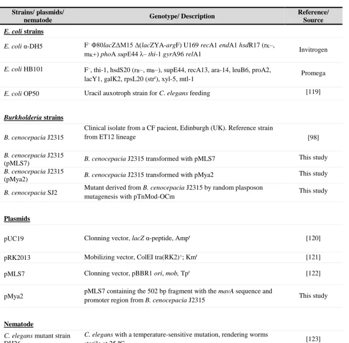
2.1 Bacterial strains, plasmids, nematode and culture conditions
All bacterial strains, plasmids and the nematode used in this work are listed in Table 2.1 B. cenocepacia and E. coli strains were maintained in Pseudomonas Isolation Agar (PIA; Benton Dickinson) and Lennox Broth (LB; Pronadisa), respectively, with 2% (w/v) agar (Iberagar). Unless otherwise stated, B.
cenocepacia and E. coli strains were cultured on solid media and incubated at 37 ºC for 24 h. In order
to maintain selective pressure, when required, solid growth media was supplemented with adequate antibiotics. Antibiotics were used as follows: 300 µg/mL chloramphenicol or 600 µg/mL trimethoprim for Burkholderia strains and 150 µg/mL ampicillin, 50 µg/mL kanamycin or 100 µg/mL trimethoprim for E. coli strains.
Table 2.1 Bacterial strains, plasmids and nematode used in this study.
Strains/ plasmids/
nematode Genotype/ Description
Reference/ Source
E. coli strains
E. coli α-DH5 F– Φ80lacZΔM15 Δ(lacZYA-argF) U169 recA1 endA1 hsdR17 (rK–,
mK+) phoA supE44 λ– thi-1 gyrA96 relA1 Invitrogen E. coli HB101 F–, thi-1, hsdS20 (rB–, mB–), supE44, recA13, ara-14, leuB6, proA2,
lacY1, galK2, rpsL20 (strr), xyl-5, mtl-1 Promega E. coli OP50 Uracil auxotroph strain for C. elegans feeding [119]
Burkholderia strains B. cenocepacia J2315
Clinical isolate from a CF pacient, Edinburgh (UK). Reference strain from ET12 lineage
[98] B. cenocepacia J2315
(pMLS7) B. cenocepacia J2315 transformed with pMLS7 This study B. cenocepacia J2315
(pMya2) B. cenocepacia J2315 transformed with pMya2 This study B. cenocepacia SJ2 Mutant derived from B. cenocepacia J2315 by random plasposon
mutagenesis with pTnMod-OCm This study
Plasmids
pUC19 Clonning vector, lacZ α-peptide, Ampr [120]
pRK2013 Mobilizing vector, ColEI tra(RK2)+; Kmr [121]
pMLS7 Clonning vector, pBBR1 ori, mob, Tpr [122]
pMya2 pMLS7 containing the502 bp fragment with the mavA sequence and
promoter region from B. cenocepacia J2315 This study
Nematode
C. elegans mutant strain DH26
C. elegans with a temperature-sensitive mutation, rendering worms


![Table 1.1 Burkholderia cepacia complex (Bcc) species, their main sources and biotechnological applications according to the List of Prokaryotic Names with Standing in Nomenclature (LPSN) [58]](https://thumb-eu.123doks.com/thumbv2/123dok_br/18561171.906672/26.892.114.778.527.1056/burkholderia-cepacia-biotechnological-applications-according-prokaryotic-standing-nomenclature.webp)