UNIVERSIDADE DO
ALGARVE
Faculdade de Ciências e Tecnologias
BREAST CANCER HER2-POSITIVE
Sequential versus concomitant
therapy
JOANA DE BRITO NEVES ROCHETA CASSIANO
Dissertation for obtaining the master’s degree inPharmaceutical Science.
The Dissertation was conducted under the guidance of Prof. Sofia Braga, MD PhD and Prof. João Rocha, PharmD MSc PhD
BREAST CANCER HER2-POSITIVE
Sequential versus concomitant therapy
Declaration of authorship of work
I declare to be the author of this work, which is original and unpublished.
Authors and works consulted are duly quoted in the text and included in the list of references.
Copyright©
The University of Algarve has the right, forever and without geographical limits, to file and advertise this work in the form of printed copies on paper or in digital form, or by any other means known or to be invented, to disclose it through and to allow it to be copied and distributed for non-commercial educational or research purposes, provided that the author and publisher are given credit.
“You become responsible, forever, for what you have tamed.” Antoine de Saint-Exupéry
Acknowledgments
The end of this long chapter of my life would only really be completed by writing a few words of appreciation to everyone who has somehow accompanied me in this battle that is about to come to its end.
First of all, I would like to thank Dr. Sofia Braga who, without knowing me, has seized my desire to know and entrusted to me with open arms a part of her life.
I also thank to Prof. João Rocha for accepting this challenge I proposed to him without blinking and for having contributed to my knowledge as almost Pharmacist that I am proud of today. Thanks, from the bottom of my heart, to my parents for all the efforts made so that today I could be here writing this that will be my last job as a master's student of pharmaceutical sciences. For all the support, for all the anxiety and support before and after exams, for all the words of encouragement in the hardest hours, for never letting me give up my dreams and for always giving me the wings I needed to fly higher, a big and sincere thank you.
To my brother, who has always been my life mate, my model of kindness and knowledge, my safe harbor. To my sister who from a young age, showed us that if we want to pursue a dream it can only be fulfilled with determination and perseverance. Thank you so much.
To my grandmother, who is my example of life, my example of love, affection and gratitude. It is a pride to be your granddaughter, and be able to walk in life beside you.
To all my family, even though some may have been less present, directly or indirectly contributed to make me a better person.
Fatima, José, Joana and Ricardo, for being like a family to me and for being by my side whenever I need it with supportive words and kindness.
To “Bárbaras” for all the moments we have experiencedand lived side by side and for showing me the real meaning of what the "family we have chosen" is.
Last but not least, my biggest thanks to my everyday companion, who holds me when I'm about to fall, who gives me wings to fly when I want to. There are no words in the world that can describe how grateful I am and at the same time in love with you. You are my safe harbor, João.
Resumo
Atualmente e segundo a Organização Mundial de Saúde (OMS), o cancro da mama é um dos oito cancros com maior taxa de prevalência e de mortalidade no mundo. Entre as mulheres, este ultrapassa em larga escala todos os outros cancros, sendo considerado a patologia com maior taxa de mortalidade e o cancro com o maior número de casos diagnosticados, com uma estimativa anual de 1,67 milhões de novos casos registados no mundo.
Este é o cancro com maior taxa de prevalência nas mulheres em todo o mundo e atualmente é o segundo cancro com maior prevalência em Portugal em ambos os sexos, sendo apenas ultrapassado pelo cancro da próstata e estando à frente do cancro do pulmão e o cancro do colón.
No nosso país, cerca de 8000 novos casos de cancro da mama são diagnosticados todos os anos; em Portugal, aproximadamente 1500 mortes anuais devem-se a este carcinoma como principal causa de morte.
Este é portanto considerado um problema grave de Saúde Pública a nível global devido à sua elevada incidência, morbilidade, mortalidade e elevado custo do tratamento.
Vários são os fatores preditivos que podem ser considerados fatores de risco para este tipo de cancro: a existência de um familiar com um grau de parentesco próximo a quem já tenha sido diagnosticado cancro da mama, a idade, a idade da menarca, uso de medicação de contraceção ou de tratamento hormonal ou idade do nascimento do primeiro filho, entre outros.
Não só o conhecimento dos fatores preditivos do cancro mas também o conhecimento dos fatores prognósticos têm sido objeto de vários estudos e determinante para a realização do melhor diagnóstico, da escolha do tratamento, da perspetiva de sobrevivência e da resposta patológica completa.
O conhecimento destes fatores têm sido alvo de estudo por médicos oncologistas e investigadores, bem como a escolha do tratamento. Hoje em dia, esta é uma área em constante crescimento não só devido à investigação mas também às variadíssimas opções de combinação de tratamento e fármacos que é possível. Uma das áreas mais investigadas
nos dias de hoje é o tratamento direcionado e específico para o tipo de cancro sendo os anticorpos monoclonais uma das abordagens mais procuradas e utilizadas.
Há vários fatores de prognóstico relevantes para caracterizar o tipo e gravidade de tumor que estão dividos em três categorias. Na categoria um encontram-se os fatores que revelam elevado valor terapêutico e de diagnóstico como é o tipo e grau histológico do cancro e o estadiamento do tumor de acordo com o sistema TNM, entre outros. Na segunda categoria encontra-se o recetor 2 do fator de crescimento epidérmico humano (HER2) que é o factor de prognóstico mais importante do nosso estudo e que é um critério de inclusão para o mesmo.
Aproximadamente 25% de todos os casos registados de cancro da mama apresentam uma sobre expressão do HER2. A amplificação ou sobre expressão, ou ambos, do HER2 (também conhecido como ERBB2), um recetor transmembranar tirosina quinase, está presente em cerca 35% dos tumores localmente avançados e metastáticos e 40% dos cancros inflamatórios da mama e está associado a doença agressiva e mau prognóstico.
Trastuzumab é um anticorpo monoclonal humanizado cujo mecanismo de ação consiste em ligar-se ao domínio extracelular do recetor HER2 inibindo a sua atividade, sendo o único aprovado na terapia adjuvante em pacientes com HER2 positivo.
Na terapêutica neoadjuvante, quando associado à quimioterapia, o trastuzumab melhorou significativamente a resposta patológica completa e reduziu o risco de recidiva, progressão da doença e morte quando comparado à quimioterapia isolada.
Gianni et al referem no ensaio clínico “Neoadjuvant herceptin” (NOAH) que, o trastuzumab obteve uma resposta significativamente positiva nas taxas de resposta patológica completa em doentes com neoplasia da mama HER-2 positiva. Outro estudo refere ainda que a inclusão do trastuzumab resulta num aumento da sobrevida, bem como da taxa de cura em esquemas adjuvantes de doença operável.
Após vários estudos que demonstraram a grande mais valia do uso desta terapêutica em Neoadjuvância, outros estudos quiseram avaliar em qual dos regimes deve ser aplicada
de sobrevivência, taxa de recidiva. Estes parâmetros foram determinados para as regiões da mama e axila individualmente e para os dois em conjunto (mama+axila).
Foram avaliadas 89 doentes em 4 Hospitais do país: Hospital Cuf Cascais, Hospital Cuf Descobertas, Hospital Vila Franca de Xira e Hospital Doutor Fernando da Fonseca. Das 89 doentes avaliadas, apenas 53 doentes possuíam o critério de inclusão no estudo (carcinoma HER2 positivo).
Num total de 53 doentes avaliadas neste estudo, 32 foram avaliadas no esquema sequencial e 21 foram avaliadas no esquema concomitante. No final, o esquema concomitante demonstrou ter um melhor resultado para resposta patológica completa mama+axila com significância estatística. No caso da resposta patológica completa para a mama e para a axila individualmente, devido ao reduzido número da amostra não foi possível obter resultados com significância estatística.
Também os efeitos adversos da terapêutica são factores que na hora da escolha do tratamento têm peso na decisão.
Como é amplamente conhecido no mundo da Oncologia, a cardiotoxicidade das antraciclinas e do trastuzumab é um problema que não se pode ignorar. Esta cardiotoxicidade está actualmente parametrizada e é agrupada em duas classes distintas: cardiotoxicidade reversível onde está inserido o anticorpo monoclonal Trastuzumab e a cardiotoxicidade irreversível onde se encontram as antraciclinas como é o caso da doxorrubicina.
Um dos fatores de risco mais determinante para este efeito adverso é a idade, o que muitas vezes pode ser um problema pois a maioria deste tipo de carcinomas surge normalmente numa idade mais avançada.
No nosso estudo provámos que existe a possibilidade de ocorrência de efeitos adversos, podendo no entanto ser controlados recorrendo a terapêutica apropriada para o efeito, nomeadamente inibidores da enzima de conversão da angiotensina.
O presente estudo, embora com uma amostra populacional reduzida e com baixa significância estatística, tem como objetivo tentar projetar alguns resultados que seriam expectáveis para um estudo realizado dentro dos mesmos moldes mas com uma maior dimensão.
Abstract
Currently, according to the World Health Organization (WHO), breast cancer is one of the eight cancers with the highest prevalence and mortality rates in the world. Among women, this large-scale surpasses all other cancers being considered the pathology with the highest mortality rate and the cancer with the largest number of diagnosed cases being the annual estimate of 1.67 million new cases registered in the world.
Approximately 25% of all registered cases of breast cancer have an overexpression of human epidermal growth factor receptor 2 (HER2). HER2 (also known as ERBB2), a transmembrane receptor tyrosine kinase receptor, is present in about 35% of locally advanced and metastatic tumors and 40% of inflammatory breast cancers and is associated with aggressive disease and poor prognosis. Trastuzumab is a humanized monoclonal antibody whose mechanism of action is to bind to the extracellular domain of the HER2 receptor inhibiting its activity, being the only one approved in adjuvant therapy in patients with HER2-positive.
In neoadjuvant therapy, when associated with chemotherapy, trastuzumab significantly improved the overall pathologic response and reduced the risk of relapse, disease progression and death when compared to chemotherapy alone.
Gianni et al reported in the "Neoadjuvant Herceptin" (NOAH) trial that trastuzumab obtained a significantly positive response in rates of complete pathological response in patients with HER-2 positive breast cancer. Another study reports that the inclusion of trastuzumab results in increased survival as well as the rate of cure in adjuvant regimens of operable disease.
Our primary objective was to compare which quimiotherapic regimen - concomitant vs sequential – is the most effective and secure for the treatment of HER-2 positive breast cancer.
The present study, although with a reduced population sample and with low statistical significance, aims to try to project some results that would be expected for a study
General Index
Acknowledgments ... II Resumo... III Abstract ... VI General Index ... VII Índex of Figures ... VIII Índex of tables ... VIII Índex of graph ... VIII Abbreviations list ... IX
I. Bibliographic revision ... 1
1. Risk Factors of Breast Cancer ... 3
2. Prognostic Factors of breast cancer ... 8
3. TNM System ... 9
4. Histopathologic types of breast cancer ... 11
5. Breast Cancer type HER2-positive ... 13
5.1 EGFR family receptors ... 13
5.2 Trastuzumab ... 15
5.3 Treatment ... 15
6. Cardiotoxicity caused by the use of anthracyclines and trastuzumab ... 17
II. Material and Methods ... 19
1. Study goals ... 19
2. Study design and settings ... 19
3. Population ... 19
4. Data analysis ... 20
5. Statistical analysis ... 20
6. Constitution of the sample ... 20
III. Results ... 21
IV. Discussion ... 25
V. Conclusion ... 29
VI. Bibliographic references ... 30
VII. Annex ... 37
1. Inclusion criterion (according to TNM system) ... 37
2. Study variables and their definition ... 38
3. Variables of clinical staging ... 39
Índex of Figures
Figure 1- Incidence rate of breast cancer (2) ... 1
Figure 2- Evolution of oncological diseases in Portugal. Adapted from National Cancer Program published by the DGS in 2015.(5) ... 2
Figure 3- Examples of mamographic density (adapted from medscape) ... 7
Figure 4- Signalling pathways activated by her2. (33) ... 14
Índex of tables
Table 1- Risk Factors of breast cancer (7) ... 4Table 2- Prognostic factors in breast cancer(21) ... 9
Table 3- Treatment groups and median and maximum ages ... 21
Table 4- Percentage of positive receptors in two different groups. ... 22
Table 5- Results of pathological complete response ... 22
Table 6- Study variables and their definitions ... 38
Table 7- Number of patients with different variables os clinical staging ... 39
Table 8- Degree of heart failure according to symptoms. Adapted from the New York Heart Association. ... 40
Índex of graph
Graph 1- TNM Staging criteria of breast cancer(21) ... 10Graph 2- Histopathologic types of the most common cancers of the breast(21) ... 12
Abbreviations list
AC- Adriamycin and Cyclophosphamide Therapy
ACE inhibitor- Angiotensin-converting-enzyme Inhibitor ADCC- Antibody Dependent Cellular Citotoxicity
AKT- Protein kinase B
AT – Anthracyclines and Trastuzumab therapy Bcl-2- B-cell Lymphoma 2
DFS- Disease Free Survival
DGS- General Health Department of Portugal DNA – Deoxyribonucleic Acid
EGFR- Epidermal Growth Factor Receptor EMA- European Medicine Agency
ERBB2- Human Epidermal Growth Factor Receptor 2 ER- Estrogen Receptor
ERK- Extracellular signal–regulated Kinases
HER 2- Human Epidermal Growth Factor Receptor 2 HF- Heart Failure
IHC- Immunohistochemistry
LVEF- Left Ventricular Ejection Fraction MAH- Marketing Authorization Holder MEK- Mitogen-activated Protein Kinase
Mib-1-Monoclonal Antibody of Cell Proliferation mTOR- Mechanistic Target of Rapamycin
OS- Overall Survival
p53- Tumor Gene Suppressor
PCR- Pathological Complete Response PI3K- Phosphoinositide 3-kinase PR- Progesterone Receptor PS2- Estrogen Response Gene
RAF- Rapid Accelerated Fibrosarcoma
RAS- A family of genes that may cause cancer when they are mutated TH- Taxol (paclitaxel) and Herceptin (Trastuzumab)
I. Bibliographic revision
According to World Health Organization (WHO), breast cancer is one of the eight most common cancers worldwide in terms of incidence and cause of death, 910 000 new cases occur yearly and the number of total deaths is around 360 000. (1)
Breast cancer is the second most common cancer in the world and, by far, the most frequent cancer among women with an estimated 1.67 million new cancer cases diagnosed in 2012 (25% of all cancers). (2)
At a global level, it is in the USA that there is the highest incidence rate of breast cancer, followed by Europe and Australia. (2)
Figure 1- Incidence rate of breast cancer (2)
Breast cancer is proved to be the second cause of death in the most developed countries where the first is lung cancer. On the contrary, in less developed countries it occupies the fifth place on the list of the most lethal cancers. (2)
The combination of a high incidence and a favourable prognosis makes breast cancer the most prevalent tumour: in the last five years approximately 4.4 million surviving women have been diagnosed breast cancer around the world. (3)
In Portugal it is also the most frequent tumour in women, with about 8000 new cases and 1500 deaths per year. (3)
According to the National Cancer Program published by the DGS in 2015, breast cancer is the second cancer with the highest gross incidence rate per 100,000 population, being only surpassed by prostate cancer. When only female sex is referred, it goes far beyond
all other carcinomas, having an almost three times higher rate than the second most recorded.
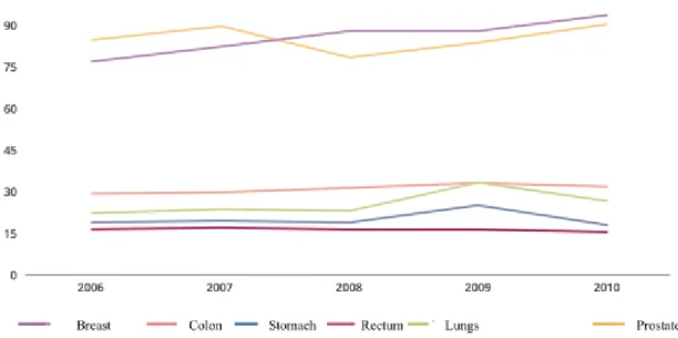
Regarding the progression of oncological diseases in Portugal, breast carcinoma also stands out among all the others because it is a pathology that since 2006 has been showing a growing progression, reaching the top of oncological diseases in 2010 in Portugal, overlapping carcinomas such as prostate, colon, lung, stomach, etc. (4)
The following graph shows the gradual growth of oncological diseases in Portugal from 2006 to 2010: the breast carcinoma occupies the most prominent place.
Figure 2- Evolution of oncological diseases in Portugal. Adapted from National Cancer Program published by the DGS in 2015.(5)
1. Risk Factors of Breast Cancer
About one-half of newly diagnosed breast cancers can be explained by already known risk factors, such as age at menarche, first live birth, menopause, and proliferative breast disease. An additional 10 percent is associated with a positive family history. In addition, risk may be modified by demographic, lifestyle, and environmental factors, although their association with breast cancer has not been clearly demonstrated. (6)
The established risk factors for breast cancer and the degree of risk associated with these factors as well as their relative risk are described in the following table:
Table 1- Risk Factors of breast cancer (7)
Established risk factors of breast cancer
Factors Low risk High risk Relative risk
Mother or sister
with breast cancer - + 2.6
Age 30 to 34 70 to 74 18.0
Age at menarche >14 <12 1.5
Age at first birth <20 >30 1.9 to 3.5
Age at menopause <45 >55 2.0
Use of
contraceptive pills Never Past/Current use 1.07 to 1.2
Hormone Replacement Therapy (estrogen
+ progesterone)
Never Current 1.2
Alcohol None 2 to 5 drinks/day 1.4
Breast density mammography
(%)
0 ≥75 1.8 to 6.0
Bone density Lowest quartile Highest quartile 2.7 to 3.5
History of a benign breast biopsy No Yes 1.7 History of atypical hyperplasia on biopsy No Yes 3.7
All factors described in the table above are correlated with a specific factor in common: the level of estrogen and progesterone. Several studies have recently provided clear evidence for the role of endogenous estrogen in the development of breast cancer. For example, a postmenopausal woman with relatively high estrogen concentrations had an approximately twofold risk of breast cancer compared with postmenopausal women with low serum concentrations. (8)
Increasing age - the older you get the greater risk of breast cancer you have. From birth
to the age of 49 the probability for a woman of developing breast cancer is of 1.9%. The previous percentage increases significantly with increasing age; the average increase is about 6.8% at the age of 70 (1 in 15 women). (9)
Menarche and menopause age - early menarche and late menopause maximize the
number of ovulatory cycles during which a woman is exposed to high levels of estrogen and progesterone. A decrease of about 20% in breast cancer risk results from each year that menarche is delayed. Regular menstrual cycles from the first year of menstruation is an important factor for the decrease of risk of breast cancer. (8)
Age at first birth - results of 47 epidemiologic studies show that the younger a woman
gives birth to her firth child, the lower the risk of breast cancer. The younger the woman gives birth for the first time, the more relative risk declines: about 3% each year. It has been proposed that full cellular differentiation, which occurs in the gland during and after pregnancy, protects the breast from breast cancer development. First birth at a later age may confere a greater risk than nulliparity because of additional proliferative stimulation of breast cells that are more likely to be fully developed and perhaps more prone to cell damage. (8,10)
Lifestyle factors - alcohol, tobacco and night shift work are three social conditions that
significantly increase the risk of developing breast cancer.
Compared to non-drinkers, daily consumption of one and two drinks was associated with a 10% and 21% increase in breast cancer risk, respectively. (11)
Although the association between alcohol and breast cancer risk is already well known, its association with tobacco was still unclear until recent studies. In a recent 2015 study, strong associations were found between the risk of developing breast cancer and exposure to tobacco (at what age you start smoking, how long you’ve been smoking, number of
cigarettes you smoke per day, approximate number of cigarettes during the exposure time). (12)
A study carried on nurses performing rotating shifts (day and night) concluded that working shifts after midnight is associated with an increased risk of breast cancer. The risk found was similar to that previously studied for female flight attendants on aircraft. (13)
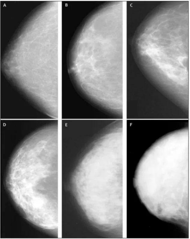
Dense breast tissue - in 1976, Wolfe described an association between a qualitative
classification of mammographic densities and the risk of breast cancer. Breast density, a measure of the extent of radiodense fibroglandular tissue in the breast, has the potential to be used as a predictor of breast cancer risk, to monitor risk- lowering interventions and as an intermediate end point in studies of breast cancer etiology. A woman whose density is greater than 75%, has 4 to 5 times more probability of developing breast cancer compared to a woman with low or no breast tissue density. (14–16)
Figure 3- Examples of mamographic density (adapted from medscape)
Legend: Panel A: 0% of density; Panel B: <10% density; Panel C: 10 to <25 % density; Panel D: 25 to <50% density; Panel E: 50 to <75% density; Panel F: >75% density.
Bone density - bone mineral density is an excellent regulator of circulating estrogen
levels. This is due to the high number of receptors of this hormone and its high selectivity to it. Women with a high bone mineral density have a higher risk of developing breast cancer. (17,18)
2. Prognostic Factors of breast cancer
The natural history of breast cancer indicates that the clinical course of the disease and survival vary from patient to patient. This variation is determined by a complex series of factors, such as the difference in the speed of tumour duplication, the potential for metastatization of the tumour and sociodemographic aspects as well as those related to the patient's immunological, hormonal and nutritional condition. These factors influence, directly or indirectly, the disease survival rate and other related mechanisms. Prognostic factors are possible parameters to be measured at the time of diagnosis and serve as a predictor of patient survival. (19,20)
The literature states an inverse relationship between tumour size and survival, whereas the presence of lymph nodes is associated with the disease recurrence in the first decade after treatment. High number of affected lymph nodes and involvement of the axillary apex and internal mammary lymph nodes indicate worse prognosis. (20)
Some prognostic factors have a dual function: besides playing their role as a prognostic factor, they also play a very important role as a predictive factor, thus allowing the establishment of specific therapies for the treatment of the tumour. (19)
According to College of American Pathologists Consensus Statement in 1999, the prognostic factors in Breast Cancer were stratified in three categories:
-Category one: includes factors proven to be of prognostic import and useful in clinical patient management. (21)
- Category two: includes factors that had been extensively studied biologically and clinically, but whose import remains to be validated in statistically robust studies. (21)
-Category three: includes all the other facts that haven’t been sufficiently studied so far to demonstrate their prognostic value. (21)
Those prognostic factors are included in the following table:
Table 2- Prognostic factors in breast cancer (21)
Category 1 Category 2 Category 3
• TNM Stage • Sentinel Lymphadenectomy • Histological Grade • Histological Type • Hormone Receptor Status
• Mitotic Figure Count
• HER2 • p53 • MIB-1
• DNA Analysis: Phase
Fraction
• DNA Ploidy Analysis • Tumour Angiogenesis • Epidermal Growth Factor Receptor • Transforming Growth Factor-α • bcl-2 • pS2 • Cathepsin D
Numerous studies carried out in many countries, have shown the value of using tumour size and nodal status to estimate prognosis in breast cancer. It is also widely accepted that age, race, histologic type, hormonal receptor status, and several other significant variables may influence an individual's prognosis. (22)
3. TNM System
The International Union Against Breast Cancer (UICC) proposed in 1954 breast cancer to be classified according the TNM system. This system characterizes the tumour according to alphanumeric codes and it is used in several malignant carcinomas according to the following codification: (21,23)
• T - describes the size of the main tumour and invades the surrounding tissue. • N - describes whether there is involvement of lymph nodes near the main tumour. • M - describes distant metastasis.
This classification is also divided into stages: 0, I, IIA / IIB, IIIA, IIIB, IIIC and IV. The following table describes the characteristics of each staging.
Graph 1- TNM Staging criteria of breast cancer (21) Stage 0 •T1 (≤2cm) •T1mic (microinvasion≤0,1cm) •T1a (0,1cm<T≤0.5cm) •T1b (0,5<T≤1cm) •T1c (1cm<T≤2cm)
•N0 (no regional lymph nodes) •M0 (no metastasis)
Stage I
•T2 (2cm<T≤5cm)
•N1 (metastasis to movable ipsilateral axillary lymph nodes) •pN1 (metastasis in 1-3 axillary lymph nodes)
Stage IIA/IIB
•T3 (>5cm)
•N2a (metastasis in ipsilateral axillary lymph nodes) •pN2 (metastasis in 4-9 axillary lymph nodes)
Stage IIIA
•T4 (any size with direct extension to chest wall or skin or both and inflammatory) •N2 (metastasis in ipsilateral axillary lymph nodes fixed or matted)
•pN2 (metastasis in 4-9 axillary lymph nodes)
•pN2b (metastasis in clinically apparent internal mammary lymph nodes in the absence of axillary lymph node metastasis)
Stage IIIB
•Any T
•N3 (metastasis in ipsilateral infraclavicular lymph nodes involvement, or in ipsilateral internal mammary lymph nodes and in the presence of clinically evident axillary lymph node metastasis or metastasis in ipsilateral infraclavicular lymph node with or without axillary or internal mammary lymph nodes involvement) •N3a (infraclavicular)
•N3b (axillary or internal mammary) •N3c (supraclavicular)
•pN3 (metastasis in ≥10 axillary nodes)
Stage IIIC
•Any T •N3 •M0
4. Histopathologic types of breast cancer
In addition to the stage classification of breast cancer, it is also classified by their histological type. There are several morphological patterns and these are important not only to define their severity but also to predict the prognosis and evolution of carcinoma. (24,25)
Most breast tumours generally occur in the ducts or lobes, and are then referred to as ductal carcinomas or lobular carcinomas. Regarding the prognosis of breast cancer, those with the worst prognosis are invasive ductal carcinoma and inflammatory carcinoma, although the later has a very low prevalence compared to other histological types. (26,27)
The following chart describes the various types of breast cancer classified according to their histological grade.
Graph 2- Histopathologic types of the most common cancers of the breast (21)
Carcinoma
In situ carcinomas
Intraductal Paget disease and
intraductal Invasive Carcinomas Ductal Inflamatory Medullary Mucinous Papillary(predominantly micropapillary pattern) Tubular Lobular
Paget disease and infiltrating Undifferentiated Squamous cell Adenoid cyst Secretory Cribiform
5. Breast Cancer type HER2-positive
5.1 EGFR family receptors
Approximately 20% to 25% of invasive primary breast cancers overexpress or have an HER2 amplification. (28)
The human epidermal growth factor receptor 2 oncogene, better known as HER2 +, belongs to the human epidermal growth factor (EGFR) family of 4 different types of tyrosine receptors (HER 1, HER 2, HER 3, HER 4). (29)
Each of these receptors consists of an extracellular domain where binding to the respective ligands occurs, a transmembrane domain and a cytoplasmic domain where intracellular signalling and development processes are activated. (30)
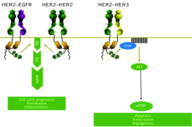
Although it is not yet known which HER2 receptor ligand is responsible for triggering the whole process, it is known that it has intracellular tyrosine kinase activity and plays a key role in the activation of intracellular signalling pathways that control cell growth and differentiation of the epithelial cells and vascularization around them (angiogenesis). (31,32)
After binding of ligand to the respective receptor, a sequence of conformational rearrangements with the formation of homodimers and heterodimers between the different receptors of the EGFR family is promoted. After this conformational rearrangement, the receptor is internalized, followed by phosphorylation and activating signalling such as the PI3K/AKT/mTOR pathway and the RAS/RAF/MEK/ERK pathway. (30)
The mechanism described above is demonstrated in the following figure:
Figure 4- Signalling pathways activated by her2 (33)
Impaired ERBB2 signalling is associated with the development of neurodegenerative diseases, such as Multiple Sclerosis and Alzheimer disease, whereas excessive ERBB2 signalling is associated with the development of cancers. (34)
5.2 Trastuzumab
Trastuzumab (whose trade name developed by Roche® is Herceptin®) is a humanized, monoclonal antibody derived from recombinant Deoxyribonucleic acid (DNA) and specifically designed to bind with high affinity to the Human Epidermal Growth Factor Receptor 2. Trastuzumab binds to ERBB2 receptor in the juxtamembrane portion of the extracellular domain. This binding prevents the receptor from activating its intrinsic tyrosine kinase, which in turn prevents the activation of several signaling pathways leading to cell proliferation. (34)
There are some mechanisms proposed for the functioning of this type of specific antibody. One of the accepted mechanisms involves the prevention of HER2-HER3 heterodimer formation, one of the most active heterodimers, and thought to have the ability to activate the signalling cascade in the absence of its true ligand. (35)
The other mechanism (antibody-dependent cellular cytotoxicity ADCC) is related to a cell-mediated autoimmune defence mechanism in which an effector cell of the immune system actively lyses a target cell whose membrane surface is covered by specific antibodies. Trastuzumab may also play an active role in the process of internalization and degradation of the HER2 receptor as well as the cleavage and release of the receptor into the extracellular domain. (34)
5.3 Treatment
The use of Trastuzumab was approved by the European Medicines Agency in August 2000 with the following indication: “Herceptin is indicated for the treatment of patients with metastatic breast cancer whose tumours overexpress HER2: as monotherapy for the treatment of those patients who have received at least two chemotherapy regimens for their metastatic disease. Prior chemotherapy must have included at least an anthracycline and a taxane unless patients are unsuitable for these treatments. Hormone receptor positive patients must also have failed hormonal therapy, unless patients are unsuitable
for these treatments.” In 2010, Trastuzumab (Herceptin®, Roche®) has been approved for use in the treatment of HER2-positive gastric cancer by the European Commission. (36,37)
Trastuzumab was initially approved to be used in the adjuvant treatment of early-stage HER2-positive breast cancer. Adjuvant therapies are used as a primary treatment (surgery), whose main goal is to increase the long-term hypothesis of disease-free survival. An example of a chemotherapy regimen is "AC + TH", which stands for Doxorubicin and Cyclophosphamide followed by Paclitaxel and Trastuzumab. (34,38)
Recently, HER2-targeted therapy has been approved by EMA to be used in the neoadjuvant setting. This appeal was proposed by Marketing Authorization Holder (MAH) and supported by the pivotal study "NOAH" which demonstrated the efficiency of Trastuzumab treatment in combination with neoadjuvant chemotherapy for locally advanced (including inflammatory) disease or tumours> 2 cm in diameter. (36,39)
Neoadjuvant therapy has an important role in patients with locally advanced and inflammatory cancers and consists of the application of pharmacological therapy prior to initiating primary therapy (surgery) with the aim of treating distant micrometastases, downstaging tumours, improving operability and to increase the chance of long-term survival disease-free period. Moreover, it facilitates breast-conserving surgery by downstaging the primary tumour and axillary nodes. Besides that, the pathologic response to neoadjuvant chemotherapy provides valuable prognostic information. (34,39)
As for the concomitant therapy (AT), it was conducted according to the NOAH protocol with IV administration and proceeded as follows:
• Doxorubicin 60 mg/m2 + Paclitaxel 150 mg/m2 + Trastuzumab (8 mg/kg on 1st administration, 6 mg/kg in subsequent administrations for 3 cycles of 21 days) • Then paclitaxel 175 mg/m2 + Trastuzumab 6mg/kg (trissemanally for 4 cycles) • Finally, Cyclophosphamide 600 mg/m2 + Methotrexate 40 mg/m2 + Fluorouracil
By contrast, the sequential therapy treatment group followed the treatment protocol 983121 as follows:
• AC + PT: Doxorubicin 60mg / m2 + Cyclophosphamide 600mg / m2 every 3 weeks for 4 cycles.
• Followed by Paclitaxel 80 mg / m2 (12 administrations weekly) + Trastuzumab at the standard dose every 3 weeks.
In both groups, treatment with Trastuzumab was completed twelve months after the surgical procedure, except for cases of disease progression.
6. Cardiotoxicity caused by the use of anthracyclines and
Trastuzumab
The cardiotoxicity caused by anthracyclines used in oncological treatments is considered to be one of the most serious adverse effects leading to a higher morbimortality rate, which may occur acutely after the end of treatment or up to two weeks later. If it only manifests up to two years after the end of treatment, it will turn chronic. (40,41) In order to diagnose the presence of cardiotoxicity it is necessary to comply with one of the following parameters:
• Presence of cardiomyopathy with reduced Left Ventricular Ejection Fraction (LVEF) with global compromise or with more evident segmental alterations in the septal region;
• Symptoms associated with cardiac failure; • Tachycardia detection;
• Reduction of LVEF by at least 5% for levels of less than 55% with signs or symptoms of heart failure, or a decrease in LVEF of at least 10% for levels of less than 55% in the absence of signs or symptoms compared to Values of the individual. (42,43)
There are several adverse effects that may occur due to the use of these drugs, including arterial hypertension, thromboembolic disease, pericardial diseases, arrhythmias and
myocardial ischemia. Heart failure with ventricular systolic dysfunction is highlighted as the most serious adverse effect. (40,41)
It was already known that the use of anthracyclines as an anti-neoplastic agent had the adverse effect of cardiomyopathy, which was irreversible, when cumulative doses of this drug were used. As time went on, it has also been found that Trastuzumab also causes cardiomyopathy. However, it is not dose-dependent and is not an irreversible effect, and the situation may reverse with the end of treatment. (40,41)
Two groups of agents causing cardiotoxicity are then classified: group I where anthracyclines such as Doxorubicin and Cyclophosphamide are inserted, which cause irreversible myocyte lesion and are dose-dependent; and group II where Trastuzumab and Sunitinib are inserted which cause reversible transient dysfunction in myocytes and are not dependent on the dose used in the treatment. (40,41)
II. Material and Methods
1. Study goals
Our primary objective was to compare which quimiotherapic regimen - concomitant vs sequential – is the most effective and secure for the treatment of HER-2 positive breast cancer.
The effectiveness was assessed by the complete pathological response which is strongly associated with favourable long-term survival rates. (44)
The degree of cardiotoxicity was also evaluated – as a benchmark of treatment security - due to the side effect of using both anthracyclines and Trastuzumab.
2. Study design and settings
This is a retrospective cohort study based on the clinical processes of a selected population of 89 breast cancer patients, diagnosed and followed between 2008 and 2016. The patients referred to above, were diagnosed and followed up at the Oncology services of four Portuguese hospitals: CUF Cascais Hospital, CUF Descobertas Hospital, Vila Franca de Xira Hospital and, Prof. Doutor Fernando da Fonseca Hospital.
This study was conducted with the approval of institutional ethics committee. No informed consent was obtained with each individual patient because it was a retrospective patient chart review-base study.
3. Population
Among the 89 patients selected with breast cancer, the inclusion criterion was applied to define the final sample: indication of the Multidisciplinary team (MDT) for Neoadjuvant chemotherapy. The criteria defining the indication for neoadjuvant chemotherapy are described in annex 1.
53 patients (59,6%) selected out of 89 patients, fulfilled the inclusion criteria in the study and from these, two treatment groups were formed: the first group, consisting of 32 patients, was treated in a sequential neoadjuvant therapeutic scheme with Anthracyclines and Trastuzumab (A-T) and the second group, consisting of 21 patients, was treated in a concomitant neoadjuvant therapeutic scheme with Anthracyclines and Trastuzumab
4. Data analysis
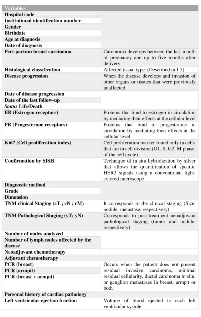
The study database consists of 35 clinicopathological variables that are described and defined in annex 2.
5. Statistical analysis
Data were analysed using SPSS software version 23.0. The comparisons between the categorical variables were performed using the chi-square test. The value of statistical significance considered was p <0.05.
6. Constitution of the sample
From the initial population of 89 patients with breast carcinoma, 53 patients were included in the study because they demonstrated the inclusion criteria. Thus, the final sample consists of 53 patients whose average age is of 52.11 years. The diagnosis and treatment of the disease occurred between 2008 and 2016.
III. Results
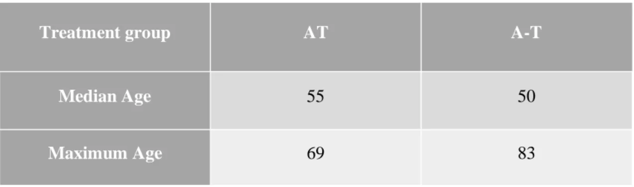
The sample population selected according to inclusion criteria (53 patients) at an average age of 52.11 years, a median of 52 and the respective standard deviation of 12.48 years, was separated into the two distinct treatment groups (AT vs A-T). In the following table, the dimensions of the groups are specified, as well as the median age and the maximum age of each:
Table 3- Treatment groups and median and maximum ages
Treatment group AT A-T
Median Age 55 50
Maximum Age 69 83
In the sequential treatment group, an 83-year-old patient was exceptionally included in the study, a too advanced age to be approved for Neoadjuvant therapy due to the complications of the cardiovascular disease that this therapy involves. This patient was accepted because of the favourable characteristics she presented: a T3N1 carcinoma (tumour with 10cm), an excellent performance status, no comorbidities at all and a very present family that was favourable family condition for the accomplishment of this type of treatment.
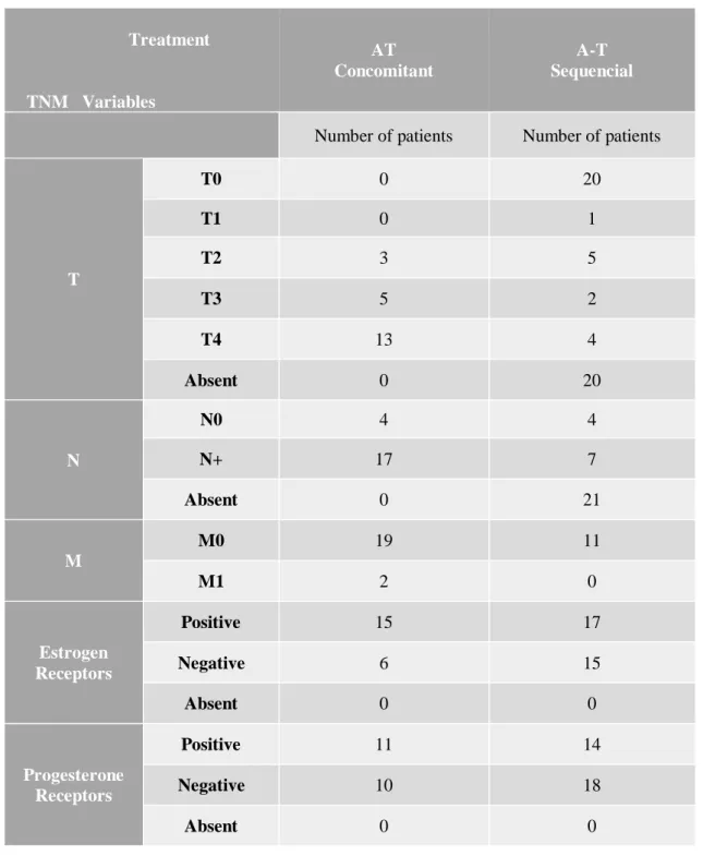
Regarding the variables of clinical staging, these are described in annex 3 but it is important to point out some important aspects such as:
• Forty-one percent of patients had a staging tumour IIIA (T4), but this was more prevalent in the concomitant treatment group (21 patients) than in the sequential treatment group (13 patients). In the sequential treatment group, there was a greater predominance of stage 2 tumours (tumour of diameter between 2 and 5 cm).
• Ninety-one percent of patients had no metastasis of the tumour at a distance; however, eighty-four percent of the patients had lymph nodes near the main
• In the total sample, there were 8 patients who did not have lymphatic nodules (N0) and were divided equally between the two groups (4 patients were in the AT group and the other 4 patients were in the A-T group).
• Only 2 patients had distant metastasis and both were treated in the concomitant treatment group.
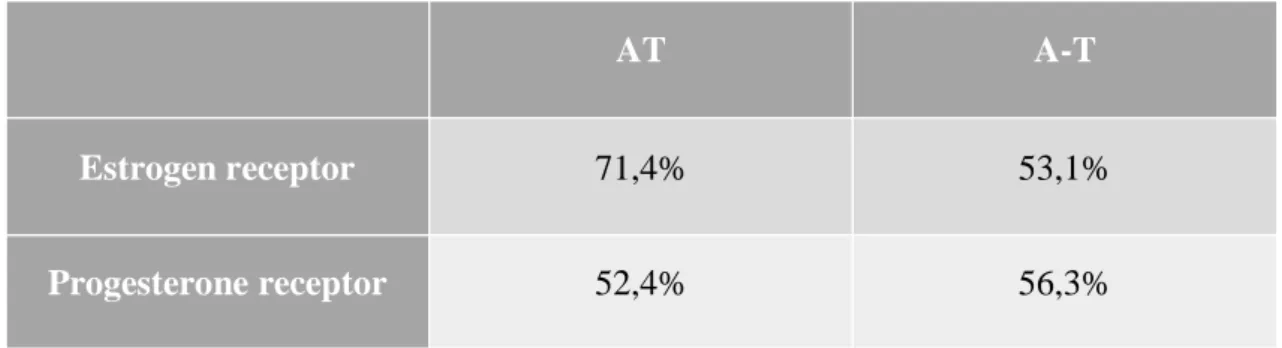
For estrogen and progesterone receptors, an immunohistochemical analysis was performed on a section of tissue removed from the biopsy and an anti-ER and anti-PgR mouse monoclonal antibody was used. Patients had the most positive levels for these receptors, as might be expected. In the following table, the percentages concerning to the positive values for the two receivers are:
Table 4- Percentage of positive receptors in two different groups.
AT A-T
Estrogen receptor 71,4% 53,1%
Progesterone receptor 52,4% 56,3%
3 different situations were evaluated concerning the pathological complete response (PCR): breast, axilla, breast + axilla. In general, better results were obtained in the case of concomitant therapy: greater percentage of positive pathological complete response as well as a lower percentage of absence of pathological complete response. The results obtained are shown in the following table:
Table 5- Results of pathological complete response
Pathological Complete
response AT A-T P
Breast Positive 56,50% 43,5% 0,055%
As for drug-induced cardiotoxicity, notably anthracyclines and Trastuzumab, out of the 53 patients included in the study only 4 showed symptoms of cardiotoxicity and all these patients belong to the concomitant treatment group. In the sequential treatment group, there were no reports about the presence of cardiotoxicity, except for 1 missing case that did not respond to the study.
Regarding the disease progression parameter, we obtained similar values in the two treatment groups. In each group, 3 cases were reported, and a total of 6 women did not continue the 12-month treatment with Trastuzumab after the surgical procedure as they presented disease progression at the end of the treatment protocols.
At the end of the study three deaths were counted, all of which from the concomitant treatment group, being these deaths related to the progression of the disease.
Concerning the possible cardiac toxicity developed by the drugs used in the treatment (anthracyclines and Trastuzumab), an evaluation of the patient’s cardiac function was made at the beginning of the study. The LVEF calculated at the beginning of the treatments had a minimum of 53% and a maximum of 75% with a median of around 62% of ventricular ejection fraction. During treatment, four patients were signalled as having a worsening degree of cardiac dysfunction, all belonging to the concomitant treatment group.
Two patients were recorded with a fall value greater than 10% of the LVEF (parameter necessary to diagnose cardiomyopathy), apparently without symptoms during treatment with adjuvant isolated Trastuzumab. Both patients stopped the treatment with Trastuzumab alone and an antihypertensive drug (ACE inhibitor) was introduced to try to reverse Trastuzumab-induced cardiomyopathy. This procedure was successfully completed and cardiomyopathy was reversed. Trastuzumab was successfully introduced again and the treatment was completed to the end with no further occurrence.
The other two patients had treatment-associated symptoms of heart failure that were included in the Class II New York Heart Association classification. (NYHA classification is listed in annex 4).
One of the patients developed heart failure even during the period of chemotherapy (Cyclophosphamide + Methotrexate + Fluorouracil + Trastuzumab). This patient
normalization resumed the treatment and was able to complete the 17 cycles of Trastuzumab foreseen in the treatment. The second patient developed heart failure already in the treatment period with Trastuzumab alone, having discontinued treatment and initiated appropriate medication for regression of symptoms. With improvement of the clinical picture, the treatment with the monoclonal antibody was started again.
Regarding the statistical results and their meaning, they were analysed according to the chi-square test. Due to the short number of cases reported, it was not possible to use the chi-square test in the following analysis parameters: status, presence of cardiotoxicity, disease progression. The results obtained and their statistical significance are described in the following graph:
Graph 3- PCR results and their statistical significance according to the chi-square test
PCR breast
showed a value of (1) = 3.687 and p> 0.05, The chi-square test for this parameter so that there was no statistical significance.PCR axilla
The chi-square test for this parameter showed a value of (1) = 1.021 and p>
0.05, so that there was no statistical significance.
PCR
breast+axilla
The chi-square test for this parameter showed a value of (1) = 6.226 and p <0.05,
where statistical significance was obtained with benefit to the concomitant scheme.
IV. Discussion
The results obtained confirmed the advantages previously demonstrated by other studies, especially in the field of neoadjuvant treatment. NOAH, a phase 3 international study in women with HER2-positive breast cancer, found that using Trastuzumab in the neoadjuvant setting almost doubled the rate of complete pathologic response and reduced the risk of relapse, disease progression and even death. (45)
According to a study aimed at HER2 receptors, approximately 22% of early breast cancers, 35% of advanced and metastatic local tumours, and 40% of breast inflammatory tumours have this amplified receptor, which is a correlated with disease aggressiveness and poor prognosis . This receptor is also overexpressed in a gastric and gastroesophageal cancer subtype, whose prevalence is approximately 18%, in lung cancer approximately 2% and in colon cancer approximately 1% . For this type of gastric cancer the use of this monoclonal antibody Trastuzumab is also approved and these are the only two indications approved by EMA. (46,47)
In the ToGA Trial, it was demonstrated the added value in the use of HER2 receptor targeted therapy in gastric and gastroesophageal tumours with a statistically significant increase in the overall survival rate (OS). (48)
The way the two types of cancer are diagnosed are different : while in breast cancer, the distribution of the antibody in the neoplastic cell is mostly circumferential, in the gastric one the distribution is generally incomplete and "U" or unilateral. Thus, the circularity of IHC staining is not a criterion for HER2 IHC in gastric cancer. Also in gastric cancer, there is an irregular pattern in the cells presenting areas with different HER2 scores which can lead to sampling errors when the biopsies are examined. In breast cancer, the pattern is usually regular and quite visible. (49,50)
Although there are significant differences between the histology of the cells and consequently the way the HER2 receptor is expressed, it is interesting to make the comparison between breast cancer and gastric / gastroesophageal cancer by both aligning therapeutics and obtaining good results with the same type of therapeutic target.
However, the adjuvant trial that tested Trastuzumab in HER2-positive gastric cancer was negative. And also the trial that tested double blockade with Pertuzumab + Trastuzumab
in metastatic HER2-positive gastric cancer was also negative. Therefore, HER2 blockade in gastric cancer has been much less efficacious than in breast cancer.
The use if anti HER2 therapy in breast cancer with Trastuzumab has been revolutionary, however we still lack knowledge about the combination with Trastuzumab and anthracyclines because of cardiotoxicity concerns. The Neoadjuvant setting is the best experimental setup for answering this question which is what we did with this study.
Neoadjuvant treatment has been the treatment of choice for patients with locally advanced breast cancer and is also generally advised in women with operable breast cancer because it is a treatment that enables breast preservation during surgery. This is a very advantageous factor for those who choose this type of treatment and it is possible due to previous chemotherapy treatments whose purpose is to minimize the lesion size.
Besides that, there is also the choice between a sequential or a concomitant treatment regimens that depends on several factors such as tumour type, adverse effects or cardiac condition of the patient.
Fornier et al. carried out a phase 2 study whose objective was to assess the responsiveness to treatment and adverse effects of 42 patients divided into 2 arms (sequential and concomitant). 42% of the patients of the concomitant treatment developed grade 3 anaemia and 63% required blood transfusion, while in the sequential arm, 23% developed grade 3 anaemia and 36% required blood transfusion. (51)
The certainties are still few as to the type of sequence to be used in the treatment. Although the results of PCR, overall survival rates and disease free survival (DFS) are similar or inconclusive when choosing one or the other, it is known that concomitant treatment entails more adverse effects than sequential treatment. One of the advantages of sequential treatment is that each drug can be administered at its maximum tolerated dose avoiding adverse effects. (52)
As shown in our study, although its statistical significance is poor since it is based on a very limited sample size, concomitant treatment demonstrated better PCR in breast and
and with little statistical significance, our study is a good mirror of what happens in large-scale studies with this type of treatment.
A phase 3 study conducted by Spanish Breast Cancer Research Group aimed at comparing sequential and concomitant therapy using the same drugs (Doxorubicin and Docetaxel) and assessing toxicity and response rates to treatment (PCR, OS, time to disease progression).
The concomitant treatment group developed a high febrile neutropenia compared to the sequential group (29.3% versus 47.8%, respectively). Although the study was not powerful enough to clearly demonstrate which treatment regimen is the most advantageous, it was useful in assessing which one triggers more toxicity. (53)
Cardiotoxicity is one of the most significant adverse effects of cancer treatment, and is responsible for considerable morbility and mortality.
Several studies have shown that the only independent risk factors for cardiac events are: age (60 years or older) and low baseline LVEF (50-55%). (54)
Recently, the American Society of Clinical Oncology has published guidelines for the prevention and monitoring of cardiac function in adult post-cancer treatment patients.
This guideline suggests three criteria for inclusion of high-risk patients, and the patients treated in our study are included in the following criteria: treatment with low doses of anthracyclines (eg. Doxorubicin <250mg / m2) followed by Trastuzumab. (55)
According to these guideline suggestions and also to the American College of Cardiology / American Heart Association, heart failure is defined in four stages (A, B, C, D). Patients in stage A are eligible for primary prevention of heart failure which includes all patients who are receiving cardiotoxic therapy such as anthracyclines and Trastuzumab as it happens in our study.
In our sample the four patients who developed cardiotoxicity were in the concomitant treatment group.
Two of them had an asymptomatic LVEF reduction greater than 10% which was reversed with cardio protective therapy (ACEI). Moreover this adverse effect occurred in the adjuvant phase of treatment when patients were receiving only Trastuzumab but they had
The other two had symptomatic heart failure class II of NYHA: in one of them cardiotoxicity developed in the neoadjuvant phase of treatment while the other had that adverse effect during the adjuvant period. However, in both cases it was reversed with appropriate ACEI therapy.
Our results are comparable with those found by Gianni et al. in the NOAH trial. In both studies patients who developed LVEF reduction were in the Trastuzumab group. Moreover in both samples most reduction was grade 1 in severity and reversible with targeted treatment. (39)
In another study conducted with a sample of 95 patients diagnosed with anthracycline-treated breast cancer followed by Trastuzumab, 19 patients developed cardiotoxicity within eight months of initiating anthracycline treatment. 13 of these 19 patients had total or partial reversibility approximately seven months after diagnosis date. (56)
In general, Trastuzumab-induced cardiotoxicity is reversible with time and ACEI appropriate treatment. However, we cite two studies one from our group of a case of sudden death by arrhythmia and another of a nearly 40% down in LVEF which never returned to baseline even after stopping Trastuzumab. (57,58)
In relation to the therapy used to control heart failure due to cardiotoxicity caused by oncological agents (stage A), the guidelines of the American College of Cardiology Foundation / American Heart Association Task Force suggest that we should use ACEI or ARB therapy. (59)
As indicated in international standards, the heart failure control reported in our study was also done with ACEI. The main objective of this medication was to control or reverse the decrease in LVEF after diagnosis with or without associated symptoms.
However, recent studies have suggested that there is a great advantage in using ACEI / -blockers in prophylaxis to control the cardiotoxicity induced in particular by Trastuzumab and anthracyclines. In Wittayanukorn et al. study, the group of patients who emphasized
V. Conclusion
At the end of this study, to our knowledge, this is one of the major studies trying to compare the concomitant and sequential therapy used in HER2-positive breast cancer.
The comparison of these two therapies involves not only the study of the therapeutic pathological complete response but also demonstrates that other factors are decisive for the choice of neoadjuvant chemotherapy regimen such as short and long term toxicity.
Our study showed that concomitant therapy was associated with increased PCR, and the one that obtained the best response to therapeutics. However, it has also been shown that it can lead to greater problems of cardiotoxicity and the possibility of heart failure, since out four patients with LVEF decrease were all in the concomitant arm.
One way of assessing heart failure determination is the low LVEF. However, several studies have demonstrated that the determination of biochemical parameters like troponin are good indicators for this type of heart failure risk assessment. These factors should not be ignored and could be a new form of diagnostic method, proving to be a more reliable and more sensitive method than LVEF. (54)
Also the way of using heart failure prevention medication is still uncertain. Although our study reveals that the use of ACEI after diagnosis of LVEF decrease is useful and reversible after stopping Trastuzumab for a few weeks, several studies have shown that there is a greater value in performing this therapy prophylactically. (60)
The present study, although with a reduced sample size and with low statistical significance, aims to try to project some results that would be expected for a study conducted in the same setting but with large sample size.
In addition, other steps must be taken to understand the benefit of choosing to use concomitant therapy with anthracyclines and Trastuzumab once it seems to be associated with increased PCR but also increased cardiotoxicity. This could be achieved, for example, with a broader follow-up of patients in order to understand which therapy achieves the disease free survival and overall survival.
VI. Bibliographic references
1. WHO | Press release. WHO. 2013;
2. GLOBOCAN WHO [Internet]. [cited 2017 Jan 31]. Available from: http://globocan.iarc.fr/Pages/fact_sheets_cancer.aspx
3. Bastos J, Barros H, Lunet N. Evolução da mortalidade por cancro da mama em Portugal (1955-2002). Acta Med Port. 2007;20(2):139–44.
4. DGS. Principais Indicadores da Saúde para Portugal. 2015;2014:2015.
5. Miranda N, Portugal C. Doenças Oncológicas em Números 2015 - Programa Nacional para as Doenças Oncológicas. 2016;5–65.
6. Wendy Y Chen, MD M. Factors that modify breast cancer risk in women [Internet]. [cited 2017 Feb 15]. Available from: http://www.uptodate.com/contents/factors-
that-modify-breast-cancer-risk-in-women?source=search_result&search=risk+factors+breast+cancer&selectedTitle =1~150
7. Clemons M, and Goss P. Estrogen and the risk of breast cancer. N Engl J Med. 2001;344(4):276–85.
8. Textbook of Breast Cancer: A Clinical Guide to Therapy eBook: Gianni Bonadonna, Gabriel N. Hortobagyi, Pinuccia Valagussa: Amazon.co.uk: Kindle Store [Internet]. [cited 2017 Feb 2]. Available from:
https://www.amazon.co.uk/Textbook-Breast-Cancer-Clinical-Therapy-
ebook/dp/B00UVBS924/ref=sr_1_1?ie=UTF8&qid=1486057102&sr=8-1&keywords=Textbook+of+Breast+Cancer%3A+a+clinical+guide+to+therapy.# reader_B00UVBS924
9. Siegel RL, Miller KD, Jemal A. Cancer Statistics , 2015. 2015;65(1):5–29.
11. Liu Y, Nguyen N, Colditz G. Links between alcohol consumption and breast cancer: a look at the evidence. Womens Health (Lond Engl) [Internet].
2015;11(1):65–77. Available from:
http://www.ncbi.nlm.nih.gov/pubmed/25581056
12. Gram IT, Park SY, Kolonel LN, Maskarinec G, Wilkens LR, Henderson BE, et al. Smoking and risk of breast cancer in a racially/ethnically diverse population of mainly women who do not drink alcohol the MEC Study. Am J Epidemiol. 2015;182(11):917–25.
13. Hansen J, Stevens RG. Case-control study of shift-work and breast cancer risk in Danish nurses: Impact of shift systems. Eur J Cancer [Internet]. 2012;48(11):1722– 9. Available from: http://dx.doi.org/10.1016/j.ejca.2011.07.005
14. Council C, Medicine S. a Risk Factor for Breast Cancer. 2002;347(12):886–94.
15. Boyd NF, Rommens JM, Vogt K, Lee V, Hopper JL, Yaffe MJ, et al. Mammographic breast density as an intermediate phenotype for breast cancer. Lancet Oncol. 2005;6(10):798–808.
16. Mccormack VA, Dos I, Silva S. Breast Density and Parenchymal Patterns as Markers of Breast Cancer Risk: A Meta-analysis. Cancer Epidemiol Biomarkers Prev. 2006;15(6):1159–69.
17. Cauley JA, Lucas FL, Kuller LH, Vogt MT, Browner WS, Cummings SR. Bone Mineral Density and Risk of Breast Cancer in Older Women. JAMA [Internet]. 1996 Nov 6 [cited 2017 Feb 17];276(17):1404. Available from: http://jama.jamanetwork.com/article.aspx?doi=10.1001/jama.1996.03540170048 031
18. Mass B. Bone Mass and the Risk of Breast Cancer Among Postmenopausal Women. 1970;611–7.
19. Buitrago F, Uemura G, Cristina M, Sena F. Fatores prognósticos em câncer de mama. 2011;69–82.
20. Fayer VA, Guerra MR, Cintra JRD, Bustamante-Teixeira MT. Sobrevida de dez anos e fatores prognósticos para o câncer de mama na região Sudeste do Brasil. Rev Bras Epidemiol [Internet]. 2016;19(4):766–78. Available from:
790X2016000400766&lng=pt&nrm=iso&tlng=en
21. Rubin P, Hansen JT. TNM Staging Atlas. Second Edi. Wolters Kluwers, editor. Philadelphia, PA USA: Lippincott Williams & Wilkins; 2012. 707 p.
22. Branch R, Cancer N, Prevention C, Branch S. Relation of Tumor Size, Lymph Node Status, and Survival in 24,740 Breast Cancer Cases. 1989;
23. De E. Estudo da sobrevivência das doentes com cancro da mama, atendidas nas Unidades de Oncologia do Centro Hospitalar Médio Ave, EPE. 2010.
24. Li CI, Uribe DJ, Daling JR. Clinical characteristics of different histologic types of breast cancer. 2005;1046–52.
25. ELSTON CW, ELLIS IO. pathological prognostic factors in breast cancer. I. The value of histological grade in breast cancer: experience from a large study with long???term follow???up. Histopathology. 1991;19(5):403–10.
26. Hlupić L, Jakić-Razumović J, Bozikov J, Corić M, Belev B, Vrbanec D. Prognostic value of different factors in breast carcinoma. Tumori [Internet]. [cited 2017 Mar
25];90(1):112–9. Available from:
http://www.ncbi.nlm.nih.gov/pubmed/15143983
27. Pinder SE, Murray S, Ellis IO, Trihia H, Elston CW, Gelber RD, et al. The importance of the histologic grade of invasive breast carcinoma and response to chemotherapy. Cancer [Internet]. 1998 Oct 15 [cited 2017 Mar 25];83(8):1529– 39. Available from: http://www.ncbi.nlm.nih.gov/pubmed/9781946
28. Ye Q, Qi F, Bian L, Zhang S-H, Wang T, Jiang Z-F. Circulating-free DNA Mutation Associated with Response of Targeted Therapy in Human Epidermal Growth Factor Receptor 2-positive Metastatic Breast Cancer. Chin Med J (Engl)
[Internet]. 2017;130(5):522. Available from:
http://www.cmj.org/text.asp?2017/130/5/522/200542
cancer cells discovered by correlative fluorescence and liquid electron microscopy. Sci Adv [Internet]. 2015;1(6):e1500165. Available from: http://www.ncbi.nlm.nih.gov/pubmed/26601217%5Cnhttp://www.pubmedcentral .nih.gov/articlerender.fcgi?artid=PMC4646781
31. Kraus MH, Aaronson SA. Human Mammary Carcinoma. :69–71.
32. Klapper LN, Glathe S, Vaisman N, Hynes NE, Andrews GC, Sela M, et al. The ErbB-2/HER2 oncoprotein of human carcinomas may function solely as a shared coreceptor for multiple stroma-derived growth factors. Proc Natl Acad Sci
[Internet]. 1999;96(9):4995–5000. Available from:
http://www.pnas.org/cgi/doi/10.1073/pnas.96.9.4995
33. Rosen LS, Ashurst HL, Chap L. Targeting Signal Transduction Pathways in Metastatic Breast Cancer: A Comprehensive Review. Oncologist. 2010;15(3):216–35.
34. Dean L. Genotype Drug : Trastuzumab. 2015;2(Md):1–8.
35. Hudis CA. Trastuzumab — Mechanism of Action and Use in Clinical Practice. 2015;
36. HERCEPTIN APPROVAL FROM EMA. 2005;(February 1999):1–61.
37. European Medicines Agency. European Public Assessment Report (EPAR) - Herceptin (trastuzumab). Eur Med Agency, Sci Med Heal [Internet].
2003;44(0):133. Available from:
http://www.ema.europa.eu/docs/pt_PT/document_library/EPAR_-_Summary_for_the_public/human/000278/WC500049819.pdf
38. Oncology J of clinical. No Title [Internet]. Available from: http://ascopubs.org/journal/jco/
39. Gianni L, Eiermann W, Semiglazov V, Manikhas A, Lluch A, Tjulandin S, et al. Neoadjuvant chemotherapy with trastuzumab followed by adjuvant trastuzumab versus neoadjuvant chemotherapy alone, in patients with HER2-positive locally advanced breast cancer (the NOAH trial): a randomised controlled superiority trial with a parallel HER. Lancet [Internet]. 2010;375(9712):377–84. Available from: http://dx.doi.org/10.1016/S0140-6736(09)61964-4
40. Cruz M, Duarte-rodrigues J, Campelo M. Cardiotoxicidade na terapêutica com antraciclinas: estratégias de prevencão. Port Soc Cardiol. 2016;35(6).
41. Adão Rui, Keulenaer Gilles, Leite-Moreira Adelino BC. Cardiotoxicity associated with cancer therapy: Pathophysiology and prevention strategies Rui. Elsevier. 2012;
42. Speyer J. Cardiac dysfunction in the trastuzumab clinical experience. J Clin Oncol. 2002;20(5):1156–7.
43. Albini A, Pennesi G, Donatelli F, Cammarota R, De Flora S, Noonan DM. Cardiotoxicity of anticancer drugs: The need for oncology and cardio-oncological prevention. J Natl Cancer Inst. 2010;102(1):14–25.
44. Pennisi A, Kieber-emmons T, Makhoul I, Hutchins L. Relevance of Pathological Complete Response after Neoadjuvant Therapy for Breast Cancer. Breast Cancer (Auckl). 2016;10:103–6.
45. Gianni L, Eiermann W, Semiglazov V, Lluch A, Tjulandin S, Zambetti M, et al. Neoadjuvant and adjuvant trastuzumab in patients with HER2-positive locally advanced breast cancer (NOAH): follow-up of a randomised controlled superiority trial with a parallel HER2-negative cohort. Lancet Oncol [Internet]. 2014 May
[cited 2016 Oct 3];15(6):640–7. Available from:
http://www.ncbi.nlm.nih.gov/pubmed/24657003
46. Ross JS, Slodkowska EA, Symmans WF, Pusztai L, Ravdin PM, Hortobagyi GN. The HER-2 receptor and breast cancer: ten years of targeted anti-HER-2 therapy and personalized medicine. Oncologist [Internet]. 2009;14(4):320–68. Available from: http://www.ncbi.nlm.nih.gov/pubmed/19346299
47. Xu W, Beeharry MK, Liu W, Yan M, Zhu Z. Preoperative Chemotherapy for Gastric Cancer: Personal Interventions and Precision Medicine. 2016;2016.
48. Bang YJ, Van Cutsem E, Feyereislova A, Chung HC, Shen L, Sawaki A, et al. Trastuzumab in combination with chemotherapy versus chemotherapy alone for