Characterization of the cyclic nucleotide phosphodiesterase
subtypes involved in the regulation of the L-type Ca
2+
current in rat
ventricular myocytes
1
Ignacio Verde,
1GreÂgoire Vandecasteele,
1Frank Lezoualc'h & *
,1Rodolphe Fischmeister
1Laboratoire de Cardiologie Cellulaire et MoleÂculaire, INSERM U-446, Universite de Paris-Sud, Faculte de Pharmacie, F-92296
ChaÃtenay-Malabry, France
1 The eects of several phosphodiesterase (PDE) inhibitors on the L-type Ca current (ICa) and
intracellular cyclic AMP concentration ([cAMP]i) were examined in isolated rat ventricular
myocytes. The presence of mRNA transcripts encoding for the dierent cardiac PDE subtypes was con®rmed by RT ± PCR.
2 IBMX (100 mM), a broad-spectrum PDE inhibitor, increased basal ICaby 120% and [cAMP]i by
70%, similarly to a saturating concentration of the b-adrenoceptor agonist isoprenaline (1 mM).
However, MIMX (1 mM), a PDE1 inhibitor, EHNA (10 mM), a PDE2 inhibitor, cilostamide
(0.1 mM), a PDE3 inhibitor, or Ro 20-1724 (0.1 mM), a PDE4 inhibitor, had no eect on basal ICa
and little stimulatory eects on [cAMP]i (20 ± 30%).
3 Each selective PDE inhibitor was then tested in the presence of another inhibitor to examine whether a concomitant inhibition of two PDE subtypes had any eect on ICaor [cAMP]i. While all
combinations tested signi®cantly increased [cAMP]i (40 ± 50%), only cilostamide (0.1 mM
)+Ro20-1724 (0.1 mM) produced a signi®cant stimulation of ICa(50%). Addition of EHNA (10 mM) to this
mix increased ICato 110% and [cAMP]ito 70% above basal, i.e. to similar levels as obtained with
IBMX (100 mM) or isoprenaline (1 mM).
4 When tested on top of a sub-maximal concentration of isoprenaline (1 nM), which increased ICa
by (&40% and had negligible eect on [cAMP]i, each selective PDE inhibitor induced a clear
stimulation of [cAMP]i and an additional increase in ICa. Maximal eects on ICa were &8% for
MIMX (3 mM), &20% for EHNA (1 ± 3 mM), &30% for cilostamide (0.3 ± 1 mM) and &50% for
Ro20-1724 (0.1 mM).
5 Our results demonstrate that PDE1-4 subtypes regulate ICa in rat ventricular myocytes. While
PDE3 and PDE4 are the dominant PDE subtypes involved in the regulation of basal ICa, all four
PDE subtypes determine the response of ICato a stimulus activating cyclic AMP production, with
the rank order of potency PDE44PDE34PDE24PDE1.
Keywords: Rat heart; L-type Ca2+ current; intracellular cyclic AMP; phosphodiesterase subtypes; phosphodiesterase
inhibitors; MIMX (PDE1 inhibitor); EHNA (PDE2 inhibitor); cilostamide (PDE3 inhibitor); Ro20-1724 (PDE4 inhibitor); b-adrenoceptor agonist
Abbreviations: EHNA, Erythro-9-[2-Hydroxy-3-nonyl]adenine; IBMX, 3-isobutyl-1-metylxanthine; ICa, L-type calcium current;
MIMX, 8-methoxymethyl-3-isobutyl-1-methylxanthine; PDE, phosphodiesterase; PDE1, Ca2+
/calmodulin-activated PDE; PDE2, cyclic GMP-stimulated PDE; PDE3, cyclic GMP-inhibited PDE; PDE4, low Km cyclic
GMP-independent PDE; PKA, cyclic AMP-dependent protein kinase
Introduction
Phosphorylation of cardiac L-type Ca2+ channels by cyclic
AMP-dependent protein kinase (PKA) plays a determinant role in the hormonal regulation of myocardial contraction. PKA increases the mean open probability of individual Ca2+
channels which results in an increase in the macroscopic L-type calcium current (ICa) (McDonald et al., 1994). Activation of
PKA usually results from an increased production of cyclic AMP by activation of membrane receptors positively coupled to adenylyl cyclase via stimulatory G proteins (Gs). The best
documented of such a regulation is the positive inotropic eect of sympathomimetic amines, such as isoprenaline (Hartzell et al., 1991; Hove-Madsen et al., 1996). However, cardiac myocytes, as most other cell types, also possess a negative feedback mechanism to adenylyl cyclase activation which is constituted of the cyclic nucleotide phosphodiesterases (PDEs), a family of enzymes that break down cyclic AMP
into 5'-AMP (Beavo, 1995). Cyclic nucleotide PDE activity, at any given location within the cell, will counterbalance the synthesis of cyclic AMP and determine the extent of PKA activation and, hence, of protein phosphorylation. In particular, at the sarcolemmal membrane, this balance between adenylyl cyclase and PDE activities will control the degree of ICa stimulation upon hormonal activation (Fischmeister &
Hartzell, 1991; Hove-Madsen et al., 1996). Other factors are involved, such as cyclic AMP compartmentation (Jurevicius & Fischmeister, 1996), PKA tethering to the membrane (Gao et al., 1997), or phosphatase activity (Wiechen et al., 1995).
Cyclic nucleotide PDEs exist in multiple molecular forms (Stoclet et al., 1995; Loughney & Ferguson, 1996) and at least four dierent subtypes have been shown to coexist in the heart muscle (Shahid & Nicholson, 1990; Bode et al., 1991; Dubois et al., 1993; Taira et al., 1993; Engels et al., 1994; Kostic et al. 1997): (1) a Ca2+/calmodulin-activated PDE (PDE1) which
hydrolyzes cyclic AMP and cyclic GMP; (2) a cyclic GMP-stimulated PDE (PDE2) which hydrolyzes cyclic AMP and
*Author for correspondence; E-mail: [email protected]
cyclic GMP; (3) a cyclic GMP-inhibited PDE (PDE3) which hydrolyzes cyclic AMP with high anity (low Km) and (4) and
a cyclic GMP-independent PDE (PDE4) which hydrolyzes selectively cyclic AMP with high anity. All these PDEs can be inhibited by xanthine derivatives, such as 3-isobutyl-1-metylxanthine (IBMX) or caeine, which have been frequently used to evaluate the role of PDEs in the metabolism of cyclic AMP in cardiac preparations (see e.g. Katano & Endoh, 1993). The use of selective or non selective PDE inhibitors has provided some information about the functional importance of PDEs in ICaregulation (Fischmeister & Hartzell, 1991). IBMX
stimulates basal ICain rat (MeÂry et al., 1991) and guinea-pig
ventricular myocytes (Levi et al., 1989; Ono & Trautwein, 1991; Shiriyama & Pappano, 1996) and enhances the stimulatory eect of cyclic AMP in frog ventricular myocytes (Fischmeister & Hartzell, 1990; MeÂry et al., 1995). Erythro-9-[2-Hydroxy-3-nonyl]adenine (EHNA), a selective PDE2 in-hibitor (MeÂry et al., 1995; Podzuweit et al., 1995), stimulates basal ICain human atrial cells (Rivet-Bastide et al., 1997), and
antagonizes the inhibitory eect of intracellular cyclic GMP or NO-donors on isoprenaline- or cyclic AMP-stimulated ICain
frog ventricular myocytes (MeÂry et al., 1995). Inhibition of PDE3 with milrinone results in an increase in basal ICa in
human atrial myocytes (Kirstein et al., 1995). Similarly, application of intracellular cyclic GMP in guinea-pig ventricular (Shiriyama & Pappano, 1996; Ono & Trautwein, 1991) or human atrial myocytes (Rivet-Bastide et al., 1997) or activation of guanylyl cyclase by NO donors in human atrial myocytes (Kirstein et al., 1995) leads to an increase in ICa
mediated by cyclic GMP inhibition of PDE3. Ro20-1724 or rolipram, two selective PDE4 inhibitors, enhance the response of ICa to cyclic AMP, isoprenaline or forskolin in frog
cardiomyocytes (Fischmeister & Hartzell, 1990; MeÂry et al., 1995).
Despite these previous studies, to our knowledge the respective contribution of any given PDE subtype to the regulation of ICaor to the control of intracellular cyclic AMP
concentration has not been examined at once in the same mammalian cardiac preparation. Thus, the aim of the present study was to get some insight into the respective roles of the four dierent PDE subtypes in the regulation of cardiac ICa
and intracellular cyclic AMP levels in freshly isolated rat ventricular myocytes. After verifying the expression of PDE1-4 subtypes in rat heart by RT ± PCR, we have examined how selective inhibition of one PDE subtype or simultaneous inhibition of two PDE subtypes modulate basal or isoprena-line-stimulated ICa and intracellular cyclic AMP. ICa was
recorded using the whole-cell patch-clamp technique and intracellular cyclic AMP was measured using radioimmunoas-say. Selective PDE inhibition was achieved using 8-methox-ymethyl-3-isobutyl-1-methylxanthine (MIMX) for PDE1, EHNA for PDE2, cilostamide for PDE3, and Ro20-1724 for PDE4 (for reviews, see Beavo, 1995; Stoclet et al., 1995).
A preliminary account of this work was presented at the XVIIIth European Section Meeting of the International Society for Heart Research, Versailles, France, July 1997 (Verde et al., 1997).
Methods
The investigation conforms with the European Community guiding principles in the care and use of animals (86/609/CEE, CE O J noL358, 18 December, 1986) and the French decree no87/748 of October 19, 1987 (J O ReÂpublique FrancËaise, 20 October, 1987, pp. 12245 ± 12248). Authorizations to perform
animal experiments according to this decree were obtained from the French MinisteÁre de l'Agriculture et de la ForeÃt (no04226, April 12, 1991).
Preparation of rat ventricular myocytes
Rat ventricular myocytes were obtained by retrograde perfusion from hearts of male Wistar rats (200 ± 250 g) as described (MeÂry et al., 1991), with slight modi®cations. Brie¯y, the rats were subjected to anaesthesia by intraperitoneal injection of penthotal and hearts were excised rapidly. The ionic composition of the Ca2+-free Ringer solution was as
follows (in mM): NaCl 117, KCl 57, NaHCO34.4, KH2PO41.5,
MgCl2 1.7, D-glucose 11.7, sodium phosphocreatine 10,
taurine 20, and HEPES 21, adjusted to pH 7.1 with NaOH at room temperature. For enzymatic dissociation, 1 mg ml71
collagenase A (Boehringer Mannhein, Mannhein, Germany) and 300 mM EGTA were added to the Ca2+-free Ringer
solution, so that the free Ca2+concentration was adjusted to
20 mM. The hearts were perfused retrogradedly at a constant
¯ow of 6 ml min71and at 378C by Ca2+-free solution during
5 min followed by 1 h of perfusion at 4 ml min71 with the
same solution containing collagenase. The ventricles were then separated from atria, chopped ®nely and agitated gently to dissociate individual cells. The resulting cell suspension was ®ltered on a gauze and the cells were allowed to settle down. The supernatant was discarded and cells resuspended four more times in Ca2+-free solution containing a progressively
increasing calcium concentration. The cells were maintained at 378C until use.
Measurement of rat PDE mRNA expression by reverse transcriptase polymerase chain reaction (RT ± PCR) Total RNA was prepared from rat isolated ventricular myocytes and rat ventricular tissue using the Trizol RNA puri®cation system (Gibco BRL, Cergy-Pontoise, France). All RNAs were checked in 1% formaldehyde agarose gel. cDNA was prepared from mRNA with random hexanucleotide primers (20 pmol mg71RNA) using MMLV reverse
transcrip-tase and the conditions recommended by the manufacturer (Clontech, Palo Alto, CA, U.S.A.). Fifty ng of cDNA were ampli®ed in 50 ml PCR reaction mixture (200 mMdNTPs ®nal
concentrations) containing 2.5 U of Taq polymerase in the buer supplied by the manufacturer (Bioprobe Sytems, Montreuil, France), and 1 mM primers. Oligonucleotide
primers designed against the C-terminal region of each PDE sub-type were as follows:
PDE1C (Genbank accession number L41045): Forward primer TTTTCTCCTCTGTGTGACCG-3', reverse primer 5'-GTGTTCCGTTGACTTGACCT-3', fragment size 505 bp; PDE2A2 (Genbank accession number U21101): Forward primer GAAGGACTATCAGCGAATGC-3', reverse primer 5'-GGATGGTGAACTTGTGGGAC-3', fragment size 461 bp; PDE3B1 (Genbank accession number Z22867): Forward primer CACCCAGGAAGAACAAATGC-3', reverse primer 5'-AAGCCAGCAGCATCATAGGA-3', fragment size 551 bp; PDE4A5 (Genbank accession number L27057): Forward primer TACAGTGGTGGAAGTGGCAG-3', reverse primer 5'-GAGCAGAGATGATGGCAGAA-3', fragment size 278 bp; PDE4 B2 (Genbank accession number L27058): Forward primer 5'-GTTCTCCTCTCTACGCCAGCA-3', reverse primer 5'-ACTTGGTAGGGTTGCTCAGGTC-3', fragment size 454 bp; PDE4 D3 (Genbank accession number U09457): Forward primer 5'-GCGTCCTCCTCCTTGATAACTATT-3', reverse
pri-mer 5'-CTGACTCGCCATCTTCCTCTAA-3', fragment size 447 bp.
The PCR products were separated on 1.7% agarose gel containing 0.01% ethidium bromide and photographed under UV irradiation at 320 nm. To assess relative quantities of cDNA from each sample, a second PCR ampli®cation was conducted with primers directed to the rat housekeeping gene glyceraldehyde 3-phosphate dehydrogenase (GAPDH) as previously described (Grimaldi et al., 1998). All PCR procedures were performed as follows: 40 cycles and 35 cycles for isolated myocytes and ventricular tissue respectively (45 s at 948C, 50 s at 508C and 1 min at 728C) and a ®nal elongation (7 min at 728C).
Cyclic AMP radioimmunoassay
Freshly isolated ventricular myocytes were prepared as described above from hearts of adult male Wistar rats weighting 200 ± 250 g. Rod-shaped myocytes were counted using a Mallassez cell and resuspended in an adequate volume of the Ca2+-free Ringer solution supplemented with
1 mM Ca2+, in order to obtain a density of 105 cell ml71.
Each incubation tube contained 500 ml of this cell suspension to which was added successively 5 ml of a 2006 stock solution of the appropriate drug and 500 ml of Ringer solution. Each condition including control (i.e. in absence of any drug) was tested in triplicate. Incubation was carried out at room temperature (20 ± 248C) and lasted 15 min, allowing the cells to settle down. After this period, the supernatant was discarded and replaced by 500 ml of cold (48C) ethanol to stop the reaction. Ethanol was evaporated (1 h at 378C and low pressure) and the pellet was homogenized in 200 ml of buer for cyclic AMP assay (cyclic AMP radioimmunoassay kit, Immunotech, Marseille, France). After centrifugation at 4500 r.p.m. for 5 min, the supernatants were diluted ten times in the same buer and cyclic AMP was assayed according to the instructions provided by the manufacturer.
Electrophysiological experiments
The whole cell con®guration of the patch-clamp technique was used to record the high-threshold L-type calcium current (ICa) on Ca2+-tolerant rat ventricular myocytes. The cell was
routinely depolarized every 8 s from 750 to 0 mV for 400 ms. The use of a holding potential of 750 mV allowed the elimination of fast sodium currents. Potassium currents were blocked by replacing all K+ions with intracellular and
extracellular Cs+. For the determination of current-voltage
relationships for ICa (see Figure 4A) and ICa inactivation
curve (see Figure 4B), a double pulse voltage clamp protocol was used (Kirstein et al., 1995). Brie¯y, every 4 s, the membrane potential of the cell, which was normally maintained at its holding value of 750 mV, experienced the following sequence of events: 750 mV for 10 ms, dierent potentials values ranging from 7100 to +100 mV for 200 ms, 750 mV for 3 ms, and 0 mV for 200 ms (see inset in Figure 4B). Voltage-clamp protocols were generated by a challenger/09-VM programmable function generator (Kinetic Software, Atlanta, GA, U.S.A.). The cells were voltage-clamped using a patch-clamp ampli®er (model RK-400; Bio-Logic, Claix, France). Currents were analogue-®ltered at 3 KHz and digitally sampled at a frequency of 10 kHz using a 12-bit analogue-to-digital converter (DT2827; Data transla-tion, Marlboro, MA, U.S.A.) connected to a PC compatible
(386/33 Systempro; Compaq Computer Corp., Houston, TX, U.S.A.). All experiments were done at room temperature (21 ± 258C).
Solutions and drugs
Control external Cs+Ringer solution contained (in mM): NaCl
107.1, CsCl 20, NaHCO34, NaH2PO40.8, MgCl2 1.8, CaCl2
1.8, D-glucose 5, sodium pyruvate 5 and HEPES 10, adjusted to pH 7.4 with NaOH. Control or drug-containing solutions were applied to the exterior of the cell by placing the cell at the opening of a 250 mm inner diameter capillary tubing from which the external solution was ¯owing at a rate of 10 ml min (MeÂry et al., 1991). Patch electrodes (0.5 ± 1 MO) were ®lled with control internal solution containing (in mM): CsCl 119.8,
EGTA 5, MgCl2 4, sodium phosphocreatine 5, Na2ATP 3.1,
Na2GTP 0.42, CaCl2(pCa 8.5) 0.062 and HEPES 10, adjusted
to pH 7.3 with CsOH.
Isoprenaline, IBMX, MIMX and EHNA were purchased from Sigma Chemical Co. (St. Louis, MO, U.S.A.). Cilostamide was purchased from Calbiochem (Meudon, France), and Ro20-1724 was gently provided by Homan-La-Roche (Switzerland).
Data analysis
The maximal amplitude of ICawas measured as the dierence
between the peak inward current and the end-pulse current (I200or I400), which was the current amplitude at the end of the
200 or 400 ms duration pulse (Kirstein et al., 1995). Currents were not compensated for capacitive and leak currents. Cell membrane capacitance and series resistance were measured by exponential analysis of current responses to 1 mV step changes in membrane potential. Membrane capacitance was 130.2+5.0 pF and series resistance 4.0+0.2 MO (n=137 dierent cells). The on-line analysis was made possible by programming a PC compatible computer in Pascal language to determine for each depolarization, peak, and steady state current value. The results are expressed as mean+s.e.mean. In each experimental condition the eects of the drugs tested on ICaor cyclic AMP levels are expressed as percent change with
respect to the values of these parameters under basal conditions, i.e., in the absence of any hormonal stimulation. The variations in ICa or cyclic AMP induced by the PDE
inhibitors were tested for statistical signi®cance by Student's t-test.
Results
Expression of PDE (1 ± 4) in rat heart
Semi-quantitative RT ± PCR analysis with primers speci®c for rat PDE genes was performed on rat ventricular tissue as well as on isolated rat ventricular myocytes in order to check for the expression of PDE1C, PDE2A, PDE3B and PDE4 (A, B and D) subtypes (Figure 1). Earlier studies have provided direct or indirect evidence for the presence of these PDE gene products in rat cardiac tissues (Bode et al., 1991; Taira et al., 1993; Engels et al., 1994; Kostic et al., 1997). Figure 1 shows that rat ventricles (Figure 1A) as well as isolated rat ventricular myocytes (Figure 1B) express signi®cant amounts of tran-scripts for each PDE subtype, suggesting the presence of these enzymes in this tissue. In addition, we found that PDE2 and PDE4 transcripts were more abundant than those coding for PDE1 and PDE3.
Eect of PDE inhibitors on basal ICa
ICawas measured in isolated rat ventricular myocytes using the
whole-cell patch-clamp technique (Hamill et al., 1981). The eects of PDE inhibitors were ®rst examined on basal ICa, i.e.
in the absence of a stimulated cyclic AMP production. Basal ICa amplitude was on average 827.3+36.8 pA at 0 mV
membrane potential, and mean ICa density, which represents
the ratio of ICa amplitude to membrane capacitance, was
6.9+0.3 pA/pF (n=137). Figure 2 shows a typical experiment in which the eect of an extracellular application of Ro20-1724 (0.3 mM), a PDE4 inhibitor, was compared with the eect of
100 mM IBMX, a broad-spectrum PDE inhibitor. As shown,
application of Ro20-1724 produced no eect on basal ICa.
Table 1 shows that similar results were obtained with MIMX (1 mM), a PDE1 inhibitor, EHNA (10 mM), a PDE2 inhibitor,
and cilostamide (0.1 mM), a PDE3 inhibitor, when the
compounds were applied under basal conditions.
The lack of eect of selective inhibitors of PDE1-4 on ICa
may indicate that none of the four PDEs is active under basal conditions. Alternatively, it may indicate that inhibition of a single subtype of PDE does not lead to a sucient increase in cyclic AMP to stimulate basal ICa. To discriminate between
these two hypotheses, we examined the eect of IBMX, a broad-spectrum PDE inhibitor, on basal ICa. The individual
experiment of Figure 2 and the summary data of Table 1 show that application of 100 mM IBMX, a concentration which inhibits all PDE subtypes, produced a 42 fold and reversible stimulation of ICa. This result indicates that, while inhibition of
any single PDE subtype is insucient to enhance basal ICa,
complete inhibition of all PDEs produces a sucient accumulation of cyclic AMP to activate basal ICa.
We next wanted to examine which PDE subtype had a dominant activity under basal condition. To do this, we tested dierent combinations of selective PDE inhibitors for their eect on basal ICa. In the experiment shown in Figure 3, the
myocyte was initially exposed to 0.1 mMcilostamide which had
no eect on basal ICa(see also Table 1). However, when
Ro20-1724 (0.1 mM) was applied to the cell in combination with
cilostamide, ICa increased by &40%. Addition of EHNA
Figure 1 Expression analysis of PDE1, PDE2, PDE3 and PDE4 transcripts in rat ventricle. RT ± PCR analysis was performed on mRNA extracted from whole ventricular tissue (A) and isolated ventricular myocytes (B) and in the presence (+RT) or in the absence (7RT) (as a negative control) of the reverse transcriptase. The PCR products were analysed on a 1.7% agarose gel and photographs of the ethidium bromide stained gels are shown. The PCR primers used for this analysis and expected length of the PCR products are described in Methods. Positive controls were performed in a second PCR using rat glyceraldehyde 3-phosphate dehydrogenase primers (GAPDH). Positions of two molecular weight markers are indicated in bp. Note that the PCR procedure was 40 cycles and 35 cycles for isolated myocytes and ventricular tissue respectively. This ®gure is representative of three separate determinations of the PDE mRNA expressions obtained by RT ± PCR.
Figure 2 Eects of Ro20-1724 and IBMX on ICain a rat ventricular
myocyte. Each symbol corresponds to a measure of ICa at 0 mV
obtained every 8 s. The cell was ®rst superfused with control Cs+
Ringer solution and then exposed to the drugs during the periods indicated by the solid lines. 0.3 mMRo20-1724 had no eect on ICa,
while application of 100 mMIBMX produced a strong and reversible
stimulation of ICaThe individual current traces shown on the upper
part were obtained at the times indicated by the corresponding letters in the bottom graph. The dotted line indicates the zero current level.
(10 mM) on top of the two other PDE inhibitors induced a
further increase in ICa. All the stimulatory eects were
completely reversible upon washout of the drugs. Figure 4 shows the current-voltage (Figure 4A) and the inactivation relationships (Figure 4B) of ICa obtained in the experiment
of Figure 3, under control condition, in the presence of cilostamide+Ro20-1724 or cilostamide+Ro20-1724+ EHNA. As shown, the combination of cilostamide and Ro20-1724 and the addition of EHNA increased ICaessentially
by the same amount at every membrane potential (Figure 4A) which indicates that the stimulatory eects of the drugs were not dependent on membrane potential. The inactivation curve of ICa(Figure 4B) was also unchanged by the presence of the
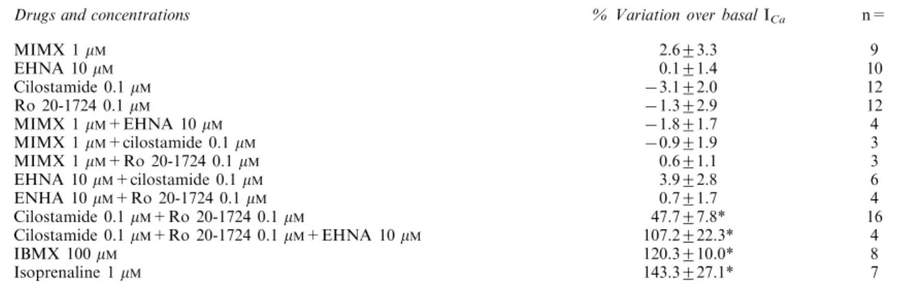
Table 1 Eect of PDE inhibitors and isoprenaline on basal ICa
Drugs and concentrations % Variation over basal ICa n=
MIMX 1 mM EHNA 10 mM Cilostamide 0.1 mM Ro 20-1724 0.1 mM MIMX 1 mM+EHNA 10 mM MIMX 1 mM+cilostamide 0.1 mM MIMX 1 mM+Ro 20-1724 0.1 mM EHNA 10 mM+cilostamide 0.1 mM ENHA 10 mM+Ro 20-1724 0.1 mM Cilostamide 0.1 mM+Ro 20-1724 0.1 mM
Cilostamide 0.1 mM+Ro 20-1724 0.1 mM+EHNA 10 mM
IBMX 100 mM Isoprenaline 1 mM 2.6+3.3 0.1+1.4 73.1+2.0 71.3+2.9 71.8+1.7 70.9+1.9 0.6+1.1 3.9+2.8 0.7+1.7 47.7+7.8* 107.2+22.3* 120.3+10.0* 143.3+27.1* 9 10 12 12 4 3 3 6 4 16 4 8 7 The result are the means+s.e.mean of the number of experiments indicated in the last column. They are expressed in % variation over basal ICaamplitude. The asterisks indicate when the eects were statistically signi®cant at the P50.01 level (*).
Figure 3 Eects of cilostamide, Ro20-1724 and EHNA on ICain a
rat ventricular myocyte. The cell was superfused for several minutes with a control solution and then challenged with dierent drugs during the periods indicated by the solid lines. While application of 0.1 mMcilostamide had no eect on basal ICa, application of 0.1 mM
Ro20-1724 on top of cilostamide or of EHNA (10 mM) on top of
cilostamide and Ro20-1724 induced a net increase in ICa. Test pulses
were 0 mV except during the recording of current-voltage (I ± V) relationships. The individual current traces shown on the upper part were obtained at the times indicated by the corresponding letters in the bottom graph. The dotted line indicates the zero current level.
Figure 4 Eects of cilostamide, Ro20-1724 and EHNA on the current-voltage and inactivation relationships of ICa in a rat
ventricular myocyte. (A) current-voltage relationships and (B) inactivation curves for ICa obtained in the experiment shown in
Figure 3 in control (squares), in the presence of 0.1 mMcilostamide
and 0.1 mM Ro20-1724 (Cil+Ro, circles), and in the presence of
0.1 mM cilostamide, 0.1 mM Ro20-1724 and 10 mM EHNA (Cil+-Ro+EHNA, triangles). Inactivation curves were obtained using a double-pulse protocol indicated in the inset and described in Methods.
drugs which indicates that neither the voltage-dependent nor the Ca-dependent inactivation process (McDonald et al., 1994) was modi®ed.
Each of the selective PDE inhibitors was tested in the presence of another inhibitor. This led to six dierent experimental conditions, and the results of these experiments are summarized in Table 1. Of all the combinations tested, only Ro20-1724 and cilostamide had a signi®cant stimulatory eect on basal ICa. The dual inhibition of PDE3 and PDE4
resulted in a &50% increase in basal ICa, an eect which
corresponded to &40% of the stimulatory eect observed in the presence of 100 mMIBMX (Table 1). At this concentration,
IBMX increased maximally ICa, since its eect was not
statistically dierent from the stimulation induced by a saturating concentration (1 mM) of isoprenaline (Table 1).
PDE1 inhibition (with 1 mMMIMX) had no eect on basal ICa
under any condition tested (data not shown). PDE2 inhibition (with 10 mM EHNA) had no eect unless PDE3 and PDE4
were simultaneously blocked (data not shown). As shown in Table 1, the concomitant inhibition of PDE2, PDE3 and PDE4 with a combination of EHNA, cilostamide and Ro20-1724 led to a similar increase in ICa as that produced by IBMX or
isoprenaline.
Eect of PDE inhibitors on isoprenaline-stimulated ICa
We next examined the respective contribution of each PDE subtype in the regulation of phosphorylated Ca2+channels. To
do this, we examined the eects of each selective PDE inhibitor on ICawhich had been stimulated by isoprenaline. However, it
was important to use a non-saturating concentration of the b-adrenoceptor agonist in order to allow ICa to be further
increased by the drugs. A concentration of 1 nMisoprenaline
was chosen, which increased ICaby 38.1+4.1% (n=82, Figure
5), i.e. which produced &30% of the maximal stimulatory eect observed with 1 mM isoprenaline or 100 mM IBMX
(Table 1).
Figure 5 shows a typical experiment in which the eects of PDE2 and PDE3 inhibition with EHNA and cilostamide, respectively, were successively tested on a rat ventricular myocyte exposed to 1 nMisoprenaline. While, as shown above, 100 nM cilostamide or 10 mM EHNA had no eect on basal
ICa, as low as 30 nMcilostamide or 3 mMEHNA induced a net
and reversible stimulation of ICaon top of that obtained with
isoprenaline alone. A similar ®nding was obtained with PDE4 inhibition by Ro20-1724. Indeed, Figure 6 shows that application of 30 nMRo20-1724 induced a net further increase
in ICain the presence of 1 nMisoprenaline, while this drug had
no eect on basal ICa when used at a 3 fold higher
concentration (Table 1).
Figure 7 summarizes the results of several similar experiments in which various concentrations of EHNA (Figure 7A), cilostamide (Figure 7B) and Ro20-1724 (Figure 7C) were tested for their eect on ICapre-stimulated by 1 nM
isoprena-line. While all these drugs induced an additional increase in ICa,
the maximal stimulatory eect was the highest with Ro20-1724 (&50% at 30 ± 100 nM), intermediate with cilostamide (&30% at 0.3 ± 1 mM) and the lowest with EHNA (&20% at 1 ±
10 mM). Under the same conditions (with 1 nM isoprenaline), PDE1 inhibition with 3 mMMIMX produced only a weak but
signi®cant (P50.05) additional stimulation of ICa(8.1+1.2%,
n=5).
Eect of PDE inhibitors on intracellular cyclic AMP concentration
To get further insights into the mechanism of action of the dierent PDE inhibitors tested on ICa, we examined their
eects on intracellular cyclic AMP concentration ([cAMP]i) in
isolated rat ventricular myocytes. Figure 8 shows the results of cyclic AMP assays performed in ventricular myocytes isolated
Figure 5 Eects of cilostamide and EHNA on ICapre-stimulated by
isoprenaline. The cell was superfused for few minutes with a control solution and then challenged with dierent drugs during the periods indicated by the solid lines. Application of 1 nMisoprenaline slightly
increased ICa. Addition of 30 nMcilostamide or of 3 or 10 mMEHNA
in the continuing presence of isoprenaline induced an additional and reversible increase in ICa. The individual current traces shown on the
upper part were obtained at the times indicated by the corresponding letters in the bottom graph. The dotted line indicates the zero current level.
Figure 6 Eects of R020-1724 on ICapre-stimulated by isoprenaline.
The cell was superfused for few minutes with a control solution and then challenged with dierent drugs during the periods indicated by the solid lines. Application of 1 nM isoprenaline increased ICa.
Addition of 30 nM Ro 20-1724 in the continuing presence of
isoprenaline induced an additional and reversible increase in ICa.
The individual current traces shown on the upper part were obtained at the times indicated by the corresponding letters in the bottom graph. The dotted line indicates the zero current level.
from three dierent rat hearts (symbols) together with the mean values (bars). Although one preparation responded by a large increase in [cAMP]iwith any of the four PDE inhibitors
tested (Figure 8A, ®lled circles), on average the eects of MIMX (1 mM), EHNA (10 mM), cilostamide (0.1 mM) and
Ro20-1724 (0.1 mM) on basal [cAMP]i were small (20 ± 30%)
and not statistically signi®cant (Figure 8A). However, when the inhibitors were used in combination to achieve a concomitant inhibition of two PDE subtypes (tested only on PDE2-4), [cAMP]iincreased by 40 ± 50% and the eect became
statistically signi®cant (P50.05, Figure 8A). [cAMP]i
in-creased further (to &80%) when all three PDE subtypes were inhibited, an eect which was similar to that obtained with a maximal concentration of IBMX (100 mM, Figure 8A). Figure
8B shows the eects of the selective PDE inhibitors on [cAMP]i
when applied in the presence of a sub-maximal concentration of isoprenaline (1 nM). Although the preparation that
responded to any single PDE inhibitor also responded by a large increase in [cAMP]i with 1 nMisoprenaline (Figure 8B,
®lled circles), the average stimulatory eect of 1 nM
isoprena-line was small (20%) and not statistically signi®cant (Figure 8B). However, in the presence of isoprenaline, any single application of PDE inhibitor (tested only on PDE2-4) induced a large (60 ± 70%) increase in [AMP]i(Figure 8B), which was
similar to the eect obtained with 1 mM isoprenaline (Figure
8B) or 100 mMIBMX (Figure 8A).
Discussion
In the recent years, it has become well established that cardiac ICaas well as myocardial contractility is modulated by cyclic
AMP-dependent phosphorylation. An increase in cyclic AMP concentration activates cyclic AMP-dependent protein kinase (PKA), which phosphorylates various proteins, including the L-type Ca2+channels, leading to an increase in the mean open
probability of individual channels (McDonald et al., 1994; Hartzell et al., 1991; Hove-Madsen et al., 1996). The cyclic AMP signal, which is generated at the level of the sarcolemmal membrane by adenylyl cyclase, is terminated by cyclic nucleotide phosphodiesterases, a class of enzymes that hydrolyse cyclic AMP into 5'-AMP (Beavo, 1995).
Figure 7 Eects of EHNA, cilostamide and R020-1724 on ICa
pre-stimulated by isoprenaline. Summary of the eects of dierent concentrations of EHNA (A), cilostamide (B) and Ro20-1724 (C) on ICapre-stimulated by 1 nMisoprenaline in rat ventricular myocytes.
The bars indicate the means and the lines the s.e.mean of the numbers of experiments indicated near the bars. The eects are expressed in % variation over the amplitude of ICain the presence of
isoprenaline alone. Statistical signi®cant dierences from ICa
amplitude in isoprenaline alone are indicated as: *P50.05; **P50.01; ***P50.005.
Figure 8 Eects of PDE inhibitors and isoprenaline on [cAMP]iin
isolated rat ventricular myocytes. The selective PDE inhibitors MIMX (1 mM), EHNA (10 mM), cilostamide (0.1 mM) and
Ro20-1724 (0.1 mM) were tested as indicated in three dierent preparations of rat ventricular myocytes (corresponding to the dierent symbols used in A and B) in the absence (A) or presence (B) of 1 nM
isoprenaline. The eects are expressed in % increase over basal cyclic AMP concentration which was, respectively, 59 (*), 52 (&) and 22 (*) pmoles per mg protein, assuming that each myocyte contained 15 ng protein. Each individual measurement was done in triplicate. The bars indicate the average response in each condition. Separate experiments were performed with IBMX (100 mM, A) and
isoprena-line (1 mM, B), and the corresponding bars indicate the mean and the
s.e.mean of the number of preparations indicated near the bars. Statistical signi®cant dierences from basal cyclic AMP concentration are indicated as: *P50.05.
At present, four dierent subtypes of PDE (PDE1-4) have been characterized in mammalian hearts (Taira et al., 1993; Engels et al., 1994; Kostic et al., 1997). Their intracellular localization and/or respective activities may vary depending on the animal species and/or the cardiac tissue (for reviews, see Beavo, 1995; Stoclet et al., 1995). In our study, we have con®rmed by RT ± PCR the presence of mRNA transcripts encoding for all four PDE subtypes in isolated rat ventricular myocytes.
Investigation of their functional role has been made possible by the development of pharmacological agents that inhibit selectively their activity. A number of compounds, such as the bypiridine derivatives amrinone and milrinone, have long been used as selective PDE3 inhibitors with therapeutic applica-tions. Among these, cilostamide is by far the most potent inhibitor of PDE3 (Reeves & England, 1990). The anti-depressants Ro-20-1724 or rolipram potentiate the positive inotropic eect of forskolin on the heart by acting as selective inhibitors of PDE4 (Muller et al., 1990). The adenosine deaminase inhibitor EHNA was recently characterised as a potent PDE2 inhibitor, which is also selective of this PDE subtype as far as other PDEs are concerned (MeÂry et al., 1995; Podzuveit et al., 1995). Potent and selective inhibitors of PDE1 are still to come, although the 8-methoxymethyl-derivative of IBMX, MIMX, has been shown to possess some selectivity at the PDE1 subtype (Wells & Miller, 1988).
In the present study, we used the broad-spectrum PDE inhibitor IBMX as well as the selective inhibitors MIMX, EHNA, cilostamide and Ro20-1724 to assess the respective roles of PDE1-4 in the regulation of basal and stimulated ICa
and [cAMP]i in rat ventricular myocytes. Some of these PDE
inhibitors have been shown earlier to increase ICaby elevating
[cAMP]i. For instance, application of IBMX leads to a large
stimulation of basal ICain guinea-pig (Mubagwa et al., 1997),
rabbit (Akita et al., 1994; Kajimoto et al., 1997) and human cardiomyocytes (Kajimoto et al., 1997). Similarly, we found that IBMX also increased basal ICain rat ventricular myocytes.
This indicates that, in all these preparations: (i) there is a substantial basal activity of adenylyl cyclase which determines a basal cyclic AMP synthesis, and (ii) there is a signi®cant basal PDE activity which limits the degree of cyclic AMP-dependent phosphorylation of L-type Ca2+-channels. The
situation in mammals diers from what was found in frog ventricular myocytes, where IBMX had no eect on ICaunless
the current had been pre-stimulated by isoprenaline, forskolin or cyclic AMP (Fischmeister & Hartzell, 1990; 1991). Thus, in frog cardiomyocytes, unlike in most mammalian species, basal adenylyl cyclase activity is insucient to generate enough cyclic AMP to lead to a stimulation of ICaeven when all PDE
activity is blocked.
For this reason, selective PDE inhibitors, such as EHNA or milrinone, have no eect on basal ICa in frog myocytes
(Fischmeister & Hartzell, 1990; MeÂry et al., 1995) while they do stimulate basal ICain human atrial myocytes (Kirstein et al.,
1995; Kajimoto et al., 1997; Rivet-Bastide et al., 1997). Similarly, PDE3 inhibitors stimulate basal ICa in rabbit
cardiomyocytes (Kajimoto et al., 1997) and exert a positive inotropic eect in guinea-pig left atria (Muller et al., 1990). However, in this study, we found that none of the selective PDE inhibitors used alone had any signi®cant eect on basal ICaor [cAMP]iin rat ventricular myocytes. Thus, inhibition of
a single PDE subtype is insucient to raise cyclic AMP concentration near Ca2+channels to the threshold
concentra-tion necessary for ICastimulation. The likely explanation for
this is that other PDE subtypes accounted for cyclic AMP hydrolysis under such condition. To test this hypothesis, we
tried to block two PDE subtypes at the same time using combinations of PDE inhibitors. The only `duo' of inhibitors that was found to enhance basal ICawas cilostamide and
Ro20-1724, indicating that PDE3 and PDE4 were the dominant PDE subtypes regulating basal cyclic AMP concentration and ICain
rat ventricular myocytes. These results are in agreement with a number of biochemical studies showing a much higher speci®c activity of PDE3 and PDE4 compared with PDE1 and PDE2 in rat heart (Shahid & Nicholson, 1990; Bode et al., 1991; Dubois et al., 1993; Picq et al., 1996). Also, functional experiments have shown that inhibition of PDE3 or PDE4 alone had no eect on the contractile activity of rat heart while a dual PDE3 and PDE4 inhibition produced a strong positive inotropic eect (Shahid & Nicholson, 1990).
Surprisingly, however, we found that not only cilostamide and Ro20-1724 but also any other `duo' combination of PDE2-4 inhibitors induced a substantial elevation in [cAMP]i
in rat ventricular myocytes. This dierence between ICa and
[cAMP]imeasurements may indicate the presence of dierent
pools of cyclic AMP inside cardiac myocytes, which are not all associated with L-type Ca2+ channels. Such a hypothesis is
supported by recent ®ndings of a strong cyclic AMP compartmentation near L-type Ca2+ channels in frog
ventricular myocytes exposed to isoprenaline (Jurevicius & Fischmeister, 1996). It is also supported by a large body of evidence for distinct intracellular localization of the dierent PDE subtypes. For instance, a substantial amount of PDE3 and PDE4 activity was found in a membrane-bound fraction of rat cardiomyocytes (Weishaar et al., 1987; Shahid et al., 1990; Kaasic & Ohisalo, 1996; Okruhlicova et al., 1996) which may accentuate the role of these PDE subtypes in the control of sarcolemmal processes, such as cyclic AMP-dependent phosphorylation of L-type Ca2+ channels. PDE3 and PDE4
have also a 5 ± 30 fold lower Km for cyclic AMP than PDE2
(Bode et al., 1991; Beavo, 1995), which should attenuate the role of PDE2 in ICa regulation when [cAMP]i near the
membrane is low. This may explain why EHNA did not aect ICaunless PDE3 and PDE4 were blocked or when their activity
was saturated by an enhanced cyclic AMP synthesis in the presence of isoprenaline.
Regulation of ICaby PDE1 in heart remains unclear. In this
study, we found that the PDE1 inhibitor MIMX (Wells & Miller, 1988) had negligible eects on basal ICaand increased
only by 8% isoprenaline stimulated ICa. However, a major
drawback of our experiments is that the solution used in the patch-pipette was adjusted to a free Ca2+concentration of pCa
8.5 using 5 mMEGTA. Although the Ca2+buering capacity
of the pipette solution should only weakly aect the Ca2+
concentration near the membrane, as evidenced by the presence of a large Ca2+-mediated inactivation of I
Ca under
these conditions, it might abolish a Ca2+-calmodulin
dependent activation of PDE1 in a remote part of the cell. Nystatin-perforated patch-clamp experiments would be neces-sary to elucidate the role of PDE1 in the control of ICa.
However, it should be mentioned that biochemical studies have shown that rat heart possesses a much lower PDE1 activity as compared to other PDE subtypes (Dubois et al., 1993, Shahid & Nicholson, 1990, Bode et al., 1991).
To the exception of MIMX, all other PDE inhibitors tested exerted a clear stimulation of ICa after application of 1 nM
isoprenaline. This low concentration of the B-adrenoceptor agonist moderately increased the activity of adenylyl cyclase localized in the membrane, bringing the cyclic AMP concentration above threshold for cyclic AMP-dependent phosphorylation of L-type Ca2+channels. In these conditions,
enhance ICa as well as [cAMP]i. Thus, unlike what was
observed under basal conditions, inhibition of any single PDE2-4 subtype stimulates Ca2+ channel activity when the
intracellular cyclic AMP level is above threshold. At their maximal eects, the rank order of potency of the dierent PDE inhibitors on isoprenaline stimulated ICa was
17244ci-lostamide4EHNA4MIMX. However, in the case of Ro20-1724, increasing the concentration above the maximal eective concentration (100 nM) produced lower stimulatory eects on ICa(Figure 7C). While, we have no clear explanation for this
eect, a similar phenomenon was observed in contractile experiments on guinea-pig heart by Muller et al. (1990) with another PDE4 inhibitor, rolipram. An interesting possibility is that the increase in cyclic AMP and PKA activity that results from PDE4 inhibition leads to phosphorylation and activation of PDE4 as has been shown recently in thyroid cells (Sette & Conti, 1996).
In summary, our results demonstrate that PDE2, PDE3 and PDE4 regulate ICa in rat ventricular myocytes. PDE3 and
PDE4 are the dominant PDE subtypes involved in the
regulation of basal ICa. Each of these two PDEs possesses a
sucient enzymatic activity to fully hydrolyse alone the cyclic AMP produced by the basal activity of adenylyl cyclase. This double hydrolytic system may prevent intracellular cyclic AMP from rising immoderately in the absence of stimulatory hormones or neuromediators. When the cell is challenged by a stimulus activating cyclic AMP production, such as a b-adrenoceptor agonist, all PDE subtypes become determinant in regulating the intracellular cyclic AMP concentration. Hence, the response of ICato a sympathetic stimulation will
be determined by the activity of each PDE subtype, with the rank order of potency PDE44PDE34PDE24PDE1.
We thank Patrick LecheÃne for excellent technical help, and Florence Lefebvre and Isabelle Paic for preparation of the myocytes. Ignacio Verde was supported by a fellowship from the Fundacion Caixa-Galicia.
References
AKITA, T., JOYNER, R.W., LU, C.B., KUMAR, R. & HARTZELL, H.C.
(1994). Developmental changes in modulation of calcium currents of rabbit ventricular cells by phosphodiesterase inhibitors. Circulation, 90, 469 ± 478.
BEAVO, J.A. (1995). Cyclic nucleotide phosphodiesterases : func-tional implications of multiple isoforms. Physiol. Rev., 75, 725 ± 748.
BODE, D.C., KANTER, J.R. & BRUNTON, L.L. (1991). Cellular distribution of phosphodiesterase isoforms in rat cardiac tissue. Circ. Res., 68, 1070 ± 1079.
DUBOIS, M., PICQ, M., NEÂMOZ, G., LAGARDE, M. & PRIGENT, A.F.
(1993). Inhibition of dierent phosphodiesterase isoforms of rat heart cytosol by free fatty acids. J. Cardiovasc. Pharmacol., 21, 522 ± 529.
ENGELS, P., FICHTEL, K., LUBBERT, H. (1994). Expression and regulation of human and rat phosphodiesterase type IV isogenes. FEBS Lett., 350, 291 ± 295.
FISCHMEISTER, R. & HARTZELL, C.(1990). Regulation of calcium current by low-Km cyclic AMP phosphodiesterases in cardiac
cells. Mol. Pharmacol., 38, 426 ± 433.
FISCHMEISTER, R. & HARTZELL, H.C. (1991). Cyclic AMP phosphodiesterases and Ca2+current regulation in cardiac cells.
Life Sci., 48, 2365 ± 2376.
GAO, T.Y., YATANI, A., DELLACQUA, M.L., SAKO, H., GREEN, S.A., DASCAL, N., SCOTT, J.D. & HOSEY, M.M. (1997). cAMP-dependent regulation of cardiac L-type Ca2+channels requires
membrane targeting of PKA and phosphorylation of channel subunits. Neuron, 19, 185 ± 196.
GRIMALDI, B., BONNIN, A., FILLION, M.P., RUAT, M., TRAIFFORT, E., FILLION, G.(1998). Characterization of 5-HT6receptor and
expression of 5-HT6mRNA in the rat brain during ontogenetic
development. Naunyn-Schmied. Arch. Pharmacol., 357, 393 ± 400.
HAMILL, O.P., MARTY, A., NEHER, E., SAKMANN, B. & SIGWORTH, F.J.(1981). Improved patch-clamp techniques for high-resolution current recording from cells and cell-free membrane patches. P¯uÈgers Arch., 391, 85 ± 100.
HARTZELL, H.C., MEÂRY, P.F., FISCHMEISTER, R. & SZABO, G.
(1991). Sympathetic regulation of cardiac calcium current is due exclusively to cAMP-dependent phosphorylation. Nature, 351, 573 ± 576.
HOVE-MADSEN, L., MEÂRY, P.-F., JUREVICIUS, J., SKEBERDIS, A.V. & FISCHMEISTER, R.(1996). Regulation of myocardial calcium channels by cyclic AMP metabolism. Basic Res. Cardiol., 91 (Suppl. 2), 101 ± 108.
JUREVICIUS, J. & FISCHMEISTER, R.(1996). cAMP compartmenta-tion is responsible for a local activacompartmenta-tion of cardiac Ca2+channels
by beta-adrenergic agonists. Proc. Natl Acad. Sci. USA, 93, 295 ± 299.
KAASIC, A. & OHISALO, J.J.(1996). Membrane-bound phosphodies-terases in rat. J. Pharm. Pharmacol., 48, 962 ± 964.
KAJIMOTO, K., HAGIWARA, N., KASANUKI, H. & HOSODA, S.
(1997). Contribution of phosphodiesterase isoenzymes to the regulation of the L-type calcium current in human cardiac myocytes. Br. J. Pharmacol., 121, 1549 ± 1556.
KATANO, Y. & ENDOH, M.(1993). Cyclic AMP metabolism in intact rat ventricular cardiac myocytes: interaction of carbachol with isoproterenol and 3-isobutyl-1-methylxanthine. Mol. Cell. Bio-chem., 119, 195 ± 201.
KIRSTEIN, M., RIVET-BASTIDE, M., HATEM, S., BENARDEAU, A., MERCADIER, J.J. & FISCHMEISTER, R. (1995). Nitric oxide regulates the calcium current in isolated human atrial myocytes. J. Clin. Invest., 95, 794 ± 802.
KOSTIC, M.M., ERDOGAN, S., RENA, G., BORCHERT, G., HOCH, B., BARTEL, S., SCOTLAND, G., HUSTON, E., HOUSLAY, M.D., & KRAUSE, E.G. (1997). Altered expression of PDE1 and PDE4 cyclic nucleotide phosphodiesterase isoforms in 7-oxo-prostacy-clin-preconditioned rat heart. J. Mol. Cell. Cardiol., 29, 3135 ± 3146.
LEVI, R.C., ALLOATI, G. & FISCHMEISTER, R.(1989). Cyclic GMP regulates the Ca-channel current in guinea pig ventricular myocytes. P¯uÈgers Arch., 413, 685 ± 687.
LOUGHNEY, K. & FERGUSON, K. (1996). Identi®cation and quanti®cation of PDE isoenzymes and subtypes by molecular biological methods. In Phosphodiesterase inhibitors. En, S., Schudt, C., Dent, G. & Rabe, K.F., ed. pp 1 ± 19. San Diego: Academic Press Inc.
MCDONALD, T.F., PELZER, S., TRAUTWEIN, W. & PELZER, D.J. (1994). Regulation and modulation of calcium channels in cardiac, skeletal, and smooth muscle cells. Physiol. Rev., 74, 365 ± 507.
MEÂRY, P.F., LOHMANN, S.M., WALTER, U. & FISCHMEISTER, R.
(1991). Ca2+ current is regulated by cyclic GMP-dependent
protein kinase in mammalian cardiac myocytes. Proc. Natl. Acad. Sci. USA, 88, 1197 ± 1201.
MEÂRY, P.F., PAVOINE, C., PECKER, F. & FISCHMEISTER, R.(1995). Erythro-9-(2-hydroxy-3-nonyl)adenine inhibits cyclic GMP-sti-mulated phosphodiesterases in isolated cardiac myocytes. Mol. Pharmacol., 48, 121 ± 130.
MUBAGWA, K., SHIRAYAMA, T., MOREAU, M. & PAPPANO, A.J.
(1997). Eects of PDE inhibitors and carbachol on the L-type Ca current in guinea pig ventricular myocytes. Am. J. Physiol., 264, H1323 ± H1363.
MULLER, B., LUGNIER, C. & STOCLET, J.C.(1990). Involvement of rolipram-sensitive cyclic AMP phosphodiesterase in the regula-tion of cardiac contracregula-tion. J. Cardiovasc. Pharmacol., 16, 796 ± 803.
OKRUHLICOVA, L., TRIVULOBA, N., ECKLY, A., LUGNIER, C., SLEZAK, J. (1996). Cytochemical distribution of cyclic AMP-dependent 3',5'-nucleotide phosphodiesterase in the rat myocar-dium. Histochem. J., 28, 165 ± 172.
ONO, K. & TRAUTWEIN, W.(1991). Potentiation by cyclic GMP of b-adrenergic eect on Ca2+current in guinea pig ventricular cells.
J. Physiol., 443, 387 ± 404.
PICQ, M., DUBOIS, M., GRYNBERG, A., LAGARDE, M. & PRIGENT, A.F.(1996). Speci®c eects of n-3 fatty acids and 8-bromo-cGMP on the cyclic nucleotide phosphodiesterase activity in neonatal rat cardiac myocytes. J. Mol. Cell. Cardiol., 28, 2151 ± 2161.
PODZUWEIT, T., NENNSTIEL, P. & MULLER, A. (1995). Isozyme selective inhibition of cGMP-stimulated cyclic nucleotide phosphodiesterases by erythro-9-(2-hydroxy-3-nonyl) adenine. Cell. Signal., 7, 733 ± 738.
REEVES, M.L. & ENGLAND, P.J.(1990). Cardiac Phosphodiesterases and the Functional Eects of Selective Inhibition. In: Cyclic nucleotide phosphodiesterases: structure, regulation and drug action, 2, pp. 299 ± 316.
RIVET-BASTIDE, M., VANDECASTEELE, G., VERDE, I., HATEM, S., BENARDEAU, A., MERCADIER, J.J. & FISCHMEISTER, R.(1997). cGMP-stimulated cyclic nucleotide phosphodiesterase regulates the basal calcium current in human atrial myocytes. J. Clin. Invest., 99, 2710 ± 2718.
SETTE, C. & CONTI, M.(1996). Phosphorylation and activation of a cAMP-speci®c phosphodiesterase by the cAMP-dependent protein kinase ± Involvement of serine 54 in the enzyme activation. J. Biol. Chem., 271, 16526 ± 16534.
SHAHID, M. & NICHOLSON, C.D. (1990). Comparison of cyclic nucleotide phosphodiesterase isoenzymes in rat and rabbit ventricular myocardium, positive inotropic and phosphodiester-ase inhibitory eects of Org 30029, milrinone and rolipram. Naunyn-Schmiedeberg's Arch. Pharmacol., 342, 698 ± 705.
SHAHID, M., NICHOLSON, C.D. & MARSHALL, R.J.(1990). Species-dependent dierences in the properties of particulate cyclic nucleotide phosphodiesterase from rat and rabbit ventricular myocardium. J. Pharm. Pharmacol., 42, 283 ± 284.
SHIRIYAMA, T. & PAPPANO, A.J. (1996). Bifasic eects of intrapipette cyclic guanosine monophosphate on L-type calcium current and contraction of guinea pig ventricular myocytes. J. Pharmacol. Exp. Ther., 279, 1274 ± 1281.
STOCLET, J.C., KERAVIS, T., KOMAS, N. & LUGNIER, C.(1995). Cyclic nucleotide phosphodiesterases as therapeutic targets in cardiovasculasr diseases. Exp. Opin. Invest. Drugs, 4, 1081 ± 1100.
TAIRA, M., HOCKMAN, S.C., CALVO, J.C., TAIRA, M., BELFRAGE, P., & MANGANIELLO, V.C.(1993). Molecular Cloning of the Rat Adipocyte Hormone-Sensitive Cyclic GMP-Inhibited Cyclic Nucleotide Phosphodiesterase. J. Biol. Chem., 268, 18573 ± 18579.
VERDE, I., VANDECASTEELE, G. & FISCHMEISTER, R. (1997). PDE4 regulates the L-type calcium current in human atrial and rat ventricular myocytes. J. Mol. Cell. Cardiol., 29, A36.
WEISHAAR, R.E., KOBYLARZ-SINGER, D.C. & KAPLAN, H.R.
(1987). Subclasses of cyclic AMP phosphodiesterase in cardiac muscle. J. Mol. Cell. Cardiol., 19, 1025 ± 1036.
WELLS, J.N. & MILLER, J.R.(1988). Methylxanthine inhibitors of phosphodiesterases. Meth. Enzymol., 159, 489 ± 496.
WIECHEN, K., YUE, D.T. & HERZIG, S. (1995). Two distinct functional eects of protein phosphatase inhibitors on guinea-pig cardiac L-type Ca2+channels. J. Physiol. (Lond.), 484, 583 ±
592.
(Received May 26, 1998 Revised January 28, 1999 Accepted February 2, 1999)


![Figure 8 Eects of PDE inhibitors and isoprenaline on [cAMP] i in isolated rat ventricular myocytes](https://thumb-eu.123doks.com/thumbv2/123dok_br/18081675.865583/7.892.68.416.99.645/figure-ects-pde-inhibitors-isoprenaline-isolated-ventricular-myocytes.webp)