Janeiro de 2015
Soraia Raquel Azevedo Pereira
Modulation of Bax by PKC :
An approach to eliminate cancer cells
U minho | 20 15 Sor aia P er eir a Modulation of Bax b y PK
Tese de Mestrado em Bioquímica Aplicada
Trabalho efectuado sob a orientação de
Doutora Susana Chaves
Professora Doutora Manuela Côrte-Real
Janeiro de 2015
Soraia Raquel Azevedo Pereira
Modulation of Bax by PKC :
Nome: Soraia Raquel Azevedo Pereira
Endereço eletrónico: [email protected] Telefone: 917743548
Nº do Bilhete de Identidade: 13764845
Título da Tese de Mestrado:
Modulation of Bax by PKC: An approach to eliminate cancer cells
Orientadores:
Doutora Susana Chaves
Professora Doutora Manuela Côrte-Real
Instituição de Acolhimento:
Centro de Biologia Molecular e Ambiental (CBMA)
Ano de Conclusão:
2015
Designação do Mestrado:
Mestrado em Bioquímica Aplicada
1. DE ACORDO COM A LEGISLAÇÃO EM VIGOR, NÃO É PERMITIDA A REPRODUÇÃO DE QUALQUER PARTE DESTA TESE/TRABALHO
Universidade do Minho, Janeiro de 2015
_____________________________________________ Soraia Pereira
iii
Agradecimentos
Ao longo deste ano pude contar com o apoio incondicional e confiança de várias pessoas. Assim, como reconhecimento da importância de todos durante este trabalho, quero expressar o meu agradecimento a todos aqueles que o tornaram possível.
Às minhas orientadoras, Doutora Susana Chaves e Professora Doutora Manuela Côrte-Real pela excecional orientação, disponibilidade e carinho, pela partilha de conhecimentos e rigor científico e por todas as sugestões diárias aos problemas que foram ocorrendo ao longo deste trabalho. Obrigada por todas as oportunidades e principalmente pela contribuição na aprendizagem de novas técnicas.
Ao CBMA e ao Departamento de Biologia e a todos os seus funcionários e docentes. Um especial à Dona Isabel e Sr. Luís por toda a boa disposição e favores prestados.
Ao FEDER e à Fundação para a Ciência e Tecnologia pelo financiamento, no âmbito dos projetos PEst-OE/BIA/UI4050/2014, FCT-ANR/BEX-BCM/0175/2012 e EXPL/BEX-BCM/0056/2012.
A todos os meus familiares, especialmente os meus pais e irmã, agradeço o incentivo durante todo este trabalho, a força, o amor e carinho e por sempre incutirem em mim a vontade de vencer e nunca desistir dos meus objetivos. Muito Obrigada.
A todos os meus colegas da Micro I por me terem proporcionado um excelente ambiente de trabalho, pelo companheirismo e bons momentos passados diariamente.
Um obrigado especial à Rita e ao António pela companhia e bons momentos passados, pela partilha de conhecimentos e por toda a ajuda prestada ao longo deste trabalho. À Lisandra, Selma e ao Dário agradeço toda a ajuda prestada.
À Andreia, minha irmã de coração, agradeço por sempre estar ao meu lado durante todo o meu percurso académico e por me apoiar e acreditar que sou sempre capaz de tudo.
Ao João, por sempre acreditar em mim e me animar nos momentos menos bons. Obrigada por todo o amor e carinho.
iv
Modulation of Bax by PKC: An approach to eliminate cancer cells
Abstract
Apoptosis is a type of cell death, known as a highly regulated program of cellular suicide; however this process is often deregulated in tumor cells. A mechanism used by tumor cells to evade apoptosis is the alteration of the different members of the Bcl-2 family. These proteins control a key step in the intrinsic apoptotic pathway: permeabilization of the outer mitochondrial membrane and release of several apoptotic factors. The pro-apoptotic members Bax and Bak are essential for this permeabilization, since deletion of either protein compromises the intrinsic apoptotic pathway. These proteins may be regulated by members of the Bcl-2 but also by other proteins, such as the protein kinase C (PKC) family.
The PKC family consists of kinases that regulate a variety of cellular functions. Of these, PKCepsilon (PKC) is involved in carcinogenesis and has been considered an oncogene. It has been shown that this protein increases the expression of anti-apoptotic members of the Bcl-2 family and inhibits the pro-anti-apoptotic members such as Bax. The interaction between Bax and PKC in mammalian cells prevents Bax translocation to the outer mitochondrial membrane, leading to a decrease in its activation. Identifying residues required for this interaction is therefore crucial to design a strategy to inhibit it.
In this work, we aimed to identify residues required for the Bax/PKC interaction. For this purpose, different domains of PKC cloned in frame with the FLAG epitope in a yeast inducible expression vector were co-expressed with Bax in
Saccharomyces cerevisiae, in order to evaluate their interaction with Bax. An
alternative approach using yeast-two-hybrid was also pursued to verify the interaction between these two proteins. Our ultimate goal is to develop specific modulators to target the PKC/Bax interaction that could open the door to new clinical opportunities for cancer treatment.
v
Modulation of Bax by PKC: An approach to eliminate cancer cells
Resumo
A apoptose é um tipo de morte celular, mais conhecida como um programa de suicídio celular altamente regulado. No entanto, em células tumorais este processo está frequentemente desregulado. Um mecanismo usado pelas células tumorais para escapar à apoptose é a alteração dos níveis de expressão dos diferentes membros da família Bcl-2. Estas proteínas controlam um passo essencial da via apoptótica intrínseca, a permeabilização da membrana mitocondrial externa e a libertação de vários fatores apoptóticos. Os membros pró-apoptóticos Bax e Bak são essenciais para esta permeabilização, pois a deleção de ambas as proteínas compromete a via apoptótica intrínseca. Estas proteínas podem ser reguladas pelos membros da família Bcl-2 mas também por outras proteínas, como as proteínas da família da cinase C PKC).
A família PKC é uma família de cinases que regulam uma grande variedade de funções celulares. Em particular, a PKCepsilon (PKC) está envolvida na carcinogénese e tem sido considerada um oncogene. Tem sido demonstrado que esta proteína aumenta a expressão dos membros anti-apoptóticos da família Bcl-2 e inibe os membros pró-apoptóticos, como a proteína Bax. A interação entre a PKC e a proteína Bax em células de mamíferos leva a que haja uma diminuição da ativação da Bax e da sua translocação para a membrana mitocondrial externa, impedindo que a Bax desempenhe a sua função na apoptose. Assim, a identificação dos resíduos responsáveis para esta interação torna-se extremamente importante para que se consiga encontrar um método de a inibir.
Neste trabalho pretendeu-se identificar os resíduos necessários para essa interação. Para este efeito, os diferentes domínios de PKC clonados como fusões com o epítopo FLAG num vector de expressão indutível em levedura, foram co-expressos com Bax em Saccharomyces cerevisiae a fim de avaliar a sua interação com Bax. Uma abordagem alternativa usando a técnica de Yeast-two-hybrid também foi efetuada para verificar a interação entre PKC e Bax. O objetivo final seria o desenvolvimento de moduladores específicos para inibir a interação PKC/ Bax de modo a abrir a porta para novas oportunidades clínicas no tratamento de cancro.
vi
Index
Agradecimentos...iii Abstract...iv Resumo...v Index...vi Abbreviations...ix Preamble……….xChapter 1- Bax modulation by PKC 1. General Introduction 1.1. Cell death...2
1.2. Apoptosis ...2
1.2.1. Apoptotic pathways...3
1.2.1.1. Major pathways and their regulation………4
1.2.2. The Bcl-2 family members……….5
1.2.2.1. Bax……….8
1.2.2.2. Bax activation and involvement in MOMP ………..9
1.2.3 The protein kinase C family ………...11
1.2.3.1. Regulation of apoptosis by PKC isoforms……….12
1.2.3.2. Protein kinase C epsilon ………..13
1.2.3.3. Regulation of Bax by PKC………..15
1.3 Yeast as a powerful model to study the regulation of Bax………...15
1.4. Aims...17
1.5. Materials and Methods ...19
1.5.1. Yeast strains and growth conditions...20
1.5.2. Plasmids...22
1.5.3.Yeast transformation by the lithium acetate method...25
1.5.4. Growth curves………26
1.5.5.Analysis of protein expression...26
1.5.5.1. Preparation of protein extracts ...26
1.5.5.2. SDS gel electrophoresis/Western Blot ...26
1.5.6. Immunoprecipitation ……….27
vii
1.6. Results...29
1.6.1. Previous Results………....30
1.6.2. Effect of expressing Bax and different PKC fragments on cell growth……….31
1.6.3. Analysis of the interaction between the Kinase domain of PKC and Bax by immunoprecippitation………35
1.6.4. Analysis of the interaction between PKC and Bax by Yeast-two-hybrid ………35
1.7. Discussion and Future perspectives...38
Chapter 2- Baxmodulation by Nt-acetyltransferase B 2. Introduction………43
2.1. Nt-acetyltransferases………43
2.1.1. Nt-acetyltransferase B………..45
2.1.2. Regulation of apoptosis by Nt-acetylation……….46
2.2. Aims...50
2.3. Materials and Methods ...52
2.3.1. Yeast strains and growth conditions...53
2.3.2. Plasmids...53
2.3.3. Growth curves and viability assay………...53
2.3.4. Accumulation of ROS (Flow Cytometry)……….54
2.3.5. Morphology and Chitin visualization………54
2.3.6. Mitochondrial and cytosolic fractionation………54
2.3.6.1. SDS gel electrophoresis/Western Blot………...55
2.4. Results...56
2.4.1. Expression of human NatB reverts the slow growth and morphology abnormality of yeast nat3Δ cells………...57
2.4.2. Expression of hNatB enhances the survival of wt and nat3Δ cells expressing Baxα and protects cells from oxidative stress...58
2.4.3. Localization of Bax and cyt c in nat3Δ cells expressing hNatB...61
viii
ix
Abbreviations
Ac-CoA- acetyl-coenzyme A AIF – Apoptosis Inducing Factor ANT - Adenine Nucleotide Translocator Apaf-1 - Apoptotic Protease Activating
Factor-1
Asn - asparagine Asp - aspartic acid
ATP - Adenosine Triphosphate
c-FLIP - Cellular-FLICE (FADD-like IL-
1β-converting enzyme)-inhibitory Protein
Caspases - Cysteine-dependent
Aspartate-specific Proteases
Cyt c – Cytochrome c DD - Death Domain
DED - Death Effector Domain
DISC - Death Inducing Signaling Complex DNA - Deoxyribonucleic Acid
DR - Death Receptors DTT - Dithiothreitol Endo G - Endonuclease G ER - Endoplasmatic Reticulum
FADD - Fas-Associated Death Domain Glu – glutamic acid
h – hours H2O- water
HtrA2/Omi - High Temperature
Requirement Protein A2
IAPs - Inhibitors of Apoptosis Proteins IMM - Inner mitochondrial membrane MAC - Mitochondrial Apoptosis-induced
Channel
Met - methionine
MOMP - Mitochondrial Outer Membrane
Permeabilization
NADH - Nicotinamide Adenine Dinucleotide NADPH - Nicotinamide Adenine
Dinucleotide Phosphate
Nat – N-terminal acetyltransferase
min - Minutes
NE – Nuclear envelope
NF-KB - Nuclear factor
kappa-light-chain-enhancer of activated B cells
OD – Optical density
OMM - Outer mitochondrial membrane PCD – Programmed Cell Death PCR - Polymerase Chain Reaction PTP - Permeability Transient Pore
RACKs - receptors for activated C kinases
ROS - Reactive Oxygen Species rpm – Rotations per minute sec - Seconds
Smac/Diablo - Second
mitochondria-derived Activator of Caspases/Direct Inhibitor of Apoptosis Protein (IAP)-Binding Protein With Low Pi
TNF- Tumor Necrosis Factor α
TNF-R - Tumor-Necrosis Factor Receptor TRADD - TNF-R-Associated Death Domain TRAIL-R - TNF-related Apoptosis-Inducing
Ligand Receptor
x
Preamble
The work developed in the scope of this thesis includes the study of Bax
modulation by PKC initially planned and presented in chapter 1, and a
study of Bax
modulation by Nt-acetyltransferase B reported in chapter 2.
Chapter1.
2
1. General Introduction
1.1. Cell death
The historical development of the concept of cell death originated in 1880 when Weigert and Cohnheim described the appearance of necrotic cell death in tissue by microscopy. In 1885, Flemming described the process chromatolysis (first name of apoptosis) (Majno & Joris, 1995), in which the nucleus of mammalian ovarian follicles broke and disappeared by a process of cell death (Kam & Ferch, 2000). Since the mid 19th century, many observations indicate that cell death plays an important role in the physiological processes of multicellular organisms, particularly during embryogenesis and metamorphosis. The term programmed cell death (PCD) was introduced in 1964 (Majno & Joris, 1995), suggesting that cell death during development is not accidental in nature, but follows a controlled sequence of steps leading to cellular destruction. In turn, the term apoptosis, a form of PCD, was first used in articles by Kerr, Wyllie and Currie in 1972 to describe a morphologically distinct form of cell death, although some features of apoptosis were explicitly described many years before (Elmore, 2007). Apoptosis was later recognized and accepted as an important characteristic mode of PCD with distinct morphologic characteristics from those found in necrosis. Nowadays, the Nomenclature Committee on Cell Death proposes the term regulated cell death (RCD) as a broader term for a genetically controlled death, in contrast to accidental cell death, and reserves the term programmed cell death for RCD instances that occur as part of a developmental program or to preserve physiologic adult tissue homeostasis. The three major pathways of RCD are: apoptotic, necrotic and autophagic (Galluzzi et
al., 2015).
1.2. Apoptosis
The term apoptosis is derived from the Greek meaning “dropping off,” and refers to the falling of leaves from trees in autumn. Since apoptosis was described by Kerr et al in the 70´s, it is used in contrast to necrosis, to describe the situation in which a cell actively pursues a course toward death upon receiving certain stimuli (Wong, 2011). This process normally occurs during differentiation, aging, and as a homeostatic mechanism to maintain populations of cells in the tissue (Elmore, 2007).
Apoptosis and necrosis are the two modes of cell death whose molecular mechanisms have been studied in most detail. Although originally thought to be mutually exclusive, recent discoveries show cellular contexts that require a balanced interaction between these two modes of cell death (Nikoletopoulou et al., 2013).
3
Autophagy is predominantly a cytoprotective process that begins with formation of autophagosomes, double membrane-bound structures surrounding cytoplasmic macromolecules and organelles destined for recycling. Autophagy has been associated with necrosis and apoptosis and plays a crucial role in cell survival and homeostasis. However, it also plays a pro-death function since excessive stress results in autophagic cell death (Nikoletopoulou et al., 2013) (Ouyang et al., 2012).
During the early process of apoptosis, cell shrinkage and pyknosis are the most visible morphological characteristics. With cell shrinkage, the cells are smaller in size, the cytoplasm is dense and the organelles are more tightly packed. Biochemical changes include chromosomal DNA cleavage into internucleosomal fragments, phosphatidylserine externalization and cleavage of a number of intracellular substrates by specific proteolysis (Ouyang et al., 2012) (Figure 1).
1.2.1. Apoptotic pathways
Similarly to other pathways, apoptosis can be initiated by activation of various internal and external factors, such as activation of death receptors at the plasma membrane or through mitochondrial membrane permeabilization.
The two most common apoptotic pathways are usually described as intrinsic (mitochondrial) and extrinsic (death receptor), which eventually can lead to a common
Figure 1. Molecular and morphological events associated with the different types of cell
death: apoptosis, autophagic cell death and necrosis (Tan et al., 2014).
Autophagic cell death
4
path; the third is less well known, and is executed through the endoplasmic reticulum (Wong, 2011) (Figure 2).
1.2.1.1. Major apoptotic pathways and their regulation
The extrinsic pathway is one of the best characterized apoptotic signaling pathways and is triggered when death ligands bind to a death receptor. Although several death receptors have been described, the best characterized are the tumor necrosis factor TNF 1 receptor (TNFR1) and Fas (CD95); its ligands are referred to as TNF and Fas (FasL), respectively (Wong, 2011). In Fas signaling, FasL binds to Fas, leading to receptor trimerization. Adaptor proteins bind to the cytosolic death domains (DD) of Fas protein (Fas-associated death domain,FADD) via their DDs. In addition, FADD contains a death effector domain (DED), to which the DED of pro-caspase-8 can bind. The complex of Fas, FasL, FADD and pro-caspase-8 is called the DISC. The procaspase-8 molecules are brought into close proximity in the DISC, so that they can transactivate one another. Active caspase-8 then can directly cleave caspase-3 or other executioner caspases, eventually leading to the apoptotic outcome. Its active form, caspase 3, will cleave the inhibitor of caspase-activated deoxyribonuclease, responsible for nuclear apoptosis (Lawen, 2003). DISC signaling can be inhibited by c-FLIP, a dominant negative caspase-8, which leads to the formation of a signaling-inactive DISC.
Figure 2. Schematic representation of death receptor (DR) and mitochondrial pathways for the induction of apoptosis (Pope, 2002).
5
The intrinsic apoptotic pathway may be activated in response to stress conditions, such as hypoxia, oncogene activation, or DNA damage caused by radiation or chemicals (Jia et al., 2012). The apoptotic signal leads to the release of cytochrome c (cyt c) from the mitochondrial intermembrane space into the cytosol, where it binds to the Apoptotic Protease Activating Factor-1 (Apaf-1).
The mitochondrial pathway of apoptosis begins with permeabilization of the mitochondrial outer membrane (MOMP). The mechanisms through which this occurs remain controversial, thought permeabilization is usually characterized as either permeability transition pore- (PTP) dependent or independent. The exact molecular composition of the PT is still unknown but the main components are the matrix protein cyclophilin D, the inner mitochondrial membrane protein adenine nucleotide translocator (ANT), and the outer mitochondrial membrane protein voltage dependent anion channel (VDAC) (Green & Kroemer, 2004).
In response to apoptotic stimuli, pro-apoptotic members of the Bcl-2 protein family (Bax and Bak) become activated and act on the mitochondria to induce the release of cyt c. Other pro-apoptotic proteins are also released by the mitochondria, including Smac/Diablo (Second Mitochondrial derived activator of Caspase/Direct IAP-Binding protein with a Low pI), the serine protease Omi/HtrA2, endonuclease G (EndoG) and apoptosis inducing factor (AIF) (Portt et al., 2011).
The cytoplasmic release of cyt c will activate caspase 3 by forming a complex known as the apoptosome, which is formed by binding of cyt c to the cytoplasmic adapter protein Apaf-1, ATP or dATP, which recruits pro-caspase 9 (cysteine protease); the heptamer formed by these apoptotic factors activates pro-caspase 3. Smac/DIABLO released from the mitochondrial intermembrane space enhances this cascade by binding to IAP (inhibitor of apoptosis protein), which subsequently leads to disruption of the interaction between IAPs and caspase 3, promoting apoptosis (Sukhanova et al., 2012).
1.2.2. The Bcl-2 family members
The Bcl-2 gene was first identified by its involvement in B-cell lymphomas, and encodes a protein of 25-26 kDa. The proteins of the Bcl-2 family do not just regulate apoptosis, they also have alternate functions in other homeostatic pathways, including glucose metabolism, cell cycle checkpoints, DNA damage and regulation of mitochondrial morphology (Danial, 2007).
The Bcl-2 family consists of at least thirty related proteins characterized by the presence of up to four conserved domains called BH (Tzifi et al., 2012). Most members
6
of the Bcl-2 family contain a transmembrane domain in their C-terminus, which encodes a stretch of hydrophobic amino acids important for targeting to intracellular membranes, closely associated with their ability to regulate apoptosis. Subcellular localization studies confirmed that proteins of the Bcl-2 family are present in the mitochondrial outer membrane (MOM), in the nuclear membrane and in the endoplasmic reticulum (Brown, 1997). The BH4 domain of Bcl-2 comprises 26 amino acids and the structure shows an amphipathic character after interaction with membranes, like antimicrobial peptides (Tzifi et al., 2012).
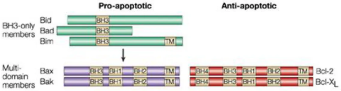
The Bcl-2 family of proteins is divided into two different sub-classes based on structural and functional characteristics: the anti-apoptotic members such as 2, Bcl-xL, Bcl-w and MCL-1 contain all four subtypes of BH domains, and promote cell survival by inhibiting the function of pro-apoptotic members. The pro-apoptotic members can be separated into two structurally distinct subfamilies: 1) "multidomain" proteins Bax and Bak, which share three BH regions and lack the BH4 domain. They are structurally similar to the anti-apoptotic proteins; 2) BH3-only proteins, including BNIP3, Nix/Bnip3L, Bid, Noxa, Puma, Bim and Bad, share only the BH3 domain and are structurally diverse (Figure 3) (Gustafsson & Gottlieb, 2007).
Expression of Bcl-2 or other related anti-apoptotic proteins, including myeloid cell leukemia-1 (Mcl-1), Bcl-XL, Bcl-w and Bcl-2-related protein A1 (Bfl-1), block cell death in response to many stimuli by preventing the activation and homooligomerization of both Bax and Bak. Anti-apoptotic proteins perform their anti-death function by sequestering BH3-only proteins or inactivating Bax and Bak. Cells that survive continuous, permanent death signaling owing to the presence of Bcl-2 are dependent on Bcl-2 for their survival (Figure 4) (Brunelle & Letai, 2009).
Figure 3. Schematic representation of members of each Bcl-2 sub-family. Bcl-2 homology (BH)
7
BH3 proteins with high affinity for binding and activating Bax and Bak are designated as “activators,” and those that only bind to antiapoptotic proteins are called “sensitizers.”The activators interact and activate Bax and Bak to promote MOMP while sensitizer proteins compete for binding with the antiapoptotic proteins releasing the activators and promoting MOMP through activation and oligomerization of Bax and Bak (Shamas-Din et al., 2013). The antiapoptotic proteins inhibit MOMP by sequestering the BH3 activators.
Figure 4. Models of Bcl-2 protein regulation in apoptosis. A: BH3-only proteins bind to and neutralize
anti-apoptotic Bcl-2 proteins, allowing Bax/Bak to become activated and initiate apoptosis. B: BH3-only proteins directly activate pro-apoptotic Bax/Bak protein. C: Anti-apoptotic Bcl-2 proteins sequester BH3-only proteins and keep them inactive. Image adapted from (Brunelle & Letai, 2009).
8
1.2.2.1. Bax
Bax resides mainly in the cytosol and translocates to the mitochondria after induction of apoptosis. The mitochondrial form of Bax found in non-apoptotic cells is a monomer of 21 kDa weakly associated with the MOM or soluble in the cytosol. After induction of apoptosis, the monomer progresses to a complex of high molecular weight (96-260 kDa), suggesting that the oligomerization of Bax occurs after insertion in the MOM (Cartron et al., 2008) (Er et al., 2006). A small portion of Bax was also found in the endoplasmic reticulum, although the function of this subpopulation remains undetermined. The Bax protein is composed of nine helices connected by short loops. Three of these helices, namely α5, α6 and α9, are probably involved in the interaction of Bax with the mitochondrial outer membrane. Indeed, the helices 5 and 6 of the Bax hydrophobic hairpin may independently form pores in lipid membranes (Walensky & Gavathiotis, 2011). The α9 helix is hydrophobic, and although it has the necessary characteristics of a transmembrane helix, experimental data suggest that it is not a typical transmembrane anchor of α-helices of the C-terminal of anti-apoptotic proteins of the Bcl-2 family (Renault & Manon, 2011).
The first nineteen residues of Bax are very mobile and data suggests that removal of these residues, leading to initiation of translation at the Met20 residue, results in a protein with a strong ability to be inserted into the MOM. Renault and Manon suggest that the N-terminal domain of Bax has the ability to lock the protein in a soluble and inactive conformation and that its movement is necessary to facilitate mitochondrial translocation of Bax; this domain is called "Apoptotic Regulation of Targeting" (ART). The ART of Bax contains two proline residues at position 8 and 13, and replacement of Pro8 or Pro13 by Gly promotes mitochondrial translocation of Bax in both yeast and human cells and in human cells undergoing apoptosis. This suggests that interactions between ART residues and residues located in other domains of Bax are central regulators of the movements of ART and subsequently the mitochondrial translocation of Bax (Renault & Manon, 2011).
Homo-oligomerization of Bax requires its BH3 death-domain (the α2 helix). Deletion of Bax segments suggests that its α2–α5 helices alone can oligomerize and that this core, together with α9, suffices for MOMP. Recent crosslinking studies suggest that homo-oligomerization of Bax starts when the BH3 domain of one monomer is exposed and engages the canonical binding groove (mainly α3–α5) of another activated monomer, forming “BH3-in-groove” dimers that multimerize by a separate interface (Czabotar et al., 2013).
9
1.2.2.2. Bax activation and involvement in MOMP
Several models of MOMP have been proposed: nonspecific MOM rupture or the formation of specific channels in the MOM.
One of the proposed models involves the rupture of the MOM after swelling of the mitochondrial matrix and opening of the PTP. The opening of this channel leads to swelling of mitochondria, which causes rupture of the MOM and the massive release of cyt c. However, several studies showed that the swelling is not an absolute pre-requisite for apoptosis in vivo and release of cyt c can occur in the absence of mitochondrial depolarization (Er et al., 2006).
Other studies have examined the ability of Bcl-2 family proteins to render lipid bilayers permeable to proteins. Tsujimoto et al reported that Bax interacts with voltage-dependent anion channel (VDAC), an abundant protein in the MOM; moreover, these investigators observed that Bax stimulated the release of cyt c, but not of larger proteins, from liposomes reconstituted with VDAC, apparently through a widening of the VDAC pore just large enough to allow efflux of cyt c (Kuwana & Newmeyer, 2003). Kuwana et al. showed that Bcl-2-family proteins can themselves permeabilize lipid bilayers, allowing the release of macromolecules considerably larger than cyt c. This group also found that the signature mitochondrial lipid cardiolipin was required not merely for the targeting of Bid to mitochondrial membranes, but also for the membrane permeabilizing activity of Bax (Kuwana & Newmeyer, 2003). Similarly to VDAC, Bax and ANT may also form some kind of protein pore. Additionally, since these two proteins play important roles in facilitating the transport of small metabolites and nucleotides across the mitochondrial membrane, binding of Bax may also contribute to the observed blockage of ATP/ADP exchange and the export of creatine phosphate during apoptosis induced by cytokine withdrawal (Wang, 2001).
Further investigations showed that Bax interacts with a component of the translocase of the MOM called TOM22, and that depleting this component from isolated mitochondria and whole cells decreases Bax translocation and the ability to trigger apoptosis. The interaction between Bax and TOM22 is transient, supporting the hypothesis that association of Bax with the TOM complex is the first step in the formation of another structure containing oligomeric Bax and possibly other mitochondrial targets (Renault & Manon, 2011) (Bellot et al., 2007).
In other models, Bax is constitutively active and therefore must be inhibited by anti-apoptotic proteins for the cell to survive. To initiate apoptosis, BH3 proteins displace Bax from the anti-apoptotic proteins to promote Bax-mediated MOMP. Because BH3 proteins selectively interact with a limited spectrum of anti-apoptotic proteins, a
10
combination of BH3 proteins is required to induce apoptosis in cells expressing multiple anti-apoptotic Bcl-2 family members (Shamas-Din et al., 2013) (Lalier et al., 2007). Other studies showed that pro-apoptotic Bax can be inactivated by Ku70, a DNA repair protein, and that Bax is regulated by Ku70-dependent deubiquitynation. In the cytoplasm of a normal cell, Bax forms a complex with Ku70, which inactivates Bax function. The release of Ku70 from the complex induces a conformational change in Bax that allows tBid to bind to Bax. The activated Bax is inserted into the MOM, destabilizing it and therefore cells proceed to apoptosis execution (Yonekawa & Akita, 2008).
Cytoplasmic Bax can undergo large but reversible conformational changes after interacting with the MOM, which increase the affinity for BH3 proteins, causing a further conformational change and allowing insertion in the mitochondrial membrane. This interaction with BH3 proteins incorporates features from two models: displacement and direct activation, because the sensitizer BH3 proteins neutralize the dual function of the anti-apoptotic proteins by displacing both the activator BH3 proteins and Bax from the membrane-embedded conformers of the anti-apoptotic proteins. Because it is the activated form of Bax that is bound to the membrane-embedded anti-apoptotic proteins, sensitizer proteins release Bax conformers competent to oligomerize and permeabilize membranes (Shamas-Din et al., 2013).
An alternative possibility is that activated Bax/Bak form pores directly in the MOM. Amphipathic α-helical peptides can permeate membranes via two separate mechanisms, termed barrel-stave or toroidal. In both models, the helices line the pore perpendicularly to the membrane. The barrel-stave model creates a proteic pore devoid of lipids, while a toroidal pore is composed of protein and lipid components, where Bax inserts three amphipathic helices (5, 6, and 9) into the MOM before oligomerization and MOMP. Electrophysiological studies identified a pore that was termed the mitochondrial apoptosis-induced channel (MAC). MAC contains oligomeric Bax or Bak, providing the first indication that these proteins can create a proteic pore (Shamas-Din et al., 2013).
Since it was identified, Bax has been the subject of many biochemical studies to identify its function at the molecular level. It is known that Bax plays a key role in apoptosis acting on mitochondrial permeability, however the mechanism of action is not yet fully known, since there are other interactions between Bax and other proteins with similar functions to the Bcl-2 family, such as the protein kinase C family.
11
1.2.3 The protein kinase C family
The protein kinase C (PKC) family was discovered in 1977 by Yasutomi Nishizuka, and is composed of protein kinases activated byseveral different substrates (Toton et
al., 2011). This family of serine/threonine kinases plays key roles in the regulation of
many cellular processes, including cell cycle, apoptosis, differentiation, angiogenesis, multi-drug resistance and senescence; however, how PKCs regulate these processes
in vivo is not evident, since all the cells or tissues express multiple isozymes of PKC
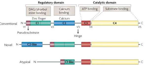
(Silva et al., 2012) (Marengo et al., 2011). The PKC family contains at least 12 kinases encoded by nine different genes. These can be classified into three different subfamilies based on their structure and cofactors required for its activation: classical or conventional PKCs (cPKCs: α, βI, βII and ) that require calcium (Ca2+), phosphatidylserine and diacylglycerol (DAG) for activation; novel PKCs (nPKCs: ,, and ) that require phosphatidylserine, DAG but not Ca2+ and atypical PKCs (aPKCs: e /1) which only require phosphatidylserine for their activation (Silva et al, 2012.).
All isozymes of this family have a conserved carboxyl-terminal tail that serves as a phosphorylation-dependent anchoring site for key regulatory molecules and a pseudosubstrate sequence that holds PKC in an inactive state. The V3 region lies between the regulatory domain and the catalytic domain, and is accessible to proteolytic cleavage by activation and conformational changes of PKCs (Fig.5). Cleavage at this site leads to the release of a constitutively active catalytic domain, suggesting that many inhibitory intramolecular interactions occur between these domains (Marengo et al., 2011).
Figure 5. Schematic representation of structure and classification of the family of PKC
12
Members of the PKC family of isozymes differ in their structure and mode of activation, but also in their tissue distribution, subcellular localization and substrate specificity. Activation of these isozymes results in changes in their subcellular localization, followed by the translocation of specific anchoring proteins, called receptors of activated C kinase (RACKS) (Mochly-Rosen & Gordon, 1998).
Other anchoring proteins have been suggested as inactivating PKC isozymes, receptors for inactive C-kinase isozymes (RICKS). Mochly-Rosen and colleagues took advantage of isoenzyme-specific interactions between PKCs and RACKS to overcome the lack of specific activators and inhibitors of PKC isozymes. They developed a number of first-generation peptides derived from PKCs or RACKS, based on their interaction sites, which interfere with PKC/RACK protein-protein interactions. By the same principle, peptides that interfere with the PKC/RICK interaction should act as specific agonists of PKCs. The advantages of using these peptides as drugs are their flexibility and fit abilty, since they are naturally selected for a particular protein interaction site, allowing them to interact more effectively and specifically with proteins and interfere with several interaction sites on a protein.
Some of these peptides, such as a PKCε-activator and a PKCδ-inhibitor have been used as pharmacological modulators of PKC activity in animal models of disease and in basic research (Wu-Zhang & Newton, 2013).
1.2.3.1. Regulation of apoptosis by PKC isoforms
The activation of PKCs can induce apoptosis in some cell types, while in other cases prevents it, making it difficult to predict the role of PKCs in the apoptotic process. In general, PKCα, ε, ζ and λ/ι are considered anti-apoptotic, promoting survival and proliferation, while PKCδ is characterized as pro-apoptotic, having a tumor suppressor role (Reyland, 2009).
Conventional PKCs prevent cell shrinkage mediated by FAS since they inhibit the TRAIL death receptor and in turn aggregation of the FAS receptor, recruitment of FADD and DISC formation. In that group, the most studied is the PKCα isoenzyme, which has been associated with the intrinsic pathway due to its ability to mediate expression and phosphorylation of Bcl-2, leading to an increase in the anti-apoptotic function of this protein. PKCα also suppresses drug-induced apoptosis by increasing the promoter activity of the multidrug resistance gene (MDR1), and thus the stability and expression of P-glycoprotein (Lønne, 2010).
The role of PKCβ in apoptosis is contradictory. The gene encoding for this protein creates two isoforms by alternatively splicing, PKCβI and PKCβII, and several studies
13
have failed to distinguish the contribution of each of these variants to apoptosis. It is believed that PKCβII is involved in the prevention of apoptosis, and expression of the oncogene v-abl causes translocation of PKCβII to the nucleus, preventing apoptosis, and indicating that PKCβII is anti-apoptotic (Gutcher et al., 2003).
The isoenzymes of the novel PKC subfamily PKCδ and PKCε are the best studied. Overexpression of PKCδ stimulates apoptosis in a wide variety of cell types via a mechanism that is not completely understood. When this protein is absent, cells are unable to respond to DNA damage-induced apoptosis, suggesting that PKCδ is required for a response to apoptotic stress (Teicher, 2006). When PKCδ is targeted to the cytosol, mitochondria, or nucleus frequently behaves as pro-apoptotic, while PKCδ targeted to the ER protected against tumor necrosis factor-related apoptosis, ligand-induced apoptosis and etoposide-ligand-induced apoptosis. PKCε increases the expression of anti-apoptotic members of the Bcl-2 family and inhibits proapoptotic members of this family, such as Bax and Bad (Sivaprasad et al., 2007). PKCε is the only isoenzyme that is considered an oncogene and regulates cancer cell proliferation through cell signaling by interacting with three major factors RhoA/C, Stat3, and Akt; however, when PKCɛ is activated it has protective roles in cardiac and brain ischemia, nociception and heat shock response (Jain & Basu, 2014) (Huang et al., 2011).
1.2.3.2. Protein kinase C epsilon
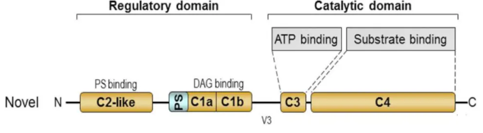
As previously mentioned, PKC protein belongs to the subfamily of the novel PKCs, whose structure comprises a regulatory N-terminus and a C-terminal catalytic domain with four conserved regions (C1-C4) and a variable region (V3) domain (Figure 6). The variable domain V3 is the region where the PKC isoforms can be cleaved by caspases (Newton, 1995).
After the discovery of PKCε, there have been many studies on the involvement of this kinase in cell survival and cell death. Though the majority of the studies suggest
14
that it favors life, some showed that activation of PKCε could contribute to apoptosis. PKCε acts as an oncogene with antiapoptotic effects when overexpressed in cancer cells, and is also oncogenic in colon epithelial cells through interference with Ras signal transduction (Yonekawa & Akita, 2008). Activation of the Ras/Raf/MAP kinase cascade results in the transcription of genes involved in cell proliferation and growth. Stimulation of Ras results in the translocation of Raf-1 from the cytosol to the plasma membrane being activated by a specific set of kinases including PKC (Toton et al., 2011).The anti-apoptotic ability of PKC also depends on the increased expression of anti-apoptotic proteins of the Bcl-2 family and the suppression of pro-anti-apoptotic members (Figure 7). Moreover, overexpression of PKC increases activation of nucleophosmin (NPM), a phosphoprotein capable of inducing carcinogenesis (Gorin & Pan, 2009). PKCε is cleaved by caspases in response to several apoptotic stimuli, including chemotherapeutic agents, starvation and TNF. In one study, cleavage of this protein was inhibited by inhibitor of caspase-3, and PKCε was cleaved by recombinant human caspase-3, suggesting that PKCε is a substrate for this caspase. The results of this study suggest that caspase-7 is the major caspase that cleaves PKCε at the Asp383 site in intact cells (Basu & Sivaprasad, 2007).
15
The subcellular distribution is an essential feature of activation of PKC. When PKC is activated, it inhibits the release of cit c from mitochondria, activation of caspases, and suppresses apoptosis. Thus, activation of PKC in mitochondria is associated with the suppression of cell death and improved cell survival (Nowak et al., 2004). To become responsive to second messengers, PKCε requires the phosphorylation of three conserved sites: Thr-566 in the activation loop, Ser-729 site in the hydrophobic C-terminus, Thr-710 and an autophosphorylation site. The subcellular localization of this kinase will depend in part on the second messenger that is connected to the C1 domain. Thus, PKC translocates from the plasma and/or membrane to the cytoskeleton in response to DAG and tridecanoic acids, or the Golgi network in response to arachidonic acid and linoleic acid (Akita, 2002).
1.2.3.3. Regulation of Bax by PKC
The anti-apoptotic properties of PKC appear to involve the regulation of several members of the Bcl-2 family, increasing the expression of anti-apoptotic members of this family and inhibiting pro-apoptotic members (Silva et al., 2012). Different studies show that PKC interacts with several proteins, one of which is pro-apoptotic Bax protein, preventing its integration into the mitochondrial membrane. Thus, PKC allows the survival of various cancer cells by inhibiting Bax conformational rearrangements that are important for its oligomerization, mitochondrial integration and release of cyt c, which inhibits cell death. PKC does not contain a similar BH3 domain, and since the crystal structure of the protein is not entirely known, it is not possible to predict if it has an appropriate interface to directly interact with Bax (McJilton et al., 2003).
The PKCε protein is an important signaling molecule, and understanding how it determines the decision of life or death of a cell will help in understanding the process of carcinogenesis and facilitate the identification of new targets for cancer therapy. In this project, we aim to understand how the interaction between this protein and Bax occurs, so that in the future new drugs to inhibit this interaction and promote cell death can be developed.
1.3. Yeast as a powerful model to study the regulation of Bax
The yeast Saccharomyces cerevisiae has already been used as a tool to understand molecular mechanisms of apoptotic pathways, such as the complexities of the function of Bcl-2 family members. The initial observation that the expression of Bax
16
confers a lethal phenotype in yeast was made during yeast-two-hybrid studies to analyze interactions between Bax and Bcl-2. These results prompted the hypothesis that the mammalian Bcl-2 proteins act on elements of a conserved endogenous yeast machinery to mediate effects on cell viability, and suggest that Bax-mediated cell death in yeast involves a regulated insertion into mitochondrial membranes and mitochondrial dysfunction leading to the release of cyt c and apoptosis, supporting the hypothesis that Bax exerts effects in yeast that are comparable to mammalian cells (Khoury & Greenwood, 2008). Manon and his team suggest that, in yeast cells, Bax induces growth arrest due to defects in the respiratory chain such as a decrease in the amount of the cyt c oxidase (COX) complex and the release of cyt c to the cytosol (Ludovico et
al., 2005). Priault et al also demonstrated that Bax-c-myc induces a massive release of
cyt c in yeast ; the observation that wild-type Bax is unable to induce the cyt c release and yeast cell death, confirm the central role of mitochondria in apoptosis (Priault et al., 2003).
Expression of Bax induces hyperpolarization of mitochondria, production of reactive oxygen species (ROS), cyt c release and mitochondrial network fragmentation reinforcing the importance of mitochondria in Bax-induced death (Silva et al., 2011)
In yeast, there are no known obvious orthologs of Bcl-2 family members; however the core components of the mammalian PTP are highly conserved in eukaryotic organisms.Almost all the studies concerning the role of PTP components in death and mitochondrial permeabilization in yeast have been performed in cells heterologously expressing the pro-apoptotic Bax protein; however, some data suggest that the PTP components do not exhibit a significant role in Bax-induced cell death (Pereira et al., 2008). In fact, was discovered that yeast cells lacking cyt c still die after Bax expression, although at a slower rate, indicating that cyt c release is not essential for Bax-induced cell death (Priault et al., 1999a).
18
The residues of PKC required for interaction with Bax were never identified. This information is crucial to direct screening of libraries of compounds with affinity for the key residues in the interaction sites to specifically disrupt the PKC /Bax interaction.
The aim of this study was to identify the PKC residues required for interaction with Bax, using the following approaches:
1. Assess which domains of PKCε interact with Bax by immunoprecipitation and Yeast-two-hybrid
2. Identify which residues could be involved in this interaction by single substitution of selected residues.
1.5. Materials and
Methods
20
1.5.1. Yeast strains and growth conditions
All strains used in this study are listed in Table I.
For Yeast-two-hybrid (YTH), the PJ69-4a strain was used (Figure 8). This strain has three reporter genes under the control of distinct GAL4 upstream activating sequences (UASs) and TATA boxes (ADE2, HIS3, and lacZ). These promoters yield strong and specific responses to GAL4. As a result, two major classes of false positives are eliminated: those that interact directly with the sequences flanking the GAL4 binding site and those that interact with transcription factors bound to specific TATA boxes.
Figure 8. The classical YTH system. The protein of interest is fused to the DNA binding domain
(DBD), a construct called bait. The potential interacting protein is fused to the activation domain (AD) and is called prey. The bait binds the upstream activator sequence (UAS) of the promoter. The interaction of bait with prey recruits the AD and thus reconstitutes a functional transcription factor, leading to further recruitment of RNA polymerase II and subsequent transcription of a reporter gene.
Table I. List of strains used in this study.
Strain Genotype Reference
XL1 blue Δ(mcrA)183 Δ(mcrCB-hsdSMR-mrr)173
endA1 supE44 thi-1 recA1 gyrA96 relA1
lac [F′ proAB lacIqZΔM15 Tn10 (Tetr)]
Stratagene
W303-1A MATa, leu2-3, ura3-1, trp1-1, his3-11,
ade2-1, can1-100
(Rothstein, 1983)
PJ69-4a
MATa trp1-∆901 leu2-3,112 901 ura3-52 his3-∆200 gal4∆ gal8∆ GAL2-ADE2 LYS2::GAL1-HIS3 met2::GAL7-lacZ (James et al., 1996) pGAD _PKC Transcription
DBD
pGBKT7 _Bax UAS PromoterAD
Reporter Gene21
W303-1A pGAD_PKC W303-1A harboring pGAD_PKC This study
XL1 blue pGBKT7_Bax XL1 blue harboring pGBKT7_Bax This study
PJ69-4a pGBKT7_Bax pGAD_PKC
PJ69-4a harboring pGBKT7_Bax and pGAD424_PKC
This study
PJ69-4a pAS2_Kap120 pGAD_PKC
PJ69-4a harboring pAS2_Kap120 and pGAD424_PKC
This study
PJ69-4a pGBKT7 pGAD_Ran PJ69-4a harboring pGBKT7 and pGAD_Ran
This study
PJ69-4a pGBKT7 pGAD_PKC PJ69-4a harboring pGBKT7 and pGAD424_PKC
This study
PJ69-4a pAS2_Kap120 pGAD_Ran
PJ69-4a harboring pAS2_Kap120 and pGAD_Ran
This study
W303-1A pYES3 pESC-His W303-1A harboring pYES3 and pESC-His This study W303-1A pYES3 pESC-His C1 W303-1A harboring pYES3 and pESC-His
C1
This study
W303-1A pYES3 pESC-His C2 W303-1A harboring pYES3 and pESC-His C2
This study
W303-1A pYES3 pESC-His Kin W303-1A harboring pYES3 and pESC-His Kin
This study
W303-1A pYES3 pESC-His PKC
W303-1A harboring pYES3 and pESC-His PKC
This study
W303-1A pYES3_Baxwt pESC-His C1
W303-1A harboring pYES3_Baxwt and pESC-His C1
This study
W303-1A pYES3_Baxwt pESC-His C2
W303-1A harboring pYES3_Baxwt and pESC-His C2
This study
W303-1A pYES3_Baxwt pESC-His Kin
W303-1A harboring pYES3_Baxwt and pESC-His Kin
This study
W303-1A pYES3_Baxwt pESC-His PKC
W303-1A harboring pYES3_Baxwt and pESC-His PKC
This study
W303-1A pYES3_Baxwt pESC-His
W303-1A harboring pYES3_Baxwt and pESC-His
This study
W303-1A pYES3 YEplac181 W303-1A harboring pYES3 and YEplac181 This study W303-1A pYES3
YEplac181_PKC
W303-1A harboring pYES3 and YEplac181_PKC
This study
W303-1A pYES3_Baxwt YEplac181
W303-1A harboring pYES3_Baxwt and YEplac181
This study
W303-1A pYES3_Baxwt YEplac181_PKC
W303-1A harboring pYES3_Baxwt and YEplac181_PKC
Silva R.
Strains were transformed with the indicated plasmids using the lithium acetate method as described in section 1.5.3. Transformants were selected on Synthetic Complete medium [SC: 0.17% (w/v) Yeast nitrogen base without aminoacids and
22
ammonium sulfate, 0.5% (w/v) ammonium sulfate, 0.14% (w/v), drop-out mixture lacking histidine, leucine, tryptophan and uracil, 0.008% (w/v) Histidine, 0.04% (w/v) Leucine, 0.008% (w/v) Tryptophan and 0.008% (w/v) Uracil] lacking the appropriate aminoacids plus 2% (w/v) of carbon source and 2% agar. Yeast strains were maintained on solid Yeast extract peptone dextrose (YPD) or Synthetic complete (SC) medium (lacking the appropriate aminoacids), grown at 30°C for 48h-72h, stored at 4°C, and refreshed every 2-3 weeks. Yeast cultures were grown aerobically in SC medium with 2% Glucose or Galactose as a carbon source or anaerobically in SC medium with 2% Lactacte pH 5.5. Strains transformed with plasmids were grown in SC medium lacking the appropriate aminoacids. Cells were incubated at 30°C with orbital shaking (200 rpm) and a liquid/air ratio of 1:5.
1.5.2. Plasmids
All the plasmids and oligonucleotides used in this study are listed in Tables II and III, respectively. The vector used to express Bax was pGBKT7 (Clontech). The pGBKT7 vector expresses proteins fused to amino acids 1–147 of the GAL4 DNA binding domain (DNA-BD). This vector also has other characteristics which make it suitable for use in the YTH system, such as two independent origins of replication in bacteria and yeast, the Kanamycin resistance for selection in E. coli and the TRP1 auxotrophic marker for selection in yeast. It also has a multiple cloning site (MCS) for insertion of the DNA fragment under consideration.
The vector used to clone PKC was pGAD424 (Clontech). pGAD424 is a shuttle vector that replicates autonomously in both E. coli and S. cerevisiae and generates a hybrid protein that contains the sequences for the GAL4 activation domain (DNA-AD). It carries Ampicillin resistance for selection in E. coli and the LEU2 auxotrophic marker that allows yeast auxotrophs carrying pGAD424 to grow on limiting synthetic medium lacking Leucine (LEU).
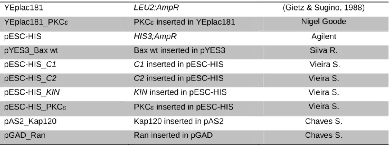
Table II. List of plasmids used in this study.
Plasmid Description Reference
pGBKT7 TRP1; KanR Clontech
pGBKT7_Bax wt Bax wt inserted in pGBKT7 This study
pGAD424 LEU2;AmpR Clontech
pGAD424_PKC PKC inserted in pGAD424 This study
23
YEplac181 LEU2;AmpR (Gietz & Sugino, 1988)
YEplac181_PKC PKC inserted in YEplac181 Nigel Goode
pESC-HIS HIS3;AmpR Agilent
pYES3_Bax wt Bax wt inserted in pYES3 Silva R.
pESC-HIS_C1 C1 inserted in pESC-HIS Vieira S.
pESC-HIS_C2 C2 inserted in pESC-HIS Vieira S.
pESC-HIS_KIN KIN inserted in pESC-HIS Vieira S.
pESC-HIS_PKC PKC inserted in pESC-HIS Vieira S.
pAS2_Kap120 Kap120 inserted in pAS2 Chaves S.
pGAD_Ran Ran inserted in pGAD Chaves S.
Table III. List of oligonucleotides used in this study.
Number Name Oligonucleotide sequence
1 pAS2-1_BaxFw 5´-AAAGACAGTTGACTGTATCGCCGGAATTCATGGACGGT TCCGGTGAACAA-3´ 2 pAS2-1_BaxRv 5´- ATTCGCCCGGAATTAGCTTGGCTGCAGTCAACCCATCT TCTTCCAGATGG-3´ 3 pGAD_PKCFw 5´-ACCCAAAAAAAGAGATCGAATTCATGGTAGTGTTCAAT GGCCTTCTTAAG-3´ 4 pGAD_PKCRv 5´-ATCTACGATTCATAGATCTCTGCAGTTAGGGCATCAGG TCTTCACCAAA-3´ 5 pAS2_seq_F 5´-TCATCGGAAGAGAGTAGTAACAAAGG-3´ 6 M13Rv 5´-TCCTGTGTGAAATTGTTATCCGCT-3´ 7 pGAD_seq_F 5´-CACTACAGGGATGTTTAATACCACTAC-3´ 8 pGAD_seq_R 5´-GTTCACTTCAACTGTGCATCGT-3´
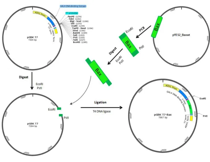
pGBKT7_Bax was constructed by standard ligation (Figure 9). Briefly, the Bax gene was amplified by PCR using pYES2_Baxwt (URA) as a template and the PCR product was purified using a DNA Clean & ConcentratorTM -5 Kit (Zymo Research) according to manufacturer’s instructions. The pGBKT7 vector and purified Bax were digested with EcoRI and PstI and the digestion product was purified using a DNA Clean & ConcentratorTM -5 Kit (Zymo Research) in order to remove the endonucleases. Ligation was performed using 1l of vector DNA, 10l of insert DNA, 2l of 10X Ligase Buffer, 1l of T4 DNA ligase and H2O to a total volume of 20l, and the reaction was
incubated at 4ºC overnight. 10l of reaction were transformed into E. coli XL1 Blue, and cells plated on Luria Bertani medium [LB; 1% (w/v) Tryptone, 0.5% (w/v) Yeast extract, 1% (w/v) NaCl and 2% (w/v) Agar] supplemented with 100 μg/mL Kanamicin. The plasmid was then extracted from E. coli using the GenElute Plasmid Miniprep kit (Sigma-Aldrich) and correct integration of the insert was confirmed by restriction analysis, PCR using oligonucleotides 5 and 6, and sequencing.
24
The pGAD-PKC clone was constructed in W303-1A by homologous recombination (Figure 10). The gene encoding the protein was amplified by Polymerase Chain Reaction (PCR) using the oligonucleotides listed in Table III (numbers 3-4), and co-transformed in yeast with cut pGAD424 through the lithium acetate method (described in section 1.5.3). After growth on SC medium (lacking the appropriate aminoacids) at 30°C for 48h, genomic DNA was extracted from putative positive clones and amplified in E. coli XL1 Blue by transformation using standard procedures and selection on LB supplemented with 100 μg/mL Ampicillin. The plasmid was then extracted from E. coli using the GenElute Plasmid Miniprep kit (Sigma-Aldrich) and correct integration of the insert was confirmed by restriction analysis and PCR using oligonucleotides 7/8 and by sequencing.
25
Figure 10. Schematic representation of the methodology used to construct pGAD424-PKC.
1.5.3. Yeast transformation by the lithium acetate method
All transformations in yeast strains were performed using the lithium acetate method. 240l of 50% (w/v) Polyethylene Glycol (PEG), 36l of 1 M LiAc, 40l of 2 mg/ml salmon sperm DNA (ssDNA) and 36l (water + plasmid DNA) were mixed, vortexed, and 50l of competent yeast cells were added. Then, the mix was incubated at 30ºC for 20 minutes, heat shocked for 30 minutes at 42°C and cooled on ice.
Cells were pelleted by centrifugation at 5000 rpm for 3 minutes, and the pellet was resuspended in 100uL of water. The total cell suspension (100L) was plated on appropriate selection medium consisting of SC medium supplemented with all essential aminoacids, except for the aminoacids of selectable markers on each plasmid and incubated at 30°C until the appearance of colonies.
26
1.5.4. Growth curves
Yeast strains 1A pYES3 pESC-His, 1A pYES3 pESC-His C1, W303-1A pYES3 pESC-His C2, W303-W303-1A pYES3 pESC-His Kin, W303-W303-1A pYES3 pESC-His PKC, W303-1A pYES3_Baxwt pESC-His C1, W303-1A pYES3_Baxwt pESC-His C2, W303-1A pYES3_Baxwt pESC-His Kin, W303-1A pYES3_Baxwt pESC-His PKC, W303-1A pYES3_Baxwt pESC-His, W303-1A pYES3 YEplac181, W303-1A pYES3 YEplac181_PKC, W303-1A pYES_Baxwt YEplac181 and W303-1A pYES_Baxwt YEplac181_PKC were first grown in SC medium with 2% of glucose lacking the appropriate aminoacids. Cells were then transferred to SC medium with 2% of Lactacte, pH 5.5, and grown to an O.D (640nm) of 0.3-0.5 (exponential phase). Then, 2% of galactose was added to induce PKC and Bax expression. The growth of yeast strains was assessed at different times. All incubations were performed at 30°C, 200 r.p.m.
1.5.5. Analysis of protein expression 1.5.5.1. Preparation of protein extracts
For detection of protein expression by Western blot of total cellular extracts, 5mL of cell culture were grown in SC medium with 2% of glucose and were then transferred to SC medium with 2% of galactose for 16h. Cells were harvested at an O.D (640nm) of 1, and resuspended in 1ml of H2O. After centrifugation at 5000 rpm for 3 min, the
pellet was suspended in 500l of H2O and 50l of a mixture of 3.5% β-mercaptoethanol
in 2M NaOH was added. After 15 min incubation on ice, proteins were precipitated with 50l of 50% (w/v) trichloroacetic acid (TCA) for 15 min on ice. Following centrifugation at 10000 g for 10 min, the pellet was solubilized in 1x Laemmli Buffer (63mM Tris-HCl pH 6.8; 10% glycerol; 0,0005% bromophenol blue; 2% SDS and 0.1M DTT) and the samples were boiled at 100ºC for 5 min in order to denature proteins.
1.5.5.2. SDS gel electrophoresis/Western blot
Total cellular extracts were separated electrophoretically in a 15% SDS polyacrylamide gel at 25 mA and transferred to a Hybond-P Polyvinylidene Difluoride Membrane (PVDF) (Hybond-ECL, GE Healthcare) at 60 mA for 1 h and 30 min. Membranes were blocked for 1h in PBS-T [80 mM Na2HPO4, 20 mM NaH2PO4 and
100 mM NaCl with 0.05% (v/v) Tween-20] containing 5% (w/v) non-fat dry milk, washed in PBS-T and then cut into strips and incubated with the primary antibodies: mouse
27
monoclonal anti-yeast phosphoglycerate kinase (PGK1) antibody (1:5000, Molecular Probes), rabbit polyclonal anti-human Bax (BAX) antibody (1:5000, Sigma), mouse anti-FLAG antibody (Sigma) and rabbit polyclonal anti-human PKC (PKC) antibody (1:100, Santa Cruz Biotechnology). Then, membranes were incubated with IgG-peroxidase secondary antibodies against mouse or rabbit (1:5000; Sigma Aldrich). Pgk1p was used as a loading control. Immunodetection of bands was revealed by chemiluminescence (Chemidoc XRS, BioRad).
1.5.6. Immunoprecipitation
Imunoprecipitation was performed using protein G-coupled Dynabeads (Invitrogen). Cells were ressuspended in resuspension buffer (10 mM Tris–malate, 0.6 M Mannitol, 1 mM EGTA, pH 6.7) supplemented with a mixture of protease inhibitors (0.4 μl/ml aprotinin, 1 μg/ml leupeptin, 1 μg/ml pepstatin, 1 mM phenylmethylsulfonyl fluoride and phosphatase inhibitors (PhosSTOP phosphatase inhibitor cocktail tablets; Roche Applied Science)). Cells were broken mechanically by vortexing with glass beads, after which 10× IP buffer (1x IP buffer: 50mM Tris-HCl, pH7.5; 100mM NaCl; 2mM EDTA) was added to the cell lysate to a final concentration of 1x, and incubated at 4 °C during 1 h. After incubation with IP buffer, 2 μg of monoclonal anti-FLAG antibody (Sigma) was added, and the lysate incubated overnight at 4 °C. Protein G-coupled Dynabeads (Invitrogen) were added and the extracts incubated for 6 h. Washing was performed with 1x IP buffer and elution was performed with Laemmli sample buffer, followed by denaturing at 65ºC for 30 min.
1.5.7. Yeast-two-hybrid
To verify the interaction between Bax and PKC proteins, pGBKT7_Bax and pGAD_PKC vectors were transformed into strain PJ69-4a, as well as various negative controls: pAS2_Kap120-pGAD424_PKC, pGBKT7-pGAD_Ran and pGBKT7-pGAD424_PKC and a positive control tested before: pAS2_Kap120-pGAD_Ran.
The cells were plated on appropriate selection medium supplemented with all essential aminoacids, except tryptophan and Leucine (SD -TRP-LEU), and incubated at 30°C until the appearance of colonies.
In order to assess whether the proteins studied are independently capable of activating transcription of the reporter genes or interact with each other, isolated colonies of the yeast strain PJ69-4a pre-transformed with the plasmids mentioned
28
above were plated on SD -TRP-LEU-HIS and SD -ADE-TRP-LEU). These colonies were also tested for the expression of -galactosidase (lacZ gene product) by X-Gal (5-bromo-4-chloro-3-indolyl-beta-D-galacto-pyranoside) filter-assay as described in (Mockli & Auerbach, 2004).
30
1.6.1. Previous Results
Before this study, the DNA sequence coding for PKCε was cloned in the pESC-HIS plasmid, in frame with the FLAG epitope, and transformed into a W303-1A strain expressing Bax. Then, Bax was immunoprecipitated from extracts from this strain with the Bax-2D2 antibody, bound proteins run on an SDS-PAGE gel, transferred to PVDF and probed with an anti-PKC antibody in order to verify whether PKC and Bax interact in yeast (Figure 11A, B).
A B
Figure 11. Immunoprecipitation of Bax-interacting proteins. Lysates from cells expressing Bax and
PKC were immunoprecipitated with Bax antibody (2D2). Bound proteins were eluted with loading buffer, denaturated at 65ºC for 30min, run on SDS-PAGE and transferred to PVDF. Western blots were performed with anti-Bax antibody (A) and anti-PKC antibody (B). C+ WB:anti-Bax was W303-1A pYES3-Baxwt and C+ WB:anti-PKC was W303-1A YEplac181-PKC pYES3-pYES3-Baxwt in order to verify expression of Bax and PKC, respectively.
However, PKC was not detected in the immunoprecipitated extract, and therefore an interaction between full length PKC and Bax could not be demonstrated in yeast. IP: α-Bax WB: anti-Bax IP: α-Bax WB: anti-PKC Bax PKC
31
0 2 4 6 8 10 12 14 16 18 20 22 24 0 1 2 3W303-1A pESC-His pYES3 W303-1A pESC-His pBax W303-1A pC2 pYES3 W303-1A pC2 pBax Time (hours) O .D 6 4 0 n m 0 2 4 6 8 10 12 14 16 18 20 22 24 0 1 2 3
W303-1A pESC-His pYES3 W303-1A pESC-His pBax W303-1A pC1 pYES3 W303-1A pC1 pBax Time (hours) O .D 6 4 0 n m
1.6.2. Effect of expressing Bax and different PKC
fragments on cell growthYeast has been considered an important tool to discover the different cellular targets of several proteins. Moreover, the preservation of functional characteristics of mammalian PKCs in yeast led to the identification and characterization of the mode of action of several PKC modulators in yeast cells expressing individual PKC isforms (Sprowl et al., 2007). It had previously been shown that expression of PKC reverted the slow growth phenotype of yeast cell expressing Bax, indicating PKC could function as an inhibitor of Bax, presumably by protein-protein interaction (R. Silva, personal communication). This could be a transient or weak interaction, and therefore not detected in the immunoprecipitation studies. We therefore sough to determine whether one or more domains of PKC had the same effect of the growth of Bax-expressing cells.
In this study, full length and individual domains of PKC (C1, C2, Kinase) cloned in pESC-His (S. Vieira) were transformed in the W303-1A yeast strain expressing Bax. We next sough to identify which of these domains could be involved in the interaction with Bax by determining if they caused a reversal of the slow growth phenotype of cells expressing Bax (Figure 12 A, B, C, D and E).
A
B
32
0 2 4 6 8 10 12 14 16 18 20 22 24 0 1 2 3W303-1A pESC-His pYES3 W303-1A pESC-His pBax W303-1A pKin pYES3 W303-1A pKin pBax
Time (hours) O .D 6 4 0 n m 0 2 4 6 8 10 12 14 16 18 20 22 24 0 1 2 3
W303-1A pESC-His pYES3 W303-1A pESC-His pBax W303-1A pPKC pYES3 W303-1A pPKC pBax Time (hours) O .D 6 4 0 n m C D E
Figure 12. Growth curves of the S. cerevisiae strain W303-1A expressing the indicated proteins
induced by galactose at T0. (A)- Growth curves for yeast strain W303-1A carrying C1 domain (pC1) and empty vector of Bax (pYES3) (); pC1 plus pBax (); empty vector of PKC (pESC-His) plus pYES3 () and pESC-His plus pBax(). (B)- Yeast strain W303-1A carrying C2 domain (pC2) and pYES3 (); pC2 plus pBax (); pESC-His plus pYES3 () and pESC-His plus pBax(). (C)- Yeast strain W303-1A carrying Kinase domain (pKin) and pYES3 (); pKin plus pBax (); pESC-His plus pYES3 () and pESC-His plus
0.00 0.05 0.10
0.15 W303-1A pESC-His pYES3
W303-1A pESC-His pBax W303-1A pC1 pYES3 W303-1A pC1 pBax W303-1A pC2 pYES3 W303-1A pC2 pBax W303-1A pKin pYES3 W303-1A pKin pBax W303-1A pPKC pYES3 W303-1A pPKC pBax S p e c if ic g ro wt h r a te ( h -1 )