The
Chlamydia trachomatis
type III secretion
substrates CT142, CT143, and CT144 are
secreted into the lumen of the inclusion
Maria da Cunha1,2, Sara V. Pais1, Joana N. Bugalhão1, Luı´s Jaime Mota1,2*
1UCIBIO—REQUIMTE, Departamento de Ciências da Vida, Faculdade de Ciências e Tecnologia, Universidade Nova de Lisboa, Caparica, Portugal,2Instituto de Tecnologia Quı´mica e Biolo´gica Anto´nio Xavier, Universidade Nova de Lisboa, Oeiras, Portugal
Abstract
Chlamydia trachomatisis a human bacterial pathogen causing ocular and genital infections. It multiplies exclusively within an intracellular membrane-bound vacuole, the inclusion, and uses a type III secretion system to manipulate host cells by injecting them with bacterially-encoded effector proteins. In this work, we characterized the expression and subcellular localization in infected host cells of theC.trachomatisCT142, CT143, and CT144 proteins, which we previously showed to be type III secretion substrates. Transcriptional analyses in
C.trachomatisconfirmed the prediction thatct142,ct143andct144are organized in an operon and revealed that their expression is likely driven by the mainσfactor,σ66. In host cells infected byC.trachomatis, production of CT142 and CT143 could be detected by immunoblotting from 20–26 h post-infection. Immunofluorescence microscopy of infected cells revealed that from 20 h post-infection CT143 appeared mostly as globular structures outside of the bacterial cells but within the lumen of the inclusion. Furthermore, immunofluo-rescence microscopy of cells infected byC.trachomatisstrains carrying plasmids producing CT142, CT143, or CT144 under the control of thect142promoter and with a C-terminal dou-ble hemagglutinin (2HA) epitope tag revealed that CT142-2HA, CT143-2HA or CT144-2HA showed an identical localization to chromosomally-encoded CT143. Moreover, CT142-2HA or CT144-2HA and CT143 produced by the same bacteria co-localized in the lumen of the inclusion. Overall, these data suggest that the CT142, CT143, and CT144 type III secretion substrates are secreted into the lumen of the inclusion where they might form a protein complex.
Introduction
Chlamydiaeare a large group of obligate intracellular bacteria including nine families [1]: Chlamydiaceae, which comprises a single genus (Chlamydia) grouping bacterial species that are pathogens of humans and other vertebrates; and 8 families of microorganisms that infect a variety of vertebrate and non-vertebrate animals as well as free-living amoebae [2]. All a1111111111 a1111111111 a1111111111 a1111111111 a1111111111 OPEN ACCESS
Citation:da Cunha M, Pais SV, Bugalhão JN, Mota LJ (2017) TheChlamydia trachomatistype III secretion substrates CT142, CT143, and CT144 are secreted into the lumen of the inclusion. PLoS ONE 12(6): e0178856.https://doi.org/10.1371/journal. pone.0178856
Editor:David M. Ojcius, University of the Pacific, UNITED STATES
Received:April 15, 2017
Accepted:May 21, 2017
Published:June 16, 2017
Copyright:©2017 da Cunha et al. This is an open access article distributed under the terms of the
Creative Commons Attribution License, which permits unrestricted use, distribution, and reproduction in any medium, provided the original author and source are credited.
Data Availability Statement:All relevant data are within the paper and its Supporting Information files.
Chlamydiaeundergo a developmental cycle involving the inter-conversion between a non-rep-licative infectious form, the elementary body (EB), and a repnon-rep-licative non-infectious form, the reticulate body (RB) [3]. Intracellular multiplication ofChlamydiaeoccurs exclusively within a membranous vacuolar compartment known as inclusion. AmongChlamydiaceae,C. tracho-matisserovars, which are divided in ocular, urogenital and lymphogranuloma venereum (LGV) strains, are the leading cause of infectious blindness in developing countries [4] and the most prevalent sexually transmitted bacteria worldwide [5].
Type III secretion (T3S) systems are present in many Gram-negative bacteria establishing pathogenic or symbiotic relationships with their hosts. They mediate the delivery of bacteri-ally-encoded effector proteins into eukaryotic host cells [6,7].Chlamydiaeuse a T3S system throughout the developmental cycle to transport several effectors across the host cell plasma membrane and the inclusion membrane [3,8]. Some of these proteins, such as TarP [9], CT694 [10], or TepP [11], are packed in EBs and are delivered into host cells during the early phases of infection to mediate invasion, by modulation of the host actin cytoskeleton [12,13], and to subvert immune signaling [11,14]. Another class of chlamydial T3S effectors, the inclu-sion membrane (Inc) proteins [15,16], insert into the inclusion membrane and interfere with several host cell processes such as cytoskeleton dynamics [17,18], vesicular and non-vesicular transport [19,20], death [21], or immune signalling [22]. OtherChlamydiaT3S effectors have been identified and shown to interfere, e.g., with host cell ubiquitination [23], histone methyla-tion [24], or the ESCRT machinery [25]. In addition,C.trachomatisT3S substrates that are enzymes for glycogen metabolism have been shown to localize in the lumen of the inclusion [26].
SeveralC.trachomatisT3S effectors have been identified but it is likely that others remain to be characterized. Before methods for transformation and regulated ectopic expression of proteins inC.trachomatishave been described [27–29], a main methodology to search and identify putative chlamydial T3S effectors relied on using heterologous host bacteria, such as Salmonella[30],Shigella[23,24,31,32], orYersinia[9,10,33–37], carrying well characterized T3S systems. While many candidate T3S substrates identified in these studies have been vali-dated in cells infected byC.trachomatis(e.g. Inc proteins, CopN, TarP, CT694, CT620, CT621, CT711, or CT737/NUE), many remain to be characterized in further detail. UsingY. enterocoliticaas a heterologous host, we previously described the identification of 10 likelyC. trachomatiscandidate T3S substrates (CT053, CT105, CT142, CT143, CT144, CT161, CT338, CT429, CT656, and CT849) [37]. Among the genes encoding these proteins, the expression of ct142,ct143andct144has been shown to be distinctly up-regulated by a protein (Pgp4) encoded in theChlamydiavirulence plasmid [38,39]. This plasmid is present in many chla-mydial species [39], and studies in animal models of infection showed it is a virulence factorin vivo[40–42].
In this work, we characterized the genetic organization ofct142,ct143andct144in theC. trachomatischromosome and showed that during infection of host cells the encoding proteins (CT142, CT143, and CT144) localize within the inclusion in globular structures that appear outside of the bacterial cells. These observations support the concept thatC.trachomatisT3S substrates can be secreted into the lumen of the inclusion.
Materials and methods
Cell lines
HeLa 229 and Vero cells (from the European Collection of Cell Culture; ECACC) were main-tained in high-glucose Dulbecco’s modified Eagle Medium (DMEM; Thermo Fisher Scientific) PT2020 Partnership Agreement
(POCI-01-0145-FEDER-007728). MdC was supported by PhD fellowship SFRH/BD/62728/2009 from FCT. SVP and JNB hold PhD fellowships PD/BD/52210/2013 and PD/BD/128214/2016, respectively, within the scope of the PhD program Molecular Biosciences (PD/00133/2012) funded by FCT. The funders had no role in study design, data collection and analysis, decision to publish, or preparation of the manuscript.
supplemented with heat-inactivated 10% (v/v) fetal bovine serum (FBS; Thermo Fisher Scien-tific) at 37˚C in a humidified atmosphere of 5% (v/v) CO2.
Bacterial strains and growth conditions
Escherichia coliTOP10 (Thermo Fisher Scientific) was used for construction and purification of plasmids,E.coliBL21 (DE3) (Novagen) for recombinant protein expression, andE.coli ER2925 (New England Biolabs) to amplify and purify plasmids for transformation ofC. tracho-matis.E.colistrains were routinely grown in liquid or solid lysogeny broth (LB) medium with the appropriate antibiotics and supplements. Plasmids were introduced intoE.coliby electroporation.
C.trachomatisLGV serovar L2 strains 434/Bu (L2/434; from ATCC) and 25667R (L2/ 2566R; [43]; kindly provided by Agathe Subtil) were propagated in HeLa 229 cells using stan-dard techniques [44]. Throughout this work we used the nomenclature of the annotatedC. tra-chomatisD/UW3 strain [45]. Transformation ofC.trachomatis, first reported by the
laboratory of Ian N. Clarke [27], was done essentially as described by Agaisse and Derre´ [28]. The optimal penicillin G concentration to select transformants was 1 U/ml. Once established, the transformed strains were cultured in the presence of 10 U/ml of penicillin and plaque puri-fied using Vero cells, as described by Nguyen and Valdivia [46].
DNA manipulation, plasmids and primers
The plasmids used in this work, their main characteristics and construction details, are described inS1 Table. The DNA primers used in their construction are listed inS2 Table. Plas-mids were constructed and purified using standard molecular biology procedures with proof-reading Phusion DNA polymerase (Thermo Fisher Scientific), restriction enzymes (Thermo Fisher Scientific), T4 DNA Ligase (Thermo Fisher Scientific), DreamTaq DNA polymerase (Thermo Fisher Scientific), DNA clean & concentratorTM-5 kit and ZymocleanTMgel DNA recovery kit (Zymo Research), and GeneElute Plasmid Miniprep kit (Sigma Aldrich) or NZY-Midiprep kit (NZYtech), according to the instructions of the manufacturers. The backbone plasmids used in this work were pGEX-4T-2 (GE Healthcare) and pMal-c (New England Bio-labs), for recombinant protein purification, and pEGFP-C1 (Clontech) for transfection of mammalian cells. Furthermore, pSVP247 (S1 FigandS1 Table), a derivative of p2TK2-SW2 [28], was the backbone to generateC.trachomatisexpression plasmids bearing genes whose transcription is halted by theincDterminator (TincD) and encoding proteins with a double
hemagglutinin epitope tag (2HA) at their C-terminus. The accuracy of the nucleotide sequence of all the inserts in the constructed plasmids was confirmed by DNA sequencing.
Infection of HeLa 229 cells with
C
.
trachomatis
For immunofluorescence analysis, 5x10⁴HeLa 229 cells were seeded per well in 24 well plates containing 13 mm glass coverslips. For immunoblotting 1x10⁵HeLa 229 cells were seeded per well in 24 well plates. Scaling-up was done as appropriate for other cell culture plates or flasks. The day after seeding, media was replaced by Hank’s balanced salt solution (HBSS) and the cells were incubated*15 min at 37˚C in a humidified atmosphere of 5% [v/v] CO2, while the
C.trachomatisinocula (previously titrated infectious particles, as described by Scidmore [44]) were prepared in either sucrose phosphate glutamate buffer (SPG; 0.2 mM sucrose, 17 mM Na2HPO4, 3 mM NaH2PO4, 5 mM L-glutamic acid) or HBSS. The buffer was then removed
and theC.trachomatisinocula were added at a multiplicity of infection (MOI) of 1–5 and incubated for 30 min at 37˚C in a humidified atmosphere of 5% [v/v] CO2(24 or 6 well-plates)
inocula were removed and replaced by DMEM supplemented with 10% (v/v) FBS and 10μg/ ml of gentamicin. This was considered the time zero of infection.
To quantify infectious progeny, HeLa cells infected byC.trachomatisstrains for different times were lysed by osmotic shock (15 min incubation in sterile H2O). Dilutions of these
lysates in HBSS were used to infect freshly seeded HeLa cells. The newly infected cells were fixed after 22–26 h,C.trachomatisbacteria were immunolabelled, and the number of inclusion forming units (IFUs) was calculated as described by Scidmore [44].
Transcription linkage analysis
HeLa cells were infected withC.trachomatisL2/434 for 20 h and RNA was isolated using NZY total RNA kit (NZYTech). cDNA was obtained by reverse transcription PCR (RT-PCR) using random hexamers and iSCRIPT (Bio-Rad). The product of a typical reverse transcription reac-tion but without iSCRIPT (cDNA-) andC.trachomatisgenomic DNA (gDNA) isolated using NZY tissue gDNA kit (NZYTech) were also used as template in control RT-PCR reactions. Specific primers-pair combinations (S2 Table) were designed in order to determine possible transcriptional linkages.
5´rapid amplification of cDNA ends (RACE)
The identification of the transcription start site ofct142inC.trachomatisL2/434 was done using a 5´/3´RACE kit (second generation; Roche). RNA was isolated from HeLa cells infected for 42 h byC.trachomatisL2/434 and RNA was isolated as described above for the transcrip-tion linkage analysis. The primers complementary toct142that were used are listed inS2 Table. Final PCR amplification of double stranded cDNA was done with Phusion DNA poly-merase (Thermo Fisher Scientific). PCR products were purified after agarose gel electrophore-sis, using High Pure PCR purification kit (Roche), and then subjected to DNA sequencing. All these manipulations were done according to instructions from the manufacturers.
Recombinant protein expression and purification
E.coliBL21(DE3) carrying a pGEX-4T-2-derived plasmid (encoding CT143 with GST fused at its N-terminus; GST-CT143) or pMal-c derived plasmids (encoding MBP, or CT142 or CT143 with MBP fused at their N-termini; MBP-CT142, MBP-CT143) were used for recombinant protein expression by auto-induction [47]. In both cases, bacterial cultures were grown for 4 h at 37˚C, with an agitation of 150 rotations per minute (rpm), and then shifted to 25˚C for an additional 24 h, with an agitation of 150 rpm. The bacterial cells were harvested by centrifuga-tion at 10,500 xgfor 15 min at 4˚C.
were centrifuged at 10,500 xgfor 30 min at 4˚C, after which the supernatants were loaded onto equilibrated amylose resin (New England Biolabs), according to the instructions of the manufacturer. The column was washed 12 times with 1 ml of ice-cold column buffer, after which the proteins were eluted with ice-cold column buffer containing 10 mM maltose.
Purified proteins were dialysed using SnakeSkin Dialysis TubingTM(Thermo Fisher Scien-tific) according to the instruction of the manufacturer.
Antibodies
Polyclonal antibodies against CT142 were generated against three synthetic peptides conju-gated to ovalbumin: peptide 1 (36–50 amino acid residues) NH2-VKSISAKESFSVKRKC-CO
OH; peptide 2 (260–274 amino acid residues) NH2-CKGGDYVDKSALSTLY-COOH; peptide
3 (126–140 amino acid residues) NH2-QKLPLIGPSRLVYQSC-COOH (Metabion, Germany).
The resulting antibodies were affinity purified on CNBr-sepharose matrix containing the respective peptides (Metabion).
Purified GST-CT143 was used to immunize New Zealand White rabbits for the production of polyclonal CT143 sera (Davids Biotechnologie, Germany). For purification, the anti-CT143 serum was firstly incubated with acetone powders obtained fromE.coliBL21 (DE3) containing pGEX-4T-2, allowing the serum to be partly depleted of the anti-GST antibodies or any contaminantE.coliprotein in purified GST-CT143. Secondly, anti-CT143 serum was affinity purified on 1 mg of MBP-CT143 immobilized on a nitrocellulose membrane. The affinity-purified antibodies were concentrated using Amicon Ultra-4 (Millipore) and quanti-fied using NanoDrop 1000 (Thermo Fisher Scientific).
The following primary antibodies were used: rabbit polyclonal anti-CT142 (this work; 1:200 dilution for immunoblotting); rabbit polyclonal anti-CT143 (this work; 1:200 dilution for immunoblotting and 1:50 dilution for immunofluorescence); mouse monoclonal anti-chla-mydial Hsp60 (A57-B9; Thermo Fisher Scientific; 1:1,000 dilution for immunoblotting and 1:200 dilution for immunofluorescence); goat polyclonal anti-MOMP ofC.trachomatis (Abcam; 1:1,000 dilution for immunoblotting and 1:500 dilution for immunofluorescence); goat anti-Chlamydia trachomatisfluorescein isothiocyanate (FITC)-conjugated polyclonal antibody (Millipore; 1:100 dilution for immunofluorescence); rat monoclonal anti-HA (3F10; Roche; 1:1,000 dilution for immunoblotting and 1:200 dilution for immunofluorescence), mouse monoclonal anti-tubulin (clone B-5-1-2; Sigma Aldrich; 1:1,000 for immunoblotting).
For immunoblotting, the secondary antibodies used were all horseradish peroxidase (HRP)-conjugated (GE Healthcare and Jackson ImmunoResearch; used at 1:10,000). For immunofluo-rescence, the following secondary antibodies were used: cyanine 5 (Cy5)-conjugated donkey anti-goat (Jackson ImmunoResearch Laboratories; 1:200), Alexa Fluor1-594 (AF594)-conju-gated AffiniPure donkey anti-goat (Jackson ImmunoResearch Laboratories; 1:200); Rhodamine Red-X-conjugated AffiniPure donkey anti-rabbit (Jackson ImmunoResearch Laboratories; 1:200); AF488-conjugated goat anti-mouse (Jackson ImmunoResearch Laboratories; 1:200); Rho-damine Red-X-conjugated anti-rat (Jackson ImmunoResearch Laboratories; 1:200); and
AF488-conjugated AffiniPure Donkey anti-rat (Jackson ImmunoResearch Laboratories; 1:200).
Immunoblotting
an incubation of 5 minutes at 100˚C, followed by incubation with benzonase (Novagen). Samples were separated by 12% (v/v) SDS-PAGE and transferred to 0.2μm nitrocellulose membranes (Bio-Rad) using Trans-Blot Turbo Transfer System (Bio(Bio-Rad). Immunoblot detection was done with SuperSignal1West Pico Chemiluminescent Substrate (Thermo Fisher Scientific) or SuperSignal1 West Femto Maximum Sensitivity Substrate (Thermo Fisher Scientific) and exposure to Amersham Hyperfilm ECL (GE Healthcare). Quantification of bands in immunoblots was performed by densi-tometry analysis using Fiji software [48].
Immunofluorescence microscopy
Infected HeLa cells were fixed in either PBS containing 4% (w/v) paraformaldehyde (PFA) for 15 min or in methanol (-20˚C) for 10 min. For immunostaining, the antibodies were diluted in PBS containing 10% [v/v] horse serum (when fixation was done with PFA, 0.1% [v/v] Triton X-100 was added to allow permeabilization of cells). After immunolabelling, the cells were consecutively washed with PBS and H2O. The coverslips were assembled using Aqua-poly/
Mount (Polysciences) on microscopy glass slides, and the cells were examined by conventional fluorescence microscopy or by confocal microscopy. Images were processed and assembled using Adobe Photoshop. Quantitative analyses of immunofluorescence images were per-formed using Fiji software [48].
Results
C
.
trachomatis ct142
, c
t143
, and
ct144
are organized in an operon
Thect142,ct143, andct144genes (namedctl0397,ctl0398, andctl0399, respectively, in strain L2/ 434) are localized adjacently in the chromosome ofC.trachomatis(Fig 1A). The CT142, CT143 and CT144 proteins are highly conserved in allC.trachomatisserovars (>95% of amino acididentity for CT142;>96% of amino acid identity for CT143; and>90% of amino acid identity
for CT144) and are also conserved inChlamydia(S3 Table). However, a PSI-BLAST analysis [49] failed to identify any significant amino acid sequence similarity with proteins from the other Chlamydiaefamilies (S3 Table). Thect142,ct143andct144genes are syntenic in the chromosome of the majority of theChlamydiaspp. (S2 Fig). InC.pneumoniaeandC.felisthe genes are located in the complementary strand, and inC.pneumoniaethe orthologue ofct143is duplicated (S3 TableandS2 Fig).
Overall, these experiments showed thatct142,ct143, andct144are organized in an operon whose transcription is directed from aσ66-like promoter (ct142promoter; Pct142) about 30
nucleotides upstream from the start codon ofct142.
CT142 and CT143 are produced during the developmental cycle of
C
.
trachomatis
To analyze the production of CT142, CT143, and CT144 byC.trachomatisduring the bacterial developmental cycle, we aimed to obtain antibodies against these proteins. We generated anti-bodies that specifically recognized CT142 and CT143 by immunoblotting when the proteins were ectopically expressed in HeLa cells (S3 Fig), but we did not succeed in generating anti-bodies recognizing CT144. We then analyzed by immunoblotting the presence of CT142 and CT143 in extracts of HeLa cells either uninfected or infected byC.trachomatisL2/434 for 2, 8, 14, 20, 26, 32, 38, or 40 h. Bands that migrated on SDS-PAGE according to the predicted molecular mass of CT142 (31.5 kDa) and CT143 (31 kDa) could be detected from 20 h post-infection (p.i.), and more obviously from 26 h p.i. (Fig 2A). At 20 h p.i., the immunoblot signal detected by each antibody was very weak and in some experiments not even visible. Transcrip-tion ofct142andct143(and also ofct144) is up-regulated byC.trachomatisplasmid-encoded Pgp4 [38]. Therefore, to further ascertain the specificity of the anti-CT142 and anti-CT143 antibodies, we analyzed by immunoblotting extracts of HeLa cells infected for 15, 20, 30 or 40 h byC.trachomatisL2/434 or L2/25667R (lacking theC.trachomatisvirulence plasmid). In Fig 1. Characterization ofct142,ct143, andct144.(A) Genetic organization ofct142,ct143andct144in the chromosome ofC.trachomatisdepicting the fragments (1–8) amplified in the transcription linkage analyses, the transcriptional start site determined by 5´RACE as well as the deduced -10 and -35σ66-like promoter regions. (B) Transcription linkage analyses inC.trachomatisL2/434: cDNA+, PCR from cDNA generated with reverse transcriptase (RT) from bacterial RNA isolated from infected HeLa cells; cDNA-, as cDNA+ but without RT; gDNA, PCR from bacterial DNA.
Fig 2. Production of CT142 and CT143 byC.trachomatisL2/434.(A) HeLa cells were either left
uninfected (UI) or infected byC.trachomatisL2/434 for 2, 8, 14, 20, 26, 32, 38 or 40 h. Whole cell lysates were analyzed by immunoblotting with antibodies against CT142 (blot on the left) or CT143 (blot on the right), C.trachomatismajor outer membrane protein (MOMP; bacterial loading control) and tubulin (loading control for host cells). The arrows indicate the position of CT142 and CT143 bands. (B) and (C) HeLa cells were either left uninfected (UI) or infected byC.trachomatisL2/434 or L2/25667 for 15, 20, 30 or 40 h, as indicated. Whole cell lysates were analyzed by immunoblotting with antibodies against CT142 (B) or CT143 (C),C. trachomatisHsp60 (bacterial loading control) and tubulin (loading control for host cells).
extracts from cells infected byC.trachomatisL2/434, bands migrating as proteins of*31 kDa were again detected with the anti-CT142 (Fig 2B) or anti-CT143 antibodies (Fig 2C). However, in extracts of HeLa cells infected byC.trachomatisL2/25667R no clear bands migrating as pro-teins of*31 kDa could be detected with any of the antibodies (Fig 2B and 2C).
In summary, these experiments confirmed that CT142 and CT143 are produced during the developmental cycle ofC.trachomatisand further illustrated that their expression is dependent on theChlamydiaplasmid.
CT143 localizes within the
C
.
trachomatis
inclusion but outside of the
bacterial cells
We previously showed that CT142, CT143, and CT144 are T3S substrates [37]. Therefore, using the anti-CT142 and anti-CT143 antibodies, we aimed to detect the presence of these proteins in the cytoplasm of infected host cells. However, the analysis could only be done for CT143 because the anti-CT142 antibody did not generate an immunofluorescence-specific signal. For this, HeLa cells were infected byC.trachomatisL2/434 for 15, 20, or 30 h. At these time-points, the cells were fixed with methanol, immunolabelled for CT143 and Hsp60 (aChlamydiacytosolic molecu-lar chaperone), and then analyzed by indirect immunofluorescence confocal microscopy. At 15 h p.i., the immunofluorescence signal for CT143 was weak and overlapped with the Hsp60 signal (Fig 3A). At 20 h p.i., the immunofluorescence signal for CT143 was more intense but still restricted to the inclusion (Fig 3A). However, the signal did not appear to overlap with the Hsp60 signal and instead we observed discrete globular structures (Fig 3A). At 30 h p.i., these anti-CT143-labelled structures were much more abundant (Fig 3A), and they were distinct from the Hsp60 signal and restricted to the inclusion (Fig 3A).
The observations described above suggested that in infected cells the majority of CT143 localizes within the inclusion but apparently outside of the bacterial cell. To ensure that the observed anti-CT143 immunofluorescence signal was specific, prior to immunolabelling of HeLa cells infected for 30 h with the L2/434 strain, the anti-CT143 antibody was incubated in the presence of an excess of purified MBP, MBP-CT142 or MBP-CT143. As control, the anti-CT143 antibody underwent a similar incubation but without any added protein. This showed that only the pre-incubation with MBP-CT143, but not with MBP or MBP-CT142, led to a clear reduction of the anti-CT143 immunofluorescence signal (Fig 3B). In addition, we com-pared the anti-CT143 immunofluorescence signal in HeLa cells infected for 30 h byC. tracho-matisstrains L2/434 or plasmidless L2/25667R. This revealed that the CT143 signal was drastically reduced in cells infected by the L2/25667R strain (Fig 3C). Overall, this indicated that the immunofluorescence signal detected with the anti-CT143 antibody was specific.
To evaluate in more detail the subcellular localization of CT143, HeLa cells infected by strain L2/434 for 30 h were immunolabelled for CT143 and Hsp60 (as before), for CT143 andC. tracho-matismajor outer membrane protein (MOMP), and for Hsp60 and MOMP (Fig 4A). This con-firmed that CT143 appeared as intra-inclusion globular structures, as the anti-CT143
immunofluorescence signal did not co-localize with the bacterial signal from the anti-MOMP or anti-Hsp60 antibodies (Fig 4A; upper and middle panels). In contrast, and as expected, the anti-MOMP signal (bacterial outer membrane) mostly encircled the anti-Hsp60 signal (bacterial cyto-plasm) (Fig 4A; lower panel). We then measured the area of each of the immunofluorescence sig-nals analyzed (CT143, Hsp60 and MOMP). This revealed that the area of the anti-CT143 signal is considerably smaller than the area of the anti-Hsp60 or anti-MOMP signals (Fig 4B).
The chlamydial developmental cycle is not significantly affected in
C
.
trachomatis
strains over-producing CT142-2HA, CT143-2HA,
CT144-2HA, or CT142, CT143, and CT144-2HA
To further validate the observations regarding the localization of CT143 and to analyze the localization in infected cells of other proteins (CT142 and CT144) encoded in the ct142-ct143-ct144operon, we transformedC.trachomatisL2/434 with four different p2TK2-SW2-derived Fig 3. Specific immunolabelling of CT143 in HeLa cells infected byC.trachomatis.HeLa cells were infected byC.trachomatisL2/434 for 15, 20 and 30 h (A), for 30 h (B), or by L2/434 orC.trachomatisL2/25667R for 30 h (C). In all cases, cells were fixed with methanol, immunolabelled with anti-CT143 and anti-Hsp60 and adequate fluorophore-conjugated secondary antibodies, and analyzed by confocal immunofluorescence microscopy. In (B), before immunolabelling, anti-CT143 antibodies were incubated in the presence of 8μg of purified MBP, MBP-CT142 or MBP-CT143, as indicated. All images are combined projections of multiple 0.2μmz-sections. In (A), images were zoomed 3-fold in the area delimited by a white box. All scale bars, 5μm.
Fig 4. CT143 localizes within in theC.trachomatisinclusion but outside of the bacterial cells.(A) HeLa 229 cells were infected withC.trachomatisL2/434 for 30 h, fixed with methanol, and immunolabelled for CT143 and anti-Hsp60 (upper panel), CT143 and MOMP (middle panel), or MOMP and Hsp60 (lower panel), and adequate fluorophore-conjugated secondary antibodies. Cells were then analyzed by confocal immunofluorescence microscopy. Images correspond to singlezsections. The images were zoomed 6-fold in the area delimited by a white box. Scale bars, 5μm. (B) Comparison of the area of the immunofluorescence signal of Hsp60, MOMP, CT143, CT142-2HA, CT143-2HA and CT144-2HA (see Fig 4). Measurements were done using Fiji software for at least 90 particles randomly chosen from independent images. P-values were calculated by a two-tailed unpaired Student’st-test and considered significant if P<0.05.
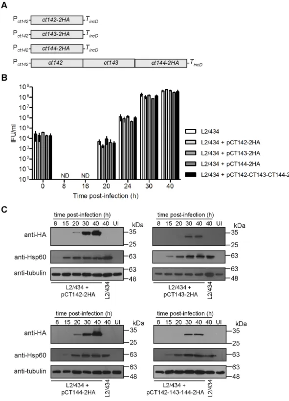
plasmids [28] encoding CT142-2HA (pCT142-2HA), CT143-2HA (pCT143-2HA), CT144-2HA (pCT144-CT144-2HA), or CT142, CT143 and CT144-CT144-2HA (pCT142-CT143-CT144-CT144-2HA) (Fig 5A). In all plasmids, expression of the genes encoding these proteins is driven by Pct142(DNA
sequence shown inFig 1Aplus additional nucleotides upstream, in a total of 165 nucleotides upstream from the start codon ofct142) and halted by the terminator of theC.trachomatis incDgene (TincD) (Fig 5A).
To test if the constructedC.trachomatisstrains were affected in their developmental cycle, we quantified the number of infectious progeny at different times of infection of HeLa cells by comparison to the parental L2/434 strain. This revealed that the kinetics of appearance of infectious particles during the developmental cycle of the four newly constructed strains was similar to the one of L2/434 (Fig 5B), indicating that the overall bacterial physiology was not affected by the plasmids or by overexpression of CT142, CT143, and/or CT144 proteins. We also followed the production of CT142-2HA, CT143-2HA, and CT144-2HA by immunoblot-ting of extracts of HeLa cells infected for 8, 15, 20, 30 or 40 h with each of the fourC. trachoma-tisstrains expressing these proteins. As for endogenous CT142 and CT143 (Fig 2B and 2C), expression of CT142-2HA, CT143-2HA and CT144-2HA could be detected from 20 h p.i. but the protein levels were much higher at 30 or 40 h p.i. (Fig 5C). For CT144-2HA encoded by plasmid pCT142-CT143-CT144-2HA, expression of CT144-2HA was only detected at 30 h p.i. (Fig 5C). We also quantified the levels of CT142-2HA, CT143-2HA and CT144-2HA produced by the four strains at 30 h p.i. While there were apparently higher levels of CT142-2HA and of CT144-2HA (encoded by pCT144-CT144-2HA) relative to CT143-CT144-2HA and CT144-CT144-2HA (encoded by pCT142-CT143-CT144-2HA), the differences were not statistically significant (Fig A and Fig B inS4 Fig). Finally, the levels of CT142-2HA or of CT143-2HA were*9-fold higher (9±1 for CT142-2HA, and 9±3 for CT143-2HA) than the levels of endogenous CT142 or CT143 in strain L2/434 (Fig A, Fig C, and Fig D inS4 Fig). Production of plasmid-encoded CT142-2HA or CT144-2HA led to an apparent increase in production of chromosomally-encoded CT142 and CT143, but the observed differences were not statistically significant (Fig A, Fig C, and Fig D inS4 Fig).
In summary, the developmental cycle of the constructedC.trachomatisstrains (Fig 5A) is not significantly different from that of the parental strain and, as expected by the predicted
*8 copies of theC.trachomatisvirulence plasmid [51], plasmid-encoded CT142-2HA and CT143-2HA proteins are produced at higher levels than chromosomally-encoded CT142 and CT143.
Plasmid-encoded CT142-2HA, CT143-2HA and CT144-2HA also
localize within the inclusion but outside of the bacteria
Fig 5. Generation and characterization ofC.trachomatisstrains producing CT142-2HA, CT143-2HA and/or CT144-2HA.(A) Schematic representation of the genes present in the plasmid of each recombinantC.trachomatis strain. Pct142,ct142promoter; TincD,incDterminator. (B) HeLa cells were infected with the indicated strains at a multiplicity of infection of 5 and recoverable inclusion forming units (IFUs) were determined at 0, 8, 16, 20, 24, 30, and 40 h p.i. Data are mean and standard error of the mean of 3 independent experiments. (C) HeLa cells were either left uninfected (UI) or infected by the indicatedC.trachomatisstrains for 8, 15, 20, 30, and 40 h. Whole cell lysates were analyzed by immunoblotting with antibodies against HA,C.trachomatisHsp60 (bacterial loading control) and tubulin (loading control for host cells).
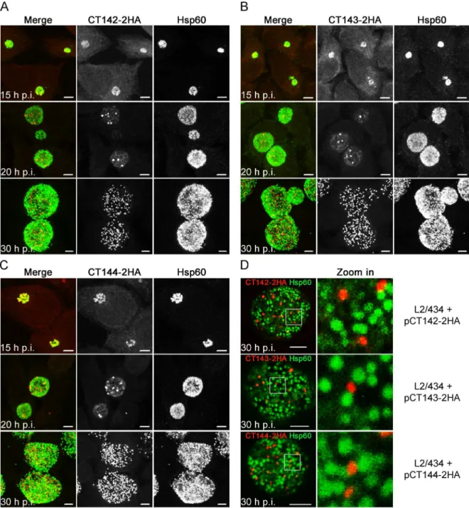
We then analyzed in more detail the HA immunofluorescence signal observed at 30 h p.i. in HeLa cells infected by theC.trachomatisstrains producing CT142-2HA, CT143-2HA, or CT144-2HA (Fig 6D). This confirmed that the globular structures revealed by the HA signal of each of the proteins did not overlap with the Hsp60 signal (Fig 6D). Additionally, the area defined by the Fig 6. CT142-HA, CT143-2HA, and CT144-2HA localize within theC.trachomatisinclusion but outside of the bacteria.
HeLa cells were infected for 15, 20 or 30 h withC.trachomatisL2/434 harbouring pCT142-2HA (A), pCT143-2HA (B), or pCT144-2HA (C). The infected cells were fixed with methanol, immunolabelled with anti-HA and anti-Hsp60 antibodies, and appropriate fluorophore-conjugated secondary antibodies, and analyzed by confocal immunofluorescence microscopy. Images are combined projections of multiple 0.2μmz-sections. Scale bars, 5μm. (D) HeLa cells infected for 30 h with the indicatedC.trachomatis strains were fixed, immunolabelled and analyzed by confocal immunofluorescence microscopy, as indicated in (A), (B), and (C). Images are singlezsections. In the area delimited by a white square, images were zoomed 5-fold. Scale bars, 5μm.
HA signal for each protein encoded by pCT142-2HA, pCT143-2HA or pCT144-2HA was signifi-cantly smaller than the area defined by the Hsp60 immunofluorescence signal (Fig 4B), but iden-tical to the area defined by the CT143 immunofluorescence signal in cells infected byC.
trachomatisL2/434 (Fig 4B).
In summary, as for chromosomally-encoded CT143, plasmid-encoded and over-expressed CT142-2HA, CT143-2HA, and CT144-2HA localize in the inclusion but outside of the bacte-ria, suggesting that from about 20 h p.i. all three proteins are secreted into the lumen of the inclusion.
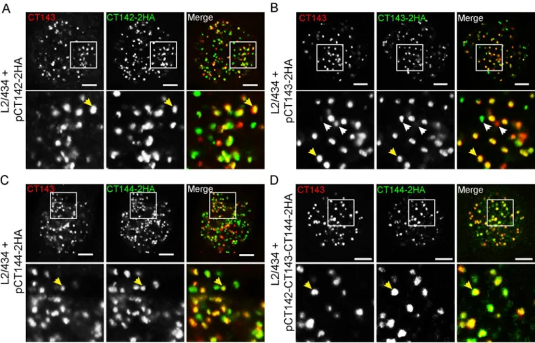
CT143 co-localizes with CT142-2HA and CT144-2HA in the lumen of the
inclusion
We next asked if the CT143 immunofluorescence signal would co-localize with the anti-HA immunofluorescence signal of CT142-2anti-HA or CT144-2anti-HA. As control, we also analyzed the co-localization between the anti-CT143 and anti-HA immunofluorescence signals of CT143 and CT143-2HA, respectively. For this, HeLa cells were infected for 30 h byC. tracho-matisL2/434-derived strains bearing 2HA, pCT143-2HA, pCT144-2HA, or pCT142-143-144-2HA. The infected cells were then fixed and immunolabelled using anti-CT143 and anti-HA antibodies. Analysis by immunofluorescence confocal microscopy of the intra-inclu-sion globular structures characteristic of CT143, CT142-2HA, CT143-2HA or CT144-2HA revealed co-localization between CT143 and CT142-2HA (Fig 7A), CT143 and CT143-2HA, as expected, (Fig 7B), and CT143 and CT144-2HA (Fig 7C and 7D). This suggested that CT142, CT143, and CT144 could be part of protein complexes within the lumen of the inclu-sion. In all cases analyzed, even for CT143 relative to CT143-2HA, the co-localization was obvious but not perfect (Fig 7). It is possible that some epitopes might be inaccessible within the putative protein complexes. Moreover, the protein levels of chromosomally-encoded CT143 relative to plasmid-encoded proteins could have interfered with the degree of co-locali-zation observed.
In summary, although the observed co-localization between CT143 and CT142-2HA or CT144-2HA does not indicate that the proteins interact, these data suggested that the globular structures revealed by immunolabelling of CT143, CT142-2HA, CT143-2HA or CT144-2HA might correspond to multi-protein complexes within the lumen of the inclusion.
Discussion
We found that in infected host cells theC.trachomatisT3S substrates CT142, CT143, and CT144 localize in apparently large globular structures within the inclusion but outside of the bacteria. This suggests that these proteins are secreted into the lumen of the inclusion. It remains possible that CT142, CT143, and CT144 might be delivered into the cytoplasm of host cells in levels too low to be detectable by immunofluorescence microscopy. However, our observations do indicate that the major proportion of these proteins is translocated across the two bacterial membranes into the lumen of the inclusion. We made identical observations for chromosomally-encoded CT143 (using an anti-CT143 antibody) and for*9-fold over-expressed, plasmid-encoded and epitope-tagged CT143-2HA. Therefore, by analogy, the presence of CT142-2HA and CT144-2HA in the lumen of the inclusion should also reflect the localization of endogenous CT142 and CT144.
mediates the transport of proteins into the lumen of the inclusion, after protein translocation only across the bacterial membranes. There are however at least two other possible scenarios explaining the predominant localization of CT142, CT143, and CT144 within the inclusion lumen. One is that, although we have shown that these proteins are T3S substrates usingY. enterocoliticaas a heterologous host [37], they are in fact secreted into the lumen of theC. tra-chomatisinclusion by another pathway. However, using another bacteria as heterologous host to study T3S signals in chlamydial proteins has been commonly used in the field [10,31,33, 52] and is normally accepted as valid. A way to further test if CT142, CT143, and CT144 are T3S substrates during the developmental cycle ofC.trachomatiscould be to use small mole-cules (salicylidene acylhydrazides) that have been described to function as inhibitors of T3S systems [53] and to disrupt the progression of theChlamydiadevelopmental cycle [54]. How-ever, the specificity of these small molecules against the T3S system is very unclear [55–59] and therefore this approach would not be the most appropriate. A T3S system-deficientC. tra-chomatismutant would be required to test without any doubt if CT142, CT143, and CT144 are type III secreted in infected cells, but such mutant would likely be non-viable because of the expected essential role of the T3S system in entry ofChlamydiainto host cells and subsequent intracellular development [3,8]. In the future, it might be possible to generate aC.trachomatis Fig 7. Co-localization between intra-inclusion structures revealed by CT143 and CT142-2HA, CT143-2HA or CT144-2HA.HeLa 229 cells were infected for 30 h byC.trachomatisL2/434 bearing pCT142-2HA (A), pCT143-2HA (B), pCT144-2HA (C), or pCT142-CT143-CT144-2HA (D). The cells fixed with methanol and immunolabelled with anti-CT143 and anti-HA antibodies and appropriate fluorophore-conjugated antibodies. Stained cells were analyzed by confocal immunofluorescence microscopy. Images are singlezsections. Yellow arrows exemplify co-localizing globular structures and white arrows highlight globular structures not showing obvious co-localization. In the area delimited by a white square (upper panels) images were zoomed 3-fold (lower panels). All scale bars, 5μm.
conditional mutant deficient in the T3S system, but this is much beyond the scope of this work. A second possibility is that the proteins are delivered into the host cell cytoplasm by the C.trachomatisT3S system and then translocated back across the vacuolar membrane into the inclusion. This seems very unlikely because it would be a rather complex transport pathway and we never detected even residual specific immunostaining of CT142-2HA, CT143 or CT143-2HA, or CT144-2HA in the host cytoplasm.
Based on the previous discussion and on observations from other studies, we do favor the pos-sibility that theC.trachomatisT3S system can transport proteins into the lumen of the inclusion [7]. First, there are precedents forC.trachomatisT3S substrates found in that localization. The glycogen metabolizing enzymes (GlgA, GlgB, GlgX, GlgP, and MalQ) have been shown to be T3S substrates usingShigella flexnerias heterologous host, and GlgA and GlgX have been immunolo-calized in the lumen of the inclusion [26,60]. GlgA was also detected in the host cytoplasm [60], and GlgX was also found in the inclusion membrane [26]. Other examples are CT620 and CT621, which were identified as T3S substrates usingS.flexnerias heterologous host and found in the lumen of the inclusion as well as in the cytoplasm of host cells [32].C.trachomatisPls1/ CT049 and Pls2/CT050 were also described to be secreted into the lumen of the inclusion [61], and also appear as globular structures that are reminiscent of those related to CT142, CT143 and CT144, but they might not be T3S substrates [61]. Second, there is no conceptual reason for why a system capable of transporting substrates across the bacterial membranes and a host cell brane would not, in some conditions, also translocate proteins only across the bacterial mem-branes. In fact, even if this might be only an experimental artifact, many bacteria possessing T3S systems can be induced to secrete protein substrates in the extracellular medium [62,63], and in Yersiniait has been proposed that different signals direct T3S substrates into distinct locations [64]. In the case ofC.trachomatis, we speculate that T3S substrates can gain direct access to the cytoplasm of host cells (e.g., TarP, Incs), be released in the lumen of the inclusion and also into the cytosol of host cells (CT620, CT621, GlgA), or simply secreted into the lumen of the inclusion (CT142, CT143, CT144 and GlgX), from where they might also reach the inclusion membrane (GlgX). Evidently, this hypothetical scenario would imply complex regulatory mechanisms of protein transport that remain to be elucidated.
using recently described methods [68–70] to inactivate thect142,ct143, orct144genes. Fur-thermore, proteomic analysis of the putative CT142-CT143-CT144 complex isolated from infected cells might identify otherC.trachomatisproteins associating with it.
We also clarified that the genes encoding CT142, CT143, and CT144 form an operon and their expression is likely driven by aσ66promoter upstream from the start codon ofct142. In a previous RNA sequencing analysis of the transcriptome ofC.trachomatisLGV serovar L2b strain UCH-1/proctitis there was no obvious definition of a transcription start site forct142 (CTLon_0393),ct143(CTLon_0394), orct144(CTLon_0395) [71]. It remains possible that in addition to the identified Pct142, transcription ofct142,ct143, orct144could be directed by
other promoters at different times or conditions of theC.trachomatisdevelopmental cycle. However, this seems unlikely as we detected no significant differences between the 3 genes in a previous real-time quantitative PCR analysis of their mRNA levels during the developmental cycle of strain L2/434 [37].
In summary, this work contributed for the characterization ofC.trachomatisT3S substrates and, in line with a recent study [26], further suggests that the chlamydial T3S system could also mediate protein secretion into the lumen of the inclusion. Additional studies are needed to solidify this intriguing hypothesis and to understand the putative regulatory processes con-trolling protein transport by the same machinery into the lumen of the inclusion and into and across the vacuolar membrane. The function of CT142, CT143, and CT144 also remains to be defined. This should be directly analyzed using recently developed methods for genetic manip-ulation ofC.trachomatis[68–70].
Supporting information
S1 Table. Plasmids used in this work. (PDF)
S2 Table. DNA primers used in this work. (PDF)
S3 Table. Identification of orthologues ofC.trachomatisCT142 (CTL0397), CT143 (CTL0398), and CT144 (CTL0399) in otherChlamydiae.
(PDF)
S1 Fig. Map of plasmid pSVP247.Details of plasmid construction are inS1 Table. The pSW2 plasmid backbone [27] is shown in black, theEscherichia coliorigin of replication (ori) in yel-low, the ampicillin resistance gene (bla) in blue, the multiple cloning site (MCS) in grey, and the double hemagglutinin (2HA) epitope tag andincDterminator in different tones of red. The DNA sequence of the MCS with unique restriction sites and the 2HA-encoding region are depicted in the box below the plasmid map.
(PDF)
S2 Fig. Genetic organization ofct142,ct143andct144orthologues inChlamydiaceae. Orthologues ofct142are depicted in green, orthologues ofct143are depicted in red, and ortho-logues ofct144are depicted in pink. The syntenic organization of the three genes is illustrated inC.trachomatisserovar A (strain A/HAR-13),C.trachomatisserovar B (strain B/Jali20/OT), C.trachomatisserovar D (strain D/UW-3/CX) as well as inC.muridarumNigg,C.abortus S26/3,C.caviaeGPIC,C.felisFe/C-56 andC.pneumoniaeCWLO29. The initial image was obtained from theChlamydiaeDB.orgwebsite (http://liferay.csb.univie.ac.at/portal/web/ chlamydiaedb) and then adapted.
S3 Fig. Characterization of antibodies against CT142 and CT143.HeLa cells were either left untransfected (NT) or transfected using jetPEI1(Polyplus-transfection) with plasmids encod-ing EGFP, EGFP-CT143 or EGFP-CT142, as indicated. Whole cell lysates were analyzed with antibodies against GFP, CT142 or CT143. Bands corresponding to EGFP-CT142 and EGFP-CT143 are indicated by an arrow.
(PDF)
S4 Fig. Quantification of CT142, CT143, and CT144 proteins produced byC.trachomatis recombinant strains.(A) HeLa cells were left uninfected (UI) or infected for 30 h byC. tracho-matisL2/434 or L2/434 carrying plasmids pCT142-2HA, pCT143-2HA, pCT144-2HA or pCT142-CT143-CT144-2HA. Whole cell lysates were analyzed by immunoblotting with anti-bodies against HA, CT142 or CT143 (as indicated),C.trachomatisHsp60 (bacterial loading control) and tubulin (loading control for host cells). The arrows indicate the position of CT142, CT142-2HA, CT143 and CT143-2HA bands. (B) The bands corresponding to HA-tagged proteins in each lane of the anti-HA blot in Fig A inS4 Fig(and in other replicates) were quantified by densitometry relative to the corresponding bands of Hsp60 and tubulin using Fiji software [48]. The calculated CT142/Hsp60/tubulin values in the graph indicate mean±standard error of the mean (SEM) from 4 independent experiments. P-values were cal-culated by a one way ANOVA and Tukey post hoc analysis and the values were not signifi-cantly different (P>0.05) between the different data sets. (C and D) The bands corresponding
to CT142 and CT142-2HA proteins (C), or the bands corresponding to CT143 and CT143-2HA proteins (D), in each lane of the anti-CT142 or CT143 blots, respectively, in Fig A inS4 Fig(and in other replicates) were quantified by densitometry relative to the corresponding bands of Hsp60 and tubulin using Fiji software [48]. The calculated HA/Hsp60/tubulin values in the graph indicate mean±SEM from 4 independent experiments, relative to the values of CT142 (in C) or CT143 (in D) in cells infected by L2/434. P-values were calculated by a one way ANOVA and Dunett post hoc analysis (relative to the L2/434 data) and there were signifi-cant differences (P<0.05) only for data corresponding to samples infected by L2/434
har-bouring pCT142-2HA or pCT142-CT143-CT144-2HA (in C) or for data corresponding to samples infected by L2/434 harbouring pCT143-2HA.
(PDF)
Acknowledgments
We are grateful to Catarina Milho and to Sofia V. Gusmão for the construction of plasmids, to Ba´rbara Figueiredo and Maria Luı´s for technical help, and to Filipe Almeida and Irina Franco for critical reading of the manuscript.
Author Contributions
Conceptualization:MdC LJM.
Formal analysis:MdC JNB LJM.
Funding acquisition:LJM.
Investigation:MdC SVP JNB LJM.
Methodology:MdC LJM.
Project administration:LJM.
Supervision:LJM.
Validation:MdC JNB LJM.
Visualization:MdC SVP LJM.
Writing – original draft:MdC LJM.
Writing – review & editing:MdC SVP JNB LJM.
References
1. Taylor-Brown A, Vaughan L, Greub G, Timms P, Polkinghorne A. Twenty years of research into Chla-mydia-like organisms: a revolution in our understanding of the biology and pathogenicity of members of the phylum Chlamydiae. Pathogens and disease. 2015; 73(1):1–15. Epub 2015/04/09.https://doi.org/ 10.1093/femspd/ftu009PMID:25854000.
2. Horn M. Chlamydiae as symbionts in eukaryotes. Annu Rev Microbiol. 2008; 62:113–31. PMID:
18473699.https://doi.org/10.1146/annurev.micro.62.081307.162818
3. Elwell C, Mirrashidi K, Engel J. Chlamydia cell biology and pathogenesis. Nat Rev Microbiol. 2016; 14 (6):385–400. Epub 2016/04/26.https://doi.org/10.1038/nrmicro.2016.30PMID:27108705; PubMed Central PMCID: PMC4886739.
4. Taylor HR, Burton MJ, Haddad D, West S, Wright H. Trachoma. Lancet. 2014. Epub 2014/07/22.
https://doi.org/10.1016/S0140-6736(13)62182-0PMID:25043452.
5. O’Connell CM, Ferone ME. Chlamydia trachomatis Genital Infections. Microb Cell. 2016; 3(9):390–403. Epub 2017/03/31.https://doi.org/10.15698/mic2016.09.525PMID:28357377; PubMed Central PMCID: PMC5354567.
6. Diepold A, Wagner S. Assembly of the bacterial type III secretion machinery. FEMS Microbiol Rev. 2014; 38(4):802–22. Epub 2014/02/04.https://doi.org/10.1111/1574-6976.12061PMID:24484471.
7. Galan JE, Lara-Tejero M, Marlovits TC, Wagner S. Bacterial type III secretion systems: specialized nanomachines for protein delivery into target cells. Annu Rev Microbiol. 2014; 68:415–38. Epub 2014/ 07/09.https://doi.org/10.1146/annurev-micro-092412-155725PMID:25002086.
8. Mueller KE, Plano GV, Fields KA. New frontiers in type III secretion biology: the Chlamydia perspective. Infect Immun. 2014; 82(1):2–9. Epub 2013/10/16.https://doi.org/10.1128/IAI.00917-13PMID:
24126521; PubMed Central PMCID: PMC3911841.
9. Clifton DR, Fields KA, Grieshaber SS, Dooley CA, Fischer ER, Mead DJ, et al. A chlamydial type III translocated protein is tyrosine-phosphorylated at the site of entry and associated with recruitment of actin. Proc Natl Acad Sci U S A. 2004; 101(27):10166–71. PMID:15199184.https://doi.org/10.1073/ pnas.0402829101
10. Hower S, Wolf K, Fields KA. Evidence that CT694 is a novel Chlamydia trachomatis T3S substrate capable of functioning during invasion or early cycle development. Mol Microbiol. 2009; 72(6):1423–37. PMID:19460098.https://doi.org/10.1111/j.1365-2958.2009.06732.x
11. Chen YS, Bastidas RJ, Saka HA, Carpenter VK, Richards KL, Plano GV, et al. The Chlamydia tracho-matis type III secretion chaperone Slc1 engages multiple early effectors, including TepP, a tyrosine-phosphorylated protein required for the recruitment of CrkI-II to nascent inclusions and innate immune signaling. PLoS Pathog. 2014; 10(2):e1003954. Epub 2014/03/04.https://doi.org/10.1371/journal.ppat. 1003954PMID:24586162; PubMed Central PMCID: PMC3930595.
12. Lane BJ, Mutchler C, Al Khodor S, Grieshaber SS, Carabeo RA. Chlamydial entry involves TARP bind-ing of guanine nucleotide exchange factors. PLoS Pathog. 2008; 4(3):e1000014. PMID:18383626.
https://doi.org/10.1371/journal.ppat.1000014
13. Jewett TJ, Miller NJ, Dooley CA, Hackstadt T. The conserved Tarp actin binding domain is important for chlamydial invasion. PLoS Pathog. 2010; 6(7):e1000997. Epub 2010/07/27.https://doi.org/10.1371/ journal.ppat.1000997PMID:20657821; PubMed Central PMCID: PMC2904776.
14. Mehlitz A, Banhart S, Maurer AP, Kaushansky A, Gordus AG, Zielecki J, et al. Tarp regulates early Chlamydia-induced host cell survival through interactions with the human adaptor protein SHC1. J Cell Biol. 2010; 190(1):143–57. Epub 2010/07/14.https://doi.org/10.1083/jcb.200909095PMID:20624904; PubMed Central PMCID: PMC2911661.
16. Subtil A, Parsot C, Dautry-Varsat A. Secretion of predicted Inc proteins of Chlamydia pneumoniae by a heterologous type III machinery. Mol Microbiol. 2001; 39(3):792–800. Epub 2001/02/13. PMID:
11169118.
17. Dumoux M, Menny A, Delacour D, Hayward RD. A Chlamydia effector recruits CEP170 to reprogram host microtubule organization. J Cell Sci. 2015; 128(18):3420–34. Epub 2015/07/30.https://doi.org/10. 1242/jcs.169318PMID:26220855; PubMed Central PMCID: PMC4582400.
18. Kokes M, Dunn JD, Granek JA, Nguyen BD, Barker JR, Valdivia RH, et al. Integrating chemical muta-genesis and whole-genome sequencing as a platform for forward and reverse genetic analysis of Chla-mydia. Cell Host Microbe. 2015; 17(5):716–25. Epub 2015/04/30.https://doi.org/10.1016/j.chom.2015. 03.014PMID:25920978; PubMed Central PMCID: PMC4418230.
19. Mirrashidi KM, Elwell CA, Verschueren E, Johnson JR, Frando A, Von Dollen J, et al. Global Mapping of the Inc-Human Interactome Reveals that Retromer Restricts Chlamydia Infection. Cell Host Microbe. 2015; 18(1):109–21. Epub 2015/06/30.https://doi.org/10.1016/j.chom.2015.06.004PMID:26118995; PubMed Central PMCID: PMC4540348.
20. Derre I, Swiss R, Agaisse H. The lipid transfer protein CERT interacts with the Chlamydia inclusion pro-tein IncD and participates to ER-Chlamydia inclusion membrane contact sites. PLoS Pathog. 2011; 7 (6):e1002092. Epub 2011/07/07.https://doi.org/10.1371/journal.ppat.1002092PMID:21731489; PubMed Central PMCID: PMC3121800.
21. Sixt BS, Bastidas RJ, Finethy R, Baxter RM, Carpenter VK, Kroemer G, et al. The Chlamydia trachoma-tis Inclusion Membrane Protein CpoS Counteracts STING-Mediated Cellular Surveillance and Suicide Programs. Cell Host Microbe. 2017; 21(1):113–21. Epub 2017/01/04.https://doi.org/10.1016/j.chom. 2016.12.002PMID:28041929; PubMed Central PMCID: PMC5233594.
22. Wolf K, Plano GV, Fields KA. A protein secreted by the respiratory pathogen Chlamydia pneumoniae impairs IL-17 signalling via interaction with human Act1. Cell Microbiol. 2009; 11(5):769–79. Epub 2009/01/23.https://doi.org/10.1111/j.1462-5822.2009.01290.xPMID:19159390; PubMed Central PMCID: PMC2927722.
23. Furtado AR, Essid M, Perrinet S, Balana ME, Yoder N, Dehoux P, et al. The chlamydial OTU domain-containing protein ChlaOTU is an early type III secretion effector targeting ubiquitin and NDP52. Cell Microbiol. 2013. Epub 2013/07/23.https://doi.org/10.1111/cmi.12171PMID:23869922.
24. Pennini ME, Perrinet S, Dautry-Varsat A, Subtil A. Histone methylation by NUE, a novel nuclear effector of the intracellular pathogen Chlamydia trachomatis. PLoS Pathog. 2010; 6(7):e1000995. Epub 2010/ 07/27.https://doi.org/10.1371/journal.ppat.1000995PMID:20657819; PubMed Central PMCID: PMC2904774.
25. Vromman F, Perrinet S, Gehre L, Subtil A. The DUF582 Proteins of Chlamydia trachomatis Bind to Components of the ESCRT Machinery, Which Is Dispensable for Bacterial Growth In vitro. Frontiers in cellular and infection microbiology. 2016; 6:123. Epub 2016/10/25.https://doi.org/10.3389/fcimb.2016. 00123PMID:27774439; PubMed Central PMCID: PMC5053991.
26. Gehre L, Gorgette O, Perrinet S, Prevost MC, Ducatez M, Giebel AM, et al. Sequestration of host metabolism by an intracellular pathogen. eLife. 2016; 5:e12552. Epub 2016/03/17.https://doi.org/10. 7554/eLife.12552PMID:26981769; PubMed Central PMCID: PMC4829429.
27. Wang Y, Kahane S, Cutcliffe LT, Skilton RJ, Lambden PR, Clarke IN. Development of a transformation system for Chlamydia trachomatis: restoration of glycogen biosynthesis by acquisition of a plasmid shuttle vector. PLoS Pathog. 2011; 7(9):e1002258. Epub 2011/10/04.https://doi.org/10.1371/journal. ppat.1002258PMID:21966270; PubMed Central PMCID: PMC3178582.
28. Agaisse H, Derre I. A C. trachomatis cloning vector and the generation of C. trachomatis strains expressing fluorescent proteins under the control of a C. trachomatis promoter. PLoS ONE. 2013; 8(2): e57090. Epub 2013/02/27.https://doi.org/10.1371/journal.pone.0057090PMID:23441233; PubMed Central PMCID: PMC3575495.
29. Bauler LD, Hackstadt T. Expression and targeting of secreted proteins from Chlamydia trachomatis. J Bacteriol. 2014; 196(7):1325–34. Epub 2014/01/21.https://doi.org/10.1128/JB.01290-13PMID:
24443531; PubMed Central PMCID: PMC3993338.
30. Ho TD, Starnbach MN. The Salmonella enterica serovar typhimurium-encoded type III secretion sys-tems can translocate Chlamydia trachomatis proteins into the cytosol of host cells. Infect Immun. 2005; 73(2):905–11. PMID:15664932.https://doi.org/10.1128/IAI.73.2.905-911.2005
31. Subtil A, Delevoye C, Balana ME, Tastevin L, Perrinet S, Dautry-Varsat A. A directed screen for chla-mydial proteins secreted by a type III mechanism identifies a translocated protein and numerous other new candidates. Mol Microbiol. 2005; 56(6):1636–47. Epub 2005/05/27. https://doi.org/10.1111/j.1365-2958.2005.04647.xPMID:15916612.
Chlamydiae. Infect Immun. 2011; 79(2):571–80. Epub 2010/11/17. https://doi.org/10.1128/IAI.00825-10PMID:21078856; PubMed Central PMCID: PMC3028825.
33. Fields KA, Hackstadt T. Evidence for the secretion of Chlamydia trachomatis CopN by a type III secre-tion mechanism. Mol Microbiol. 2000; 38(5):1048–60. PMID:11123678.
34. Chellas-Gery B, Linton CN, Fields KA. Human GCIP interacts with CT847, a novel Chlamydia tracho-matis type III secretion substrate, and is degraded in a tissue-culture infection model. Cell Microbiol. 2007; 9(10):2417–30. PMID:17532760.https://doi.org/10.1111/j.1462-5822.2007.00970.x 35. Pais SV, Milho C, Almeida F, Mota LJ. Identification of novel type III secretion chaperone-substrate
complexes of Chlamydia trachomatis. PLoS ONE. 2013; 8(2):e56292. Epub 2013/02/23.https://doi.org/ 10.1371/journal.pone.0056292PMID:23431368; PubMed Central PMCID: PMC3576375.
36. Hovis KM, Mojica S, McDermott JE, Pedersen L, Simhi C, Rank RG, et al. Genus-optimized strategy for the identification of chlamydial type III secretion substrates. Pathogens and disease. 2013. Epub 2013/ 07/23.https://doi.org/10.1111/2049-632X.12070PMID:23873765.
37. da Cunha M, Milho C, Almeida F, Pais SV, Borges V, Mauricio R, et al. Identification of type III secretion substrates of Chlamydia trachomatis using Yersinia enterocolitica as a heterologous system. BMC Microbiol. 2014; 14:40. Epub 2014/02/19.https://doi.org/10.1186/1471-2180-14-40PMID:24533538; PubMed Central PMCID: PMC3931295.
38. Song L, Carlson JH, Whitmire WM, Kari L, Virtaneva K, Sturdevant DE, et al. Chlamydia trachomatis plasmid-encoded Pgp4 is a transcriptional regulator of virulence-associated genes. Infect Immun. 2013; 81(3):636–44. Epub 2013/01/16.https://doi.org/10.1128/IAI.01305-12PMID:23319558; PubMed Cen-tral PMCID: PMC3584862.
39. Zhong G. Chlamydial Plasmid-Dependent Pathogenicity. Trends Microbiol. 2017; 25(2):141–52. Epub 2016/10/08.https://doi.org/10.1016/j.tim.2016.09.006PMID:27712952; PubMed Central PMCID: PMC5272858.
40. Kari L, Whitmire WM, Olivares-Zavaleta N, Goheen MM, Taylor LD, Carlson JH, et al. A live-attenuated chlamydial vaccine protects against trachoma in nonhuman primates. J Exp Med. 2011; 208(11):2217– 23. Epub 2011/10/12.https://doi.org/10.1084/jem.20111266PMID:21987657; PubMed Central PMCID: PMC3201208.
41. Olivares-Zavaleta N, Whitmire W, Gardner D, Caldwell HD. Immunization with the attenuated plasmid-less Chlamydia trachomatis L2(25667R) strain provides partial protection in a murine model of female genitourinary tract infection. Vaccine. 2010; 28(6):1454–62. Epub 2009/12/17.https://doi.org/10.1016/j. vaccine.2009.11.073PMID:20004265; PubMed Central PMCID: PMC2821993.
42. Sigar IM, Schripsema JH, Wang Y, Clarke IN, Cutcliffe LT, Seth-Smith HM, et al. Plasmid deficiency in urogenital isolates of Chlamydia trachomatis reduces infectivity and virulence in a mouse model. Patho-gens and disease. 2014; 70(1):61–9. Epub 2013/09/12.https://doi.org/10.1111/2049-632X.12086
PMID:24022847; PubMed Central PMCID: PMC4300952.
43. Peterson EM, Markoff BA, Schachter J, de la Maza LM. The 7.5-kb plasmid present in Chlamydia tra-chomatis is not essential for the growth of this microorganism. Plasmid. 1990; 23(2):144–8. Epub 1990/ 03/01. PMID:2362949.
44. Scidmore MA. Cultivation and Laboratory Maintenance of Chlamydia trachomatis. Curr Protoc Micro-biol. 2005;Chapter 11:Unit 11A 1. PMID:18770550.https://doi.org/10.1002/9780471729259. mc11a01s00
45. Stephens RS, Kalman S, Lammel C, Fan J, Marathe R, Aravind L, et al. Genome sequence of an obli-gate intracellular pathogen of humans: Chlamydia trachomatis. Science. 1998; 282(5389):754–9. PMID:9784136.
46. Nguyen BD, Valdivia RH. Forward genetic approaches in Chlamydia trachomatis. Journal of visualized experiments: JoVE. 2013;(80):e50636. Epub 2013/11/07.https://doi.org/10.3791/50636PMID:
24192560.
47. Studier FW. Protein production by auto-induction in high density shaking cultures. Protein Expr Purif. 2005; 41(1):207–34. Epub 2005/05/26. PMID:15915565.
48. Schindelin J, Arganda-Carreras I, Frise E, Kaynig V, Longair M, Pietzsch T, et al. Fiji: an open-source platform for biological-image analysis. Nat Methods. 2012; 9(7):676–82. Epub 2012/06/30.https://doi. org/10.1038/nmeth.2019PMID:22743772; PubMed Central PMCID: PMC3855844.
49. Altschul SF, Madden TL, Schaffer AA, Zhang J, Zhang Z, Miller W, et al. Gapped BLAST and PSI-BLAST: a new generation of protein database search programs. Nucleic Acids Res. 1997; 25 (17):3389–402. Epub 1997/09/01. PMID:9254694; PubMed Central PMCID: PMC146917.
51. Ferreira R, Borges V, Nunes A, Borrego MJ, Gomes JP. Assessment of the load and transcriptional dynamics of Chlamydia trachomatis plasmid according to strains’ tissue tropism. Microbiological research. 2013; 168(6):333–9. Epub 2013/04/18.https://doi.org/10.1016/j.micres.2013.02.001PMID:
23590987.
52. Dehoux P, Flores R, Dauga C, Zhong G, Subtil A. Multi-genome identification and characterization of chlamydiae-specific type III secretion substrates: the Inc proteins. BMC genomics. 2011; 12:109. Epub 2011/02/18.https://doi.org/10.1186/1471-2164-12-109PMID:21324157; PubMed Central PMCID: PMC3048545.
53. Nordfelth R, Kauppi AM, Norberg HA, Wolf-Watz H, Elofsson M. Small-molecule inhibitors specifically targeting type III secretion. Infect Immun. 2005; 73(5):3104–14. Epub 2005/04/23.https://doi.org/10. 1128/IAI.73.5.3104-3114.2005PMID:15845518; PubMed Central PMCID: PMC1087345.
54. Wolf K, Betts HJ, Chellas-Gery B, Hower S, Linton CN, Fields KA. Treatment of Chlamydia trachomatis with a small molecule inhibitor of the Yersinia type III secretion system disrupts progression of the chla-mydial developmental cycle. Mol Microbiol. 2006; 61(6):1543–55. PMID:16968227.https://doi.org/10. 1111/j.1365-2958.2006.05347.x
55. Prantner D, Nagarajan UM. A role for chlamydial type III secretion apparatus in host cytokine expres-sion. Infect Immun. 2008. PubMed PMID: 18852236.https://doi.org/10.1128/IAI.00963-08
56. Slepenkin A, Enquist PA, Hagglund U, de la Maza LM, Elofsson M, Peterson EM. Reversal of the antic-hlamydial activity of putative type III secretion inhibitors by iron. Infect Immun. 2007; 75(7):3478–89. PMID:17470544.https://doi.org/10.1128/IAI.00023-07
57. Engstrom P, Nguyen BD, Normark J, Nilsson I, Bastidas RJ, Gylfe A, et al. Mutations in hemG mediate resistance to salicylidene acylhydrazides, demonstrating a novel link between protoporphyrinogen oxi-dase (HemG) and Chlamydia trachomatis infectivity. J Bacteriol. 2013; 195(18):4221–30. Epub 2013/ 07/16.https://doi.org/10.1128/JB.00506-13PMID:23852872; PubMed Central PMCID: PMC3754756.
58. Wang D, Zetterstrom CE, Gabrielsen M, Beckham KS, Tree JJ, Macdonald SE, et al. Identification of bacterial target proteins for the salicylidene acylhydrazide class of virulence-blocking compounds. J Biol Chem. 2011; 286(34):29922–31. Epub 2011/07/05.https://doi.org/10.1074/jbc.M111.233858
PMID:21724850; PubMed Central PMCID: PMC3191033.
59. Martinez-Argudo I, Veenendaal AK, Liu X, Roehrich AD, Ronessen MC, Franzoni G, et al. Isolation of Salmonella mutants resistant to the inhibitory effect of Salicylidene acylhydrazides on flagella-mediated motility. PLoS ONE. 2013; 8(1):e52179. Epub 2013/01/10.https://doi.org/10.1371/journal.pone. 0052179PMID:23300965; PubMed Central PMCID: PMC3534715.
60. Lu C, Lei L, Peng B, Tang L, Ding H, Gong S, et al. Chlamydia trachomatis GlgA Is Secreted into Host Cell Cytoplasm. PLoS ONE. 2013; 8(7):e68764. Epub 2013/07/31.https://doi.org/10.1371/journal. pone.0068764PMID:23894341; PubMed Central PMCID: PMC3722199.
61. Jorgensen I, Valdivia RH. Pmp-like proteins Pls1 and Pls2 are secreted into the lumen of the Chlamydia trachomatis inclusion. Infect Immun. 2008; 76(9):3940–50. PMID:18591235.https://doi.org/10.1128/ IAI.00632-08
62. Bahrani FK, Sansonetti PJ, Parsot C. Secretion of Ipa proteins by Shigella flexneri: inducer molecules and kinetics of activation. Infect Immun. 1997; 65(10):4005–10. PMID:9316999.
63. Michiels T, Cornelis GR. Secretion of hybrid proteins by the Yersinia Yop export system. J Bacteriol. 1991; 173(5):1677–85. PMID:1999387.
64. Lee VT, Mazmanian SK, Schneewind O. A program of Yersinia enterocolitica type III secretion reactions is activated by specific signals. J Bacteriol. 2001; 183(17):4970–8. PMID:11489848.https://doi.org/10. 1128/JB.183.17.4970-4978.2001
65. Yang C, Starr T, Song L, Carlson JH, Sturdevant GL, Beare PA, et al. Chlamydial Lytic Exit from Host Cells Is Plasmid Regulated. mBio. 2015; 6(6):e01648–15. Epub 2015/11/12.https://doi.org/10.1128/ mBio.01648-15PMID:26556273; PubMed Central PMCID: PMC4659467.
66. Carlson JH, Whitmire WM, Crane DD, Wicke L, Virtaneva K, Sturdevant DE, et al. The Chlamydia tra-chomatis plasmid is a transcriptional regulator of chromosomal genes and a virulence factor. Infect Immun. 2008; 76(6):2273–83. Epub 2008/03/19.https://doi.org/10.1128/IAI.00102-08PMID:
18347045; PubMed Central PMCID: PMC2423098.
67. Gordon FB, Quan AL. Occurence of Glycogen in Inclusions of the Psittacosis-Lymphogranuloma Vene-reum-Trachoma Agents. J Infect Dis. 1965; 115:186–96. PMID:14308365.
68. Mueller KE, Wolf K, Fields KA. Gene Deletion by Fluorescence-Reported Allelic Exchange Mutagenesis in Chlamydia trachomatis. mBio. 2016; 7(1):e01817–15. Epub 2016/01/21.https://doi.org/10.1128/ mBio.01817-15PMID:26787828; PubMed Central PMCID: PMC4725004.
70. Fischer A, Harrison KS, Ramirez Y, Auer D, Chowdhury SR, Prusty BK, et al. Chlamydia trachomatis-containing vacuole serves as deubiquitination platform to stabilize Mcl-1 and to interfere with host defense. eLife. 2017; 6. Epub 2017/03/30.https://doi.org/10.7554/eLife.21465PMID:28347402; PubMed Central PMCID: PMC5370187.