REVIEW published: 06 October 2020 doi: 10.3389/fncel.2020.584277
Edited by: Guei-Sheung Liu, University of Tasmania, Australia
Reviewed by: Spyros Petrakis, Institute of Applied Biosciences (INAB), Greece Xiao-Jiang Li, Emory University, United States
*Correspondence: Catarina O. Miranda [email protected] Luís Pereira de Almeida [email protected]
†Present address: Adriana Marcelo, Algarve University, Faro, Portugal Teresa P. Silva, IBB-Institute for Bioengineering and Biosciences and Department of Bioengineering, Instituto Superior Técnico, Universidade de Lisboa, Lisbon, Portugal João Barata, Institute for Genome Stability in Ageing and Disease, Cologne Cluster of Excellence in Cellular Stress Response in Aging-Associated Diseases (CECAD), University Hospital Cologne, Cologne, Germany Specialty section: This article was submitted to Cellular Neuropathology, a section of the journal Frontiers in Cellular Neuroscience
Received: 16 July 2020 Accepted: 03 September 2020 Published: 06 October 2020
Citation: Barros I, Marcelo A, Silva TP, Barata J, Rufino-Ramos D, Pereira de Almeida L and Miranda CO (2020) Mesenchymal Stromal Cells’ Therapy for Polyglutamine Disorders: Where Do We Stand and Where Should We Go? Front. Cell. Neurosci. 14:584277. doi: 10.3389/fncel.2020.584277
Mesenchymal Stromal Cells’ Therapy
for Polyglutamine Disorders: Where
Do We Stand and Where Should
We Go?
Inês Barros1,2,3,Adriana Marcelo1,2†,Teresa P. Silva1,2†,João Barata1,2†,
David Rufino-Ramos1,2,4,Luís Pereira de Almeida1,2,4,5* andCatarina O. Miranda1,2,3* 1
CNC—Center for Neuroscience and Cell Biology, University of Coimbra, Coimbra, Portugal,2
CIBB—Center for Innovative Biomedicine and Biotechnology, University of Coimbra, Coimbra, Portugal,3
III—Institute for Interdisciplinary Research, University of Coimbra, Coimbra, Portugal,4
Faculty of Pharmacy, University of Coimbra, Coimbra, Portugal,5
Viravector—Viral Vector for Gene Transfer Core Facility, University of Coimbra, Coimbra, Portugal
Polyglutamine (polyQ) diseases are a group of inherited neurodegenerative disorders caused by the expansion of the cytosine-adenine-guanine (CAG) repeat. This mutation encodes extended glutamine (Q) tract in the disease protein, resulting in the alteration of its conformation/physiological role and in the formation of toxic fragments/aggregates of the protein. This group of heterogeneous disorders shares common molecular mechanisms, which opens the possibility to develop a pan therapeutic approach. Vast efforts have been made to develop strategies to alleviate disease symptoms. Nonetheless, there is still no therapy that can cure or effectively delay disease progression of any of these disorders. Mesenchymal stromal cells (MSC) are promising tools for the treatment of polyQ disorders, promoting protection, tissue regeneration, and/or modulation of the immune system in animal models. Accordingly, data collected from clinical trials have so far demonstrated that transplantation of MSC is safe and delays the progression of some polyQ disorders for some time. However, to achieve sustained phenotypic amelioration in clinics, several treatments may be necessary. Therefore, efforts to develop new strategies to improve MSC’s therapeutic outcomes have been emerging. In this review article, we discuss the current treatments and strategies used to reduce polyQ symptoms and major pre-clinical and clinical achievements obtained with MSC transplantation as well as remaining flaws that need to be overcome. The requirement to cross the blood-brain-barrier (BBB), together with a short rate of cell engraftment in the lesioned area and low survival of MSC in a pathophysiological context upon transplantation may contribute to the transient therapeutic effects. We also review methods like pre-conditioning or genetic engineering of MSC that can be used to increase MSC survival in vivo, cellular-free approaches—i.e., MSC-conditioned medium (CM) or MSC-derived extracellular vesicles (EVs) as a way of possibly replacing the use of MSC and methods required to standardize the potential of
Barros et al. MSC for PolyQ Disorders
MSC/MSC-derived products. These are fundamental questions that need to be addressed to obtain maximum MSC performance in polyQ diseases and therefore increase clinical benefits.
Keywords: mesenchymal stromal cells, polyglutamine disorders, neurodegenerative disorders, cell therapy, extracellular vesicles, secretome
INTRODUCTION
Polyglutamine (polyQ) diseases are a group of nine inherited neurodegenerative disorders including dentatorubral pallidoluysian atrophy, spinal bulbar muscular atrophy (SBMA), Huntington’s disease (HD), and spinocerebellar ataxias (SCAs) type 1, 2, 3, 6, 7 and 17. All of them are autosomal dominant, apart from SBMA, which is X-linked (Orr and Zoghbi, 2007).
PolyQ disorders are associated with the unstable expansion of the cytosine-adenine-guanine (CAG) trinucleotide repeat in the respective causative gene. Such mutation encodes for extended glutamine (Q) tract, which occurs in different proteins according to the disorder, thus having unrelated functions and distinct cellular and subcellular locations. Nonetheless, all polyQ diseases share common features suggesting that the polyQ stretch directly contributes to the toxic properties of these proteins through a ‘‘toxic gain of function.’’
Although these disorders express distinct symptoms, all of them greatly impact a patient’s quality of life by leading to both physical and psychological complications. Some of the shared common features include: onset typically during midlife; the inverse correlation between the number of repeats and the age of onset; degeneration of specific neuronal subpopulations that slowly progress over 10–20 years after the onset, culminating in premature death (Maciel et al., 1995; Ranum et al., 1995; Dürr et al., 1996; Zoghbi and Orr, 2000). The cerebellum, basal ganglia, brainstem nuclei, and spinal motor nuclei are some of the regions of the nervous system that are transversely affected in polyQ diseases (Zoghbi and Orr, 2000; Ross et al., 2003). The neuropathological alterations are the result of cellular changes such as accumulation of polyQ proteins in the cytoplasm and nucleus, the formation of inclusions, disturbance of the quality control systems of the cell, mitochondrial dysfunction, axonal transport disruption, synaptic activity decline, and neuroinflammation (Shao and Diamond, 2007; Havel et al., 2009). Additionally, the generation of toxic fragments resulting from the proteolytic cleavage has also been described in a few polyQs. The existence of common pathological mechanisms opens windows for the design of possible common therapies. In that sense, cell therapies and cellular-derived treatments are good candidates to be used for that purpose.
Currently, besides palliative care, treatments for polyQ disorders are only able to suppress or reduce specific symptoms. Levodopa or a dopamine agonist have been successful in cases displaying parkinsonian features, with dystonia and bradykinesia also being reduced through this approach (Tuite et al., 1995; Paulson, 2007). Furthermore, gait symptoms plus dysarthria can be improved by occupational and physical therapy, and dysphagia might be ameliorated by implementing
strategies on what and how patients eat to avoid complications (Paulson, 2007). On the other hand, since pre-symptomatic testing already exists, medical and ethical guidance regarding treatment and parenthood counseling can be given, not only to post-symptomatic patients but also to pre-symptomatic individuals (Sequeiros et al., 1998; Drüsedau et al., 2004; Bettencourt and Lima, 2011; Schuler-Faccini et al., 2014).
Despite the efforts that have been made to develop effective therapies, there is still no strategies that can prevent, cure, or delay the progression of polyQ disorders.
Promising molecular and cellular therapies have been investigated in the last decades. Regarding gene therapy, the most frequently used method consists of silencing the defective protein resulting from the CAG expansion in a given gene (depending on the polyQ), mostly using RNA interference. However, there are challenges regarding delivery to the central nervous system and the risk of off-target effects. Significant progress has been made regarding this approach and such strategies will probably be implemented soon. Still, since the genetic defects differ from disorder to disorder, the strategy will require adaptations for each disease. As this subject is not the main focus of this review, we will not discuss it in detail, but this subject is reviewed in
Matos et al. (2018).
Cellular therapies, on the other hand, may exert a pan effect on the common defective mechanisms that polyQ disorders share, as described above. For that, neural precursor and neural stem cells, embryonic stem cells, induced pluripotent stem cells, and mesenchymal stem cells (MSC) can be envisioned. Naturally, each source of the referred stem cells has its advantages, drawbacks, and success upon usage in polyQ diseases, as summarized elsewhere (Mendonça et al., 2018). Presently, MSC is not the first choice for cellular therapies in neurodegenerative conditions. Nonetheless, they are undoubtedly the safest kind among the different stem cells that can be used for therapeutic purposes since they are adult cells, and thus, not prone to develop tumors in a stable organism. Therefore, they have been widely used in studies of neurodegenerative disorders, including polyQs. However, despite some promising results, some aspects need to be improved to amplify MSCs healing effects and achieve translational significance. These concerns will be discussed in the present review article.
MESENCHYMAL STROMAL CELLS (MSC):
PROMISING TOOLS FOR THE
TREATMENT OF PolyQ DISORDERS?
Mesenchymal stromal cells (MSC) are adult multipotent progenitor cells capable of giving rise to tissues from the
Barros et al. MSC for PolyQ Disorders
mesenchymal lineage. They were described in the 70’s decade by Friedenstein et al. (1970, 1974) and since the discovery of their multipotency in 1997 (Prockop, 1997), their use in cellular therapies for neurodegenerative disorders suffered a steadfast evolution, due to the high potential/low-risk balance (Figure 1).
In neuronal context, their role was initially thought to be associated with the replacement of aged and damaged cells (Wakitani et al., 1995; Pittenger et al., 1999) but, currently, there is consensus that this is not their principal mode of action. Cell fusion was also described to occur by some authors. Of note, this mechanism was described in a SCA1 mouse model, where Purkinje cells (PCs) of the host animals fused with MSC (Huda et al., 2016). However, this mechanism is rare in the literature and does not appear to account significantly for MSC’s overall outcomes. So MSC’s main effects are probably mainly mediated by influencing neighboring cells, through secretion of bioactive factors, or by inducing secretion of these factors in host cells, thus being able to modulate the immune system and to promote tissue repair (Gao et al., 2001; Tremain et al., 2001). The establishment of strong communication through chemical signals between MSC and surrounding cells, which can be either other MSC, neurons, astrocytes, or glia, is thought to be the anchor for MSC’s therapeutic capabilities. Indeed, MSC can respond to stress ‘‘signals’’ of the nearby cell by modulating their molecular pathways towards the production of specific factors, which can support these cells under stress (Millard and Fisk, 2013). As an example, a mechanism called ‘‘bio bridge’’ in which MSC seem to form a stream of extracellular matrix (bridge) between the lesion site and neurogenic niches was described for some neurological contexts such as traumatic brain injury and stroke, and could occur in other neurological disorders (Tajiri et al., 2013; Sullivan et al., 2015). This bridge is concomitant with higher expression levels of metalloproteinases (MMP) such as MMP9, leading to better communication between MSC and endogenous neuronal precursors and increment in neurogenesis post-lesion. Precursors cells from
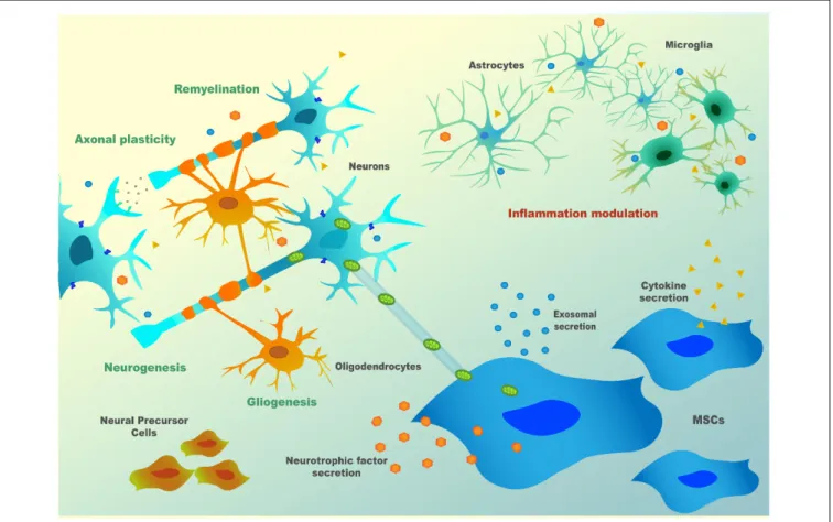
neurogenic niches then reach the brain-lesioned tissue, replacing injured cells. For all these mechanisms, there is evidence of bidirectional communication exerted through bioactive soluble molecules [neurotrophins, cytokines, chemokines, microRNAs (miRNAs), etc.], extracellular vesicles (EVs; that can contain proteins, RNAs, or even DNA) or by direct contact (e.g.,via tunneling nanotubes or through mechanotransduction). Therefore, depending on the defects in the host damaged tissue, MSC may: (1) modulate inflammatory processes; (2) reduce oxidative stress, either by inducing survival pathways or by the direct transfer of healthy mitochondria to the host cells (via nanotubes); (3) favor neurogenesis by the secretion of neurotrophins and by the formation of ‘‘bio bridges’’; (4) induce gliogenesis and remyelination; and (5) increase axonal survival and plasticity, thus inducing synaptogenesis (Paul and Anisimov, 2013; Figure 2). These exquisite cross-talks lead to a wide evaluation of MSC for the therapy of neurological diseases in preclinical and clinical models.
From Alzheimer’s (AD) to Parkinson’s (PD) or HD, the encouraging effects of MSC in a few pre-clinical studies prompted clinicians to perform preliminary clinical trials to evaluate their safety and/or effectiveness. However, this process started before fundamental issues were properly addressed at the pre-clinical level, which led to some disappointing results relative to the ones expected. Due to the initial lack of information, strategies did not contemplate solutions for problems such as the challenge of surpassing the blood-brain barrier (BBB), the low rate of cells engraftment in the lesioned tissue, the low survival of MSC, or the unidentified mechanisms involved in MSC’s positive effects. Finally, the standardization of MSC source of cells or even of methods capable of evaluating their potential, are imperative to make translation possible.
The investigation in this field is therefore currently aiming at resolving these difficulties and giving answers to the urgent need of efficacious therapies for neurodegenerative disorders for which therapeutic tools are presently scarce.
FIGURE 1 | Timeline of mesenchymal stromal cells (MSC)’s history and their use in neurodegenerative disorders (Friedenstein et al., 1970;Owen and Friedenstein, 1988;Caplan, 1991; Prockop, 1997; Chopp et al., 2000; Mazzini et al., 2003; Bonafede et al., 2016).
Barros et al. MSC for PolyQ Disorders
FIGURE 2 | MSC’s paracrine mechanism(s) in neuronal cells.
This review gives an overview of this subject with a particular focus on polyQ disorders, which besides HD, are scarcely referred to in the literature.
Pre-clinical Studies Assessing MSC’s
Therapeutic Potential in PolyQs
Several pre-clinical studies have investigated the therapeutic efficiency of MSC isolated from different sources, including bone marrow (BM-MSC), adipose tissue (AD-MSC), and umbilical cord (UC-MSC), in rodent models of HD. HD is the polyQ disease with the highest prevalence worldwide affecting about 1 in 7,500 individuals (Evans et al., 2013; Fisher and Hayden, 2014). HD causes brain atrophy in several regions such as the striatum, thalamus, cerebellum, brain stem, and cortex (Harper et al., 2005; Hassel et al., 2008; Labbadia and Morimoto, 2013; Chao et al., 2017) leading to progressive motor dysfunction and incoordination, cognitive impairment and psychiatric symptoms.
Over the last decade, it has been demonstrated that MSC can relieve phenotype and neuropathology of HD in both transgenic (Lee et al., 2009; Im et al., 2010; Snyder et al., 2010; Lin et al., 2011; Yu-Taeger et al., 2019) and chemically-induced models (Lee et al., 2009; Edalatmanesh et al., 2011; Rossignol et al., 2011, 2015; Hosseini et al., 2015; Ebrahimi et al., 2018). These studies show that animals treated with
MSC displayed improved behavioral performance, cognitive functions, and, in the excitotoxic Quinolinic Acid (QA)-induced HD rats, reduction of apomorphine-induced rotation.Hosseini et al. (2015)also showed that MSC was able to reduce anxiety levels in treated QA-induced HD rats. Also, the administration of MSC was able to increase the survival of the R6/2 mouse model (Lee et al., 2009; Lin et al., 2011; Yu-Taeger et al., 2019). Importantly, one of these studies pointed out the importance of well-dosing the number of MSC administered. The authors compared two different doses of MSC (2 × 10⁵ and 4 × 10⁵) and surprisingly only the group treated with the lowest dose presented motor improvements (Rossignol et al., 2011). The authors concluded that a high number of MSC may cause tissue damage to striatal architecture.
Regarding neuropathological improvement, MSC preserved the volume of the striatum, induced neurogenesis, and differentiation of the host endogenous neural stem cells, and reduced neuronal death (Snyder et al., 2010). Lin et al.
(2011) saw a decrease in the levels of Bax and caspase-3
as well as an up-regulation in the expression of Erk1/2, which suggests that MSC can inhibit apoptosis and promote neuronal survival. Furthermore, this study points out the strong capability of MSC for recruiting microglia, neuroblasts, and bone marrow-derived cells to the lesion site which helps tissue regeneration (Lin et al., 2011). MSC administration has also
Barros et al. MSC for PolyQ Disorders
FIGURE 3 | Schematic representation of exosome biogenesis and molecular composition. Exosomes have an endocytic origin and are released to the extracellular milieu upon the fusion of MVB with the plasma membrane. Other MVB may fuse with lysosomes leading to their degradation. Overall, exosomes surface markers, cytoskeleton, and cellular metabolism proteins are common to those of the cell of origin. Plus, exosomes also include signaling proteins, DNA, several types of RNA, and lipids. Abbreviations: ESCRT, Endosomal Sorting Complex Required for Transport; MVB, Multivesicular bodies; mRNA, messenger RNA, miRNA, microRNA.
led to the reduction of the number of huntingtin aggregates (Lee et al., 2009).
Notably, Yu-Taeger et al. (2019) showed that MSC could ameliorate disruptions in the dopaminergic signaling cascade, increasing the expression of DARPP-32 and tyrosine hydroxylase in the striatum, known to be reduced in R6/2 HD mice (Bibb et al., 2000). This study also evidenced the immunomodulatory potential of MSC as treated mice restored the expression of Iba1, and microglia morphology in the striatum, plus inducing the downregulation of several inflammatory modulators (Yu-Taeger et al., 2019).
The feasibility of performing autologous transplants in HD has also been assessed. Researchers compared the efficacy of AD-MSC isolated from the subcutaneous adipose tissue of an HD patient and a healthy donor in the treatment of YAC128 transgenic mice, a long-lived HD model (Im et al., 2010). Even though both kinds of cells expressed the same cell surface markers and showed a similar secretory potential in terms of neurotrophic factors, normal AD-MSC could reduce striatal atrophy when they were transplanted into 8 month-old YAC128 mice, whereas HD AD-MSC failed to promote such benefits. Nonetheless, the injection of cells in this time-window did not produce improvements in any of the treated groups regarding rotarod performance or body weight. On the contrary, when transplanting normal AD-MSC at 12 months of age, there were significant improvements in motor performance. These results show that AD-MSC could modify the course of HD in long-lived mice, though their
effect is dependent on the donor condition and time window of delivery.
More recently, two studies assessing how the number of passages of MSC inex vivo cultures can affect their therapeutic effects were performed in R6/2 transgenic mice (Fink et al., 2013; Rossignol et al., 2015). In the first study, BM-MSC isolated from mice were grown for a low (3–8) or high (40–50) number of passages, and no neuronal or glial differentiation was observed. The striatum of R6/2 mice transplanted with BM-MSC at higher passage showed increased metabolic activity compared with non-treated R6/2 mice and significantly-improved behavioral sparing compared to either untreated R6/2 mice or R6/2 mice receiving low-passage BM-MSC. Thus, the passage number of BM-MSC expansion could alter their efficacy.
In the second study, they compared the therapeutic potential of low- (3–8) and high-passage (40–50) UC-MSC in the R6/2 transgenic HD mouse model (Fink et al., 2013). Interestingly, high-passage UC-MSC showed a higher survival ratein vivo, but contrary to the previous study, both transplanted groups showed increased metabolic activity and reduced brain damage in the striatal area. Moreover, no major differences were observed in the motor and non-motor behavioral tests compared with non-treated HD mice. In contrast, Ebrahimi and collaborators showed that the administration of UC-MSC at a low passage in 3-NP lesioned rat models led to significant improvement of motor performance and muscle activity. Plus, they also observed neuropathological ameliorations as UC-MSC were able to decrease gliosis and increase both the striatal volume
Barros et al. MSC for PolyQ Disorders
and dendritic length of striatal cells (Ebrahimi et al., 2018). So, as a conclusion, the time of MSC expansion and concomitant number of passages is an important parameter and should be further and carefully addressed in future assessments.
The relief of phenotypical and neuropathological defects in HD models has been associated with MSC trophic effect. Several studies reported that MSC transplantation increased the expression of both messenger RNA (mRNA) and protein levels of brain-derived neurotrophic factor (BDNF) in the striatum (Snyder et al., 2010). Additionally, Snyder et al. (2010)reported that BM-MSC transplantation led to increased expression of the neurotrophic factors fibroblast growth factor (FGF), ciliary neurotrophic factor (CNTF), vascular endothelial growth factor (VEGF), and nerve growth factor (NGF). Over-expression of collagen type-I and fibronectin in the striatum of MSC-treated animals has also been observed which contributes to the stabilization of the extracellular matrix, helping tissue regeneration (Rossignol et al., 2011).
Regarding other polyQ disorders, few preclinical studies have been performed in mouse models of SCA1, SCA2, and SCA3. Despite the causative mutation being located in different genes, an expansion of the CAGs in ATXN1, ATXN2, and ATXN3 (also known as MJD1), respectively, many symptoms and major mechanisms involved in disease progression are similar among SCAs (Paulson, 2009; Klockgether et al., 2019; Sullivan et al., 2019). Over time, patients develop motor incoordination as the main progressive clinical feature, along with tremors, muscle rigidity, dysarthria, dysphagia, peripheral neuropathy, oculomotor problems (such as double vision, nystagmus or ophthalmoplegia), and parkinsonism in some cases (Paulson, 2009). Cognitive impairments have also been pointed out to occur.
MSC’s effect has shown to be equally promising in these groups of polyQs, demonstrating that they can alleviate motor function deterioration in SCAs. A pre-symptomatic study was performed in a transgenic mouse model of SCA-1 (SCA1-Tg mice). Intrathecal (IT) injection of an MSC line (KUM10) in SCA1-Tg mice led to neuropathological improvements. MSC was shown to migrate towards the cerebellum and mitigate neuronal disorganization. The cerebellum of these SCA1 transgenic mice had ectopically-located PCs bodies that resulted in a multilayer of PC, and mice showed atrophy of the cerebellar molecular layer (ML). In the referred study, MSC-treated mice displayed a single layer of PC and the ML was thicker than that of the untreated mice. Motor function assessed by the rotarod test was also shown to be improved in treated animals (Matsuura et al., 2014).
Additionally, MSC showed to mitigate motor functions deterioration in a SCA2 transgenic mouse model (Chang et al., 2011). The study compared the efficacy of repeated intravenous (IV) or intracranial (IC) injections of human BM-MSC and concluded that IV infusions effectively delayed diseases’ onset and improved motor performance by rescuing cerebellar PC, whereas IC injections failed to improve motor skills. In this study, more grafted BM-MSC were found in the IV transplantation group, located in the cerebellum and cerebral cortex (CC). The authors explained the higher success of IV delivered BM-MSC due to the extravasation capacity as cells migrated
from intracranial vessels through white matter (WM) into several regions of the cerebellum.
Our group has recently demonstrated that mouse BM-MSC transplantation can ameliorate both phenotypical and neuropathological MJD symptoms in Machado–Joseph disease (MJD)/SCA3 (Oliveira Miranda et al., 2018). In agreement with the previous study, our investigation showed that the administration of several consecutive IV injections of BM-MSC sustainably ameliorated motor impairments, when delivered post-symptomatically. Using intracerebroventricular administration (ICV), a single injection could only alleviate MJD symptoms transiently in the same Tg-ATXN3-69Q transgenic mouse model of MJD (Torashima et al., 2008). This suggests that repeated treatment through the IV route led to better outcomes. Notably, our results were in line withLi et al. (2018)
findings, which showed that repeated systemic treatment of pre-symptomatic transgenic homozygous MJD mice [SCA3-YAC-84Q; (Cemal et al., 2002)] with human UC-MSC was able to alleviate motor impairments and neuropathology of MJD.
These findings were corroborated with low survival and poor engraftment in host tissues, which represent a common problem in these therapies. In our study, we demonstrated that after ICV transplantation, despite cells surviving up to 8 weeks after transplantation, the SPION-labeled MSC signal was drastically reduced from 1 to 8 weeksin vivo. Moreover, BM-MSC delivered IV was detected in the brain of mice 30 min after transplantation, but completely disappeared from circulation before completion of 7 days (Oliveira Miranda et al., 2018). Similarly, other studies in HD animal models also observed that MSC remained only for a short time in the lesion site (Snyder et al., 2010), or in other cases, although the authors could detect cells in the brain, only a small percentage of the implanted cells could survive (Lee et al., 2009; Im et al., 2010; Snyder et al., 2010; Lin et al., 2011; Yu-Taeger et al., 2019). A concise review of these studies is given in (Table 1).
Interestingly, these studies in SCAs also relate the action of MSC to neuronal survival/activity pathways, which is following the previous studies performed in HD rodent models. In the study of Li et al. (2018), the neuroprotective action of MSC was linked with the upregulation of the insulin-like growth factor 1/heat shock protein 70 (IGF-1/HSP70) pathway. In our study, we demonstrated that MSC protected GABAergic and Glutamatergic neurons, as by proton magnetic resonance spectroscopy (1H-MRS) we detected an increased expression of Gaba and the complex Glutamine-Glutamate upon a treatment, along with increased levels of mRNA for the receptors Gabrb2 and Grm1 in the cerebellum, denoting a higher synaptic activity (Oliveira Miranda et al., 2018).
In conclusion, despite the general agreement on the fact that MSC promotes beneficial outcomes in polyQ diseases, the different severity grades of the rodent models used as well as the ability to simulate human diseases make the interpretation of the potency scale of the treatments difficult. The development of models that better recapitulate polyQ diseases neuropathology in humans would be essential. Thus, knock-in models of larger mammals like the HD CRISPR-Cas9 knock-in pig model that shows strong neurodegeneration patterns (Yan et al., 2018) could
Barr os et al. MSC for PolyQ Disor ders
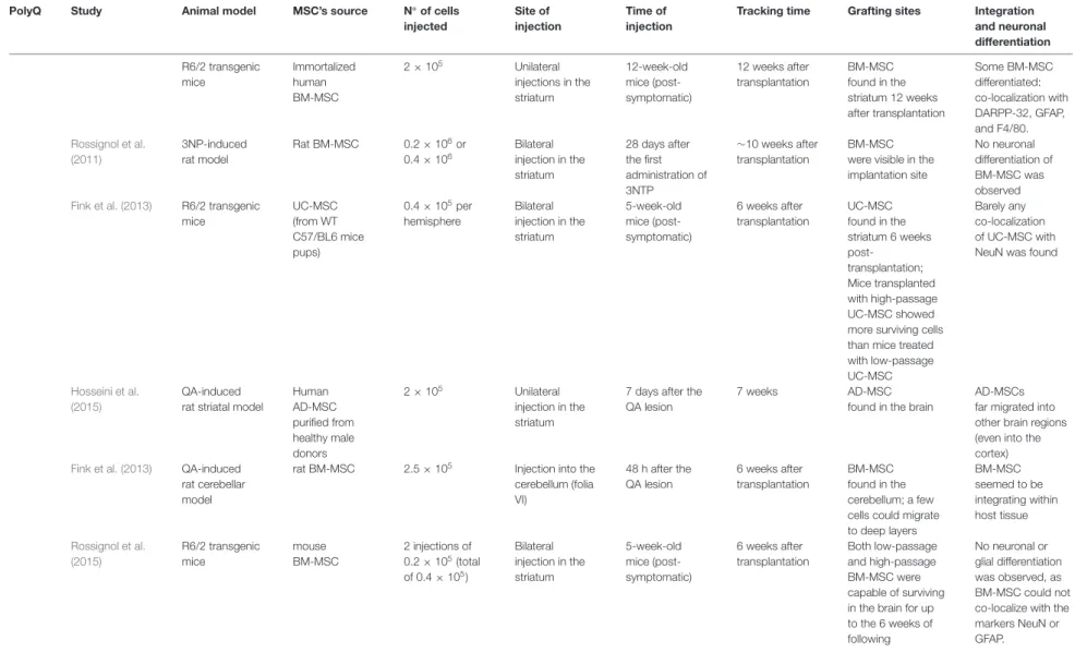
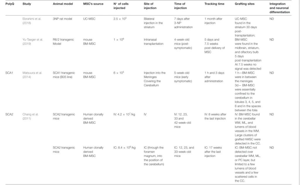
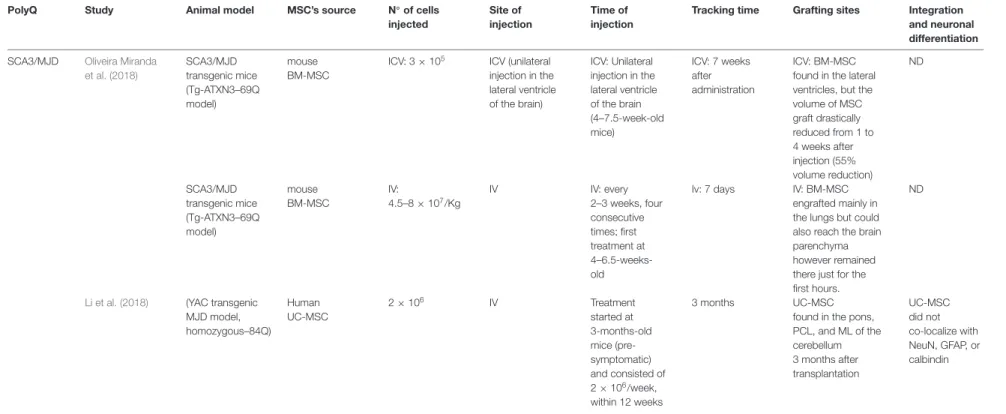
TABLE 1 | Sum up of the survival, grafting, and integration of mesenchymal stromal cells (MSC) in pre-clinical studies in animal models of polyglutamine (polyQ) disorders.
PolyQ Study Animal model MSC’s source N◦of cells
injected
Site of injection
Time of injection
Tracking time Grafting sites Integration and neuronal differentiation
HD Bantubungi et al. (2008)
QA-induced rat striatal model
rat BM-MSC 1 × 10⁵ Unilateral injection in the striatum 1-week after the QA lesion 8 weeks after transplantation 1.65 ± 0.18 × 10⁵ BM-MSC found at the lesion site in the striatum at 3 and 8 weeks
BM-MSC remained undifferentiated
Lee et al. (2009) QA-induced rat striatal model
human AD-MSC 1 × 10⁶ Unilateral
injection in the striatum immediately after the QA injection 4 weeks after transplantation AD-MSC found near the initial transplantation foci, forming a lump Co-labeling with BDNF, calbindin, GABA, and GAD R6/2 transgenic mice*
human AD-MSC 0.5 × 10⁶ Bilateral injection in the striatum 8.5-weeks-old mice (post-symptomatic treatment) 4 weeks after transplantation Few AD-MSC migrated toward the dorsolateral subventricular area, or into the striatum; mean survival rate: 13.5 ± 1.4% (n = 3)
low rate of in vivo proliferation of AD-MSC; expression of nestin (∼70%), doublecortin (∼80%), Tuj-1 (∼25%), and GAD (∼80%)
Im et al. (2010) YAC128 transgenic mice human AD-MSC isolated from an HD patient and a healthy donor 5 × 10⁵ Bilateral injection in the striatum 8-months-old (pre-symptomatic) and 12-months-old (symptomatic) 4 weeks after transplantation No cells detected extremely low % of cell survival AD-MSC found in the striatum and in the periventricular area ND Snyder et al. (2010) N171–82Q transgenic mice human BM-MSC isolated from healthy donors 0.1 × 10⁵ Unilateral injection in the striatum 8-week-old mice (pre-symptomatic) 2–3 weeks after transplantation Grafted BM-MSC do not survive long in the striatum (disappeared over 3–15 days); Only 15.1% survived in the first 24 h (15.063 ± 3.776) BM-MSC did not proliferate, but induced neurogenesis and neural differentiation of the host endogenous neural stem cells
Lin et al. (2011) QA-induced mouse striatal model Immortalized human BM-MSC 2 × 10⁵ Unilateral injections in the striatum
7 days after the QA lesion 8 weeks after transplantation BM-MSC found in the striatum; a small amount of BM-MSC survived for 16 weeks Some BM-MSC co-localized with GFAP, NeuN, and DARPP-32 (Continued) Fr ontiers in Cellular Neur oscience | www .fr ontiersin.org 7 October 2020 | V olume 14 | Article 584277
Barr os et al. MSC for PolyQ Disor ders TABLE 1 | Continued
PolyQ Study Animal model MSC’s source N◦of cells
injected
Site of injection
Time of injection
Tracking time Grafting sites Integration and neuronal differentiation R6/2 transgenic mice Immortalized human BM-MSC 2 × 10⁵ Unilateral injections in the striatum 12-week-old mice (post-symptomatic) 12 weeks after transplantation BM-MSC found in the striatum 12 weeks after transplantation Some BM-MSC differentiated: co-localization with DARPP-32, GFAP, and F4/80. Rossignol et al. (2011) 3NP-induced rat model Rat BM-MSC 0.2 × 10⁶or 0.4 × 10⁶ Bilateral injection in the striatum 28 days after the first administration of 3NTP ∼10 weeks after transplantation BM-MSC were visible in the implantation site
No neuronal differentiation of BM-MSC was observed
Fink et al. (2013) R6/2 transgenic mice UC-MSC (from WT C57/BL6 mice pups) 0.4 × 10⁵per hemisphere Bilateral injection in the striatum 5-week-old mice (post-symptomatic) 6 weeks after transplantation UC-MSC found in the striatum 6 weeks post-transplantation; Mice transplanted with high-passage UC-MSC showed more surviving cells than mice treated with low-passage UC-MSC
Barely any co-localization of UC-MSC with NeuN was found
Hosseini et al. (2015)
QA-induced rat striatal model
Human AD-MSC purified from healthy male donors 2 × 10⁵ Unilateral injection in the striatum
7 days after the QA lesion
7 weeks AD-MSC
found in the brain
AD-MSCs far migrated into other brain regions (even into the cortex)
Fink et al. (2013) QA-induced rat cerebellar model
rat BM-MSC 2.5 × 10⁵ Injection into the cerebellum (folia VI) 48 h after the QA lesion 6 weeks after transplantation BM-MSC found in the cerebellum; a few cells could migrate to deep layers BM-MSC seemed to be integrating within host tissue Rossignol et al. (2015) R6/2 transgenic mice mouse BM-MSC 2 injections of 0.2 × 10⁵(total of 0.4 × 10⁵) Bilateral injection in the striatum 5-week-old mice (post-symptomatic) 6 weeks after transplantation Both low-passage and high-passage BM-MSC were capable of surviving in the brain for up to the 6 weeks of following No neuronal or glial differentiation was observed, as BM-MSC could not co-localize with the markers NeuN or GFAP. (Continued) Fr ontiers in Cellular Neur oscience | www .fr ontiersin.org 8 October 2020 | V olume 14 | Article 584277
Barr os et al. MSC for PolyQ Disor ders TABLE 1 | Continued
PolyQ Study Animal model MSC’s source N◦of cells
injected
Site of injection
Time of injection
Tracking time Grafting sites Integration and neuronal differentiation
Ebrahimi et al. (2018)
3NP rat model UC-MSC 2.5 × 10⁵ Bilateral
injection in the striatum 7 days after 3-NP administration 1 month after injection UC-MSC found in the striatum 30 days post-transplantation; ND Yu-Taeger et al. (2019) R6/2 transgenic Model mouse BM-MSC 1 × 10⁶ Intranasal transplantation 4-week-old mice (post-symptomatic) 5 days and 7.5 weeks post-delivery of MSC BM-MSC were found in the midbrain, striatum, and olfactory bulb 5 days
post-transplantation At 7.5 weeks no signal was detected
ND
SCA1 Matsuura et al. (2014)
SCA1 transgenic mice (B05 line)
mouse BM-MSC
6 × 10⁵ Injection into the Meninges Covering the Cerebellum 5-week-old mice (early symptomatic) 1 h and 3 days after administration 1 h—BM-MSC were in between the meninges 3d— BM-MSC were essentially confined to the cerebellum in lobules 3, 4, 5, and 6 and in the spaces between the folia
ND
SCA2 Chang et al. (2011) SCA2 transgenic mice Human clonally derived BM-MSC IV: 4.2 × 10⁷/kg IV IV: 12, 23, 33 and 42-week-old mice
IV: 8 weeks after the last injection
IV: BM-MSC found in the cerebellar WM, ML, and lumens of blood vessels in the WM. Large clusters of grafted hMSC were detected in the CC. ND SCA2 transgenic mice Human clonally derived BM-MSC
IC: 8.4 × 10⁶/kg IC (through the foramen magnum, into the position of the cerebellum) IC: 12, 23, and 33-week-old mice IC: 17 weeks after the last injection IC: BM-MSC not detected over cerebellar WM, ML, or PC layer, but limited to a few lumens of blood vessels and a few scattered cells in the CC. ND (Continued) Fr ontiers in Cellular Neur oscience | www .fr ontiersin.org 9 October 2020 | V olume 14 | Article 584277
Barr os et al. MSC for PolyQ Disor ders TABLE 1 | Continued
PolyQ Study Animal model MSC’s source N◦of cells
injected
Site of injection
Time of injection
Tracking time Grafting sites Integration and neuronal differentiation
SCA3/MJD Oliveira Miranda et al. (2018) SCA3/MJD transgenic mice (Tg-ATXN3–69Q model) mouse BM-MSC
ICV: 3 × 10⁵ ICV (unilateral injection in the lateral ventricle of the brain) ICV: Unilateral injection in the lateral ventricle of the brain (4–7.5-week-old mice) ICV: 7 weeks after administration ICV: BM-MSC found in the lateral ventricles, but the volume of MSC graft drastically reduced from 1 to 4 weeks after injection (55% volume reduction) ND SCA3/MJD transgenic mice (Tg-ATXN3–69Q model) mouse BM-MSC IV: 4.5–8 × 10⁷/Kg IV IV: every 2–3 weeks, four consecutive times; first treatment at 4–6.5-weeks-old
Iv: 7 days IV: BM-MSC
engrafted mainly in the lungs but could also reach the brain parenchyma however remained there just for the first hours.
ND
Li et al. (2018) (YAC transgenic MJD model, homozygous–84Q) Human UC-MSC 2 × 10⁶ IV Treatment started at 3-months-old mice (pre-symptomatic) and consisted of 2 × 10⁶/week, within 12 weeks 3 months UC-MSC
found in the pons, PCL, and ML of the cerebellum 3 months after transplantation UC-MSC did not co-localize with NeuN, GFAP, or calbindin
List of abbreviations: HD, Huntington’s disease; QA, quinolinic acid; BM-MSC, Bone marrow-derived mesenchymal stromal cells; AD-MSC, adipose tissue-derived mesenchymal stromal cells; BDNF, Brain-derived neurotrophic factor; GABA, gamma-aminobutyric acid; GAD, glutamic acid decarboxylase; YAC, yeast artificial chromosome; ND, not determined; GFAP, glial fibrillary acidic protein; NeuN, neuronal nuclei; DARP-32, dopamine- and cAMP-regulated neuronal phosphoprotein; F4/80, major macrophage marker; UC-MSC, umbilical cord-derived mesenchymal stromal cells; 3-NP, 3-nitropropionic acid; SCA1, Spinocerebellar ataxia type-1; SCA2, Spinocerebellar ataxia type-2; IV, Intravenous injections; IC, Intracerebral injection; WM, white matter; ML, molecular layer; PCL, Purkinje cell layer; CC, cerebral cortex; SCA3/MJD, Spinocerebellar ataxia type-3 or Machado-Joseph disease; ICV, Intracerebroventricular injection.
Fr ontiers in Cellular Neur oscience | www .fr ontiersin.org 10 October 2020 | V olume 14 | Article 584277
Barros et al. MSC for PolyQ Disorders
be an important contribution to the field to validate findings achieved with rodent models and test a possible replacement of degenerated neurons promoted by MSC.
Furthermore, different cell sources, heterogeneous methods of isolation, discrepancies in MSC origins (i.e., from healthy/disease tissues) and characteristics (cell passage number, conditions used during cell growth) have been used throughout the studies, which makes it difficult to reach a correct comprehension and make assertive conclusions with enough confidence to implement these therapies in the clinics.
Clinical Studies in PolyQ Patients:
Achievements and Flaws
Despite the extensive number of unanswered issues from the pre-clinical assessments herein referred, the promising results obtained from research on the use of MSC in polyQ soon led to the investigation of their therapeutic application in clinical studies. The clinical trials, mostly phase I/II, aiming at testing the safety and effectiveness of MSC in the treatment of polyQ disorders (completed and ongoing clinical trials— ClinicalTrials.gov Identifiers: NCT02728115, NCT03252535, NCT04219241, NCT03378414 for HD and NCT01360164, NCT01489267, NCT01649687, NCT02540655 for other polyQs/SCAs) are summarized in Table 2.
Data collected from four of these clinical trials have demonstrated that the therapeutic application of MSC is safe, does not produce severe side effects and might delay the progression of disease symptoms (Dongmei et al., 2011; Jin et al., 2013; Miao et al., 2015; Tsai et al., 2017). In one of these studies, fourteen patients diagnosed with SCA and ten with multiple system atrophy-cerebellar type C (MSA-C) were treated weekly with IT injections of UC-MSC at a dose of 1 × 10⁶/kg during four weeks (except for three patients who received two courses of treatment, all the other patients received only one course). Furthermore, the neuronal function and quality of daily life were assessed through the International Cooperative Ataxia Rating Scale (ICARS) and Activity of Daily Living Scale (ADL). Improvements in ICARS and ADL scores were observed after 1 month of treatment (Dongmei et al., 2011). In another study, Jin et al. (2013) demonstrated that MSC treatment could alleviate SCA symptoms for at least half a year. In this study, 16 patients diagnosed with SCA1, 2 or 3, received IV or IT injections of UC-MSC (first treatment: 4 × 10⁷ UC-MSC were infused iv.; following three treatments: 2 × 10⁷UC-MSC were infused IV and simultaneously 2 × 10⁷ UC-MSC were infused by IT administration; all patients received only one-course treatment). Results showed that most patients obtained improved scores on the Berg Balance Scale (BBS), which evaluates patients’ balance, and ICARS at 3 and 6 months after treatment (Jin et al., 2013). A different clinical trial using UC-MSC was performed to evaluate the technical difficulties underlying IT UC-MSC injectionvia lumbar puncture and assess the effects of the cell in different neurodegenerative disorders, including eight SCA patients (Miao et al., 2015). The UC-MSC was injected 4–6 times between the L4 and L5 interspace within 5–7 days (single course of treatment). All patients were followed-up for more than 1 year for clinical status. Three of
the eight patients showed motor function improvement after treatment. Recently,Tsai et al. (2017)intravenously administered (5–7 × 10⁷) allogeneic MSC derived from the adipose tissue (AD-MSC, single injection) into six patients with MJD and one with MSA, who showed a slight trend of improvement and stabilization of the disease according to the Scale for Assessment and Rating of Ataxia (SARA) and the sensory organization testing (SOT).
Importantly, no major side effects were reported, besides dizziness, headache, back pain in case of IT injections, or fever (Dongmei et al., 2011; Jin et al., 2013; Miao et al., 2015; Tsai et al., 2017). Consistently with these results, no adverse effects were reported so far in any of the pre-clinical studies. Moreover, in our study, we assessed for the levels of hepatic aminotransferases, which were not increased when compared to non-treated or WT mice, denoting the absence of liver toxicity in mice treated repeatedly with MSC (Oliveira Miranda et al., 2018).
Altogether, the studies reinforce the safety and tolerability of MSC. Furthermore, they demonstrate that treatment with allogenic UC-MSC may delay disease progression, improving motor performance, and patients’ quality of life without severe adverse reactions. However, the regression of some patients to the status before the treatment was reported (Dongmei et al., 2011; Jin et al., 2013; Miao et al., 2015; Tsai et al., 2017; summarized in Table 2). This was also following the need for performing several treatments suggested by some of the pre-clinical studies (Chang et al., 2011; Li et al., 2018; Oliveira Miranda et al., 2018). Nonetheless, in the clinical context, periodic MSC injections may not be feasible as they could lead to unwanted side effects. Indeed, some studies highlighted the long-term risk that could emerge from the repeated therapeutic transplantation of these cells, namely maldifferentiation, immunosuppression (increasing the risk of opportunistic infections), liver and lung accumulation, and malignant tumor growth promotion in patients with pre-existing malignancy (Sundin et al., 2006; Dierks et al., 2007; Kunter et al., 2007; Ning et al., 2008). On the other hand, as previously discussed, the local administration of MSC may cause brain damage if administered above a certain number (Rossignol et al., 2011). Therefore, it would be important to further clarify the mechanism involved in MSC therapeutic effects, uncovering new potential treatments that do not require the infusion of cells.
On the other hand, these clinical trials often mix patients with different conditions/stages of the disease and frequently include all disorders when reporting results, making reports not as informative as desired. Future clinical studies should focus not only on the feasibility of repeated treatments for sustained benefits but also on the appropriate timing, dosage and best method of injection to be used, while also performing longer follow-ups of all patients to evaluate treatment efficacy and safety, and with placebo-controls being used. For that purpose, established cohorts, well-defined and characterized must be performed so that clinical studies can be well-designed and as instructive as possible. An example of such a cohort has been implemented by the European Consortium ESMI (‘‘European Spinocerebellar Ataxia Type 3/Machado-Joseph
Barr os et al. MSC for PolyQ Disor ders
TABLE 2 | Concise information on the outcomes obtained from clinical trials performed with MSCs in patients with PolyQ diseases.
Trial identification Disease Type of MSC Route of administration Outcomes
Dongmei et al. (2011) SCA and MSA-C UC-MSC IT ICARS and ADL (treatment. Unstable walking and standing, slow movement, fine motor disordersKatona et al. (2008)) scores significantly decreased 1 month after
of the upper limbs and writing difficulties, and dysarthria were greatly improved except for 1 patient, who had no response.
Ten cases (42%) remained stable for half a year or longer, while 14 cases (58%) had regressed to the status before the treatment within 1–14 months (an average of 3 months).
NCT01360164Jin et al. (2013)
SCA1, SCA2, and SCA3
UC-MSC IV and IT After 1 year of treatment: 44% of the patients exhibited improved Berg Balance Scale (BBS) over baseline, and only 31% of the patients suffered from disease aggravation; 63% of the patients exhibited improved ICARS over baseline, and only 25% of the patients suffered from disease exacerbation.
Miao et al. (2015) Several neurodegenerative disorders, including SCAs
UC-MSC IT Patients were followed-up for more than 1 year. No significant side effects were reported. Three of the 8 patients with SCAs (37.5%) showed motor function improvement 1 year after the treatments.
NCT01489267 SCA1 UC-MSC IT Unknown
NCT01649687Tsai et al. (2017)
SCA3 or MSA-C AD-MSC IV Patients were monitored for 1 year with no reported adverse events. They showed
an initial trend toward brief improvement, followed by a slight stabilization and a progression of the disease in later stages.
At 6 months follow-up, SARA scores were improved in 2 (33.3%), unaltered in 2 (33.3%), and worsened in 2 (33.3%) patients as compared with the baseline. At 1-year follow-up, SARA scores were improved in 1 (16.6%), unaltered in 3 (50%), and worsened in 2 (33.3%) patients as compared with the baseline.
NCT02540655 Cerebellar Ataxias AD-MSC
(Stemchymalr)
IV Unknown.
NCT02728115 HD Cellavita* IV Active, not recruiting
NCT03252535 HD Cellavita IV Active, not recruiting
NCT04219241 HD Cellavita IV This study is not yet open for participant recruitment.
NCT03378414 SCA1, SCA2,
SCA3 and SCA6
UC-MSC IV Unknown
List of abbreviations: SCA, Spinocerebellar ataxia; MSA-C, and multiple system atrophy-cerebellar types; SCA1, Spinocerebellar ataxia type-1; SCA2, Spinocerebellar ataxia type-2; SCA3, Spinocerebellar ataxia type-3 or Machado-Joseph disease; SCA6, Spinocerebellar ataxia type-6; UC-MSC, umbilical cord-derived mesenchymal stromal cells; AD-MSC, adipose tissue-derived mesenchymal stromal cells; IT, Intrathecal injection; IV, Intravenous injections; ICARS, International Cooperative Ataxia Rating Scale; ADL, Activity of Daily Living; BBS, Berg Balance Scale. *Stem cells isolated from the dental pulp.
Fr ontiers in Cellular Neur oscience | www .fr ontiersin.org 12 October 2020 | V olume 14 | Article 584277
Barros et al. MSC for PolyQ Disorders
Disease Initiative,’’ Joint Programme on Neurodegenerative disorders), which put together eight European cohorts that over the last 4 years integrated all the existing data in a common database to allow for standardized and quality-controlled protocols in the forthcoming studies.
Major Questions to be Addressed to
Increase Clinical Benefits
From the clinical studies referred so far, we can conclude that the major weakness of the cellular therapies using MSC (or another adult/progenitor stem cell source) is that their effect is not prolonged throughout time. However, the cause of these transitory effects of MSCin vivo remains elusive. In our study, we could determine that MSC survival was limited in time after administration, which is consistent with other pre-clinical studies (as shown in Table 1). This may result from the inhospitable environment they face—the high oxidative stress of the damaged tissue, together with the low glucose availability—that leads MSC to choose an anerobic metabolic pathway, forcing cells to rapidly undergo apoptosis (Moya et al., 2018). Methods for increasing MSC fitness are described in the following section.
Another important question is whether MSC can effectively pass through the BBB upon intravenous injection. Several pre-clinical studies reported the finding of MSC in the central nervous system, including ours, as referred in Table 1. However, whether MSC crosses this barrier by mechanisms that are similar to leukocytes such as transmigration (Chamberlain et al., 2007), or whether this barrier needs to be impaired to let MSC pass-through remains undetermined. BBB leakage was already described for HD (Drouin-Ouellet et al., 2015). Due to the strong presence of inflammatory mechanisms in polyQ disorders, it can be speculated that this is also the case for the remaining polyQs. In accordance, we recently reported evidence that the BBB is damaged in MJD (Lobo et al., 2020). Still, there are no indications that the BBB opening is mandatory for MSC to reach the brain parenchyma. Studies accessing MSC biodistribution inside the nervous system in non-neurodegenerative rodents (wild type animals), should be performed to answer this intriguing question. In case MSC cannot cross the BBB when it is fully preserved, then other strategies should be explored such as the expression of specific receptors on the MSC surface that would allow their passage, or consider the use of MSC products (either naïve or engineered) that can effectively reach brain cells.
Finally, the prophylactic administration of this cellular treatment is also an important issue to be discussed. Since these disorders are monogenic and can be passed to the offspring, the patients can choose to know whether they carry the disease mutation before the manifestation of disease symptoms. Therefore, successful treatment could be implemented in pre-symptomatic patients. Accordingly, there is evidence that this would favor a later appearance of symptoms (Snyder et al., 2010; Li et al., 2018), supporting such early intervention as a feasible clinical approach for polyQs patients in the future. Nonetheless, Im and collaborators showed that MSC only modified the course of the disease when administered after its phenotypic expression in long-lived HD transgenic mice (Im
et al., 2010). Therefore, the time window of delivery should be further investigated.
Overall, both pre-clinical and clinical studies show the therapeutic relevance of MSC for polyQ but also highlight the need to design better clinical approaches and more adjusted to the real biology and behavior of cells upon transplantation. Possible solutions are discussed in the following sections.
METHODS TO INCREASE MSC’s
EFFICACY IN VIVO
Strategies to overcome some of these shortcomings and improve MSC’s efficiency have been explored and will be described next, such as pre-conditioning of cells, the use of adjuvant factors, or the genetic enhancement of MSC. A concise review of that is given in Table 3.
Pre-conditioning of MSC
Knowing that the secretion of soluble and insoluble factors is an adaptive mechanism, which allows MSC to regulate intracellular stress and influence their surroundings, researchers have investigated whether the pre-conditioning of MSC could improve their therapeutic abilities. Pre-conditioning is nothing more than manipulating cells, so they perform a specific function or undergo a desirable differentiation pathway. Some studies showed that pre-conditioning of MSC using hypoxia, different culture conditions, biomolecules (cytokines and growth factors), or pharmacological compounds, may lead to greater MSC survival in vivo and enhanced regenerative and immunomodulatory effects (Ferreira et al., 2018; Noronha et al., 2019).
Pre-conditioning: Hypoxia
Oxygen tension under standard cell culture conditions (21%) is much higher than physiological oxygen tension in tissues, which can vary from 1% to 12% depending on vascularization and metabolic activity (Das et al., 2010). Notably, when we speak of hypoxia in the context of cell culture, usually it means that oxygen availability ranges from 0 to 10% to better mimic physiological conditions. MSC is usually found in tissues with low oxygen tension (1% to 7%), is naturally able to endure in in vivo hypoxic environments (Deschepper et al., 2011).
When cultured under hypoxic conditions, MSC increases their fitness to adapt to adverse circumstances which will make them better prepared to face inhospitable environments. Indeed, under hypoxia MSC changes their metabolism, increasing their glucose consumption, decreasing the production of oxygen species, and shortening telomeres (Das et al., 2010). In these conditions, MSC presents a greater proliferation rate and increased secretion of soluble factors without changing their multipotency, which improves MSC capacity to survive in damaged tissues upon transplantation (Das et al., 2010; Bader et al., 2015; Lee et al., 2017). Accordingly, Lee et al. (2017)
showed that culturing MSC in hypoxic conditions leads to the up-regulation of Hypoxia-inducible factor (HIF)-α, which induces survival and proliferation rate.
Barr os et al. MSC for PolyQ Disor ders
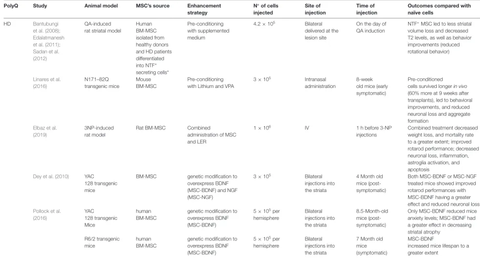
TABLE 3 | Summary of results using modified-MSCs in PolyQ diseases in comparison to naive cells—evidence from pre-clinical studies.
PolyQ Study Animal model MSC’s source Enhancement
strategy N◦ of cells injected Site of injection Time of injection
Outcomes compared with naïve cells HD Bantubungi et al. (2008); Edalatmanesh et al. (2011); Sadan et al. (2012) QA-induced rat striatal model
Human BM-MSC isolated from healthy donors and HD patients differentiated into NTF+ secreting cells* Pre-conditioning with supplemented medium 4.2 × 10⁵ Bilateral delivered at the lesion site On the day of QA induction
NTF+MSC led to less striatal volume loss and decreased T2 levels, as well as behavior improvements (reduced rotational behavior) Linares et al. (2016) N171–82Q transgenic mice Mouse BM-MSC Pre-conditioning with Lithium and VPA
3 × 10⁵ Intranasal
administration
8-week old mice (early symptomatic)
Pre-conditioned cells survived longer in vivo (60% more at 9 weeks after transplants), led to behavioral improvements, and reduced neuronal loss and aggregate formation Elbaz et al. (2019) 3NP-induced rat model Rat BM-MSC Combined administration of MSC and LER 1 × 10⁶ IV 1 h before 3-NP injections
Combined treatment decreased weight loss, and mortality rate to a greater extent; improved rotarod performance; decreased neuronal loss, inflammation, astroglia activation, and apoptosis
Dey et al. (2010) YAC 128 transgenic mice BM-MSC genetic modification to overexpress BDNF (MSC-BDNF) and NGF (MSC-NGF) 3 × 10⁵ Bilateral injections into the striata 4 Month old mice (post-symptomatic) Both MSC-BDNF or MSC-NGF treated mice showed improved rotarod performances with MSC-BDNF having a greater effect and reduced neuronal loss
Pollock et al. (2016) YAC 128 transgenic Mice human BM-MSC genetic modification to overexpress BDNF (MSC-BDNF) 5 × 10⁵per hemisphere Bilateral injections into the striata 8.5-Month-old mice (post-symptomatic)
Only MSC-BDNF reduced mice anxiety levels; MSC-BDNF had a greater effect in decreasing striatal atrophy R6/2 transgenic mice human BM-MSC genetic modification to overexpress BDNF (MSC-BDNF) 5 × 10⁵per hemisphere Bilateral injections into the striata 7 Month old mice (symptomatic) MSC-BDNF
increased mice lifespan to a greater extent
List of abbreviations: HD, Huntington’s disease; QA, quinolinic acid; VPA, valproic acid; AD-MSC, adipose tissue-derived mesenchymal stromal cells; BM-MSC, Bone marrow-derived mesenchymal stromal; LER, lercanidipine; BDNF, Brain-derived neurotrophic factor; NTF, neurotrophic factors; NGF, Nerve growth factor IV, Intravenous injections; *NTF secreting cells produce and secrete large amounts of neurotrophic factors like BDNF and GDNF.
Fr ontiers in Cellular Neur oscience | www .fr ontiersin.org 14 October 2020 | V olume 14 | Article 584277
Barros et al. MSC for PolyQ Disorders
Hence, UC-MSC pre-conditioned with hypoxia (MSC-h) showed to have neuroprotective, anti-apoptotic, and anti-inflammatory actions in a rat model of spinal cord injury. When compared with MSC cultured under standard conditions, MSC-h exhibited increased expression of neuroprotective trophic factors, such as hepatocyte growth factor (HGF), BDNF, and VEGF. Rats transplanted with MSC-h into the spinal cord immediately after lesion induction showed increased axonal preservation, even higher as compared to MSC not submitted to hypoxia. MSC-h also reduced the number of caspase-3 (apoptotic marker) positive cells, microglia, and macrophage infiltration (Zhilai et al., 2016).
Furthermore, Wang J. W. et al. (2017) demonstrated that hypoxia pre-conditioning enhanced the migration and integration of MSC upon transplantation into a rat model of cerebral ischemia, induced by cardiac arrest. These actions were linked with the activation of PI3K/AKT pathways and increased expression of HIF-1α and C-X-C chemokine receptor type 4. Plus, the IV injection of MSC-h was able to diminish neuronal death and inflammation in the cortex.
Even though these studies suggest hypoxia pre-conditioning may improve MSC neuroprotective effect and increase MSC survival after transplant, we found no studies that investigated the potential use of this approach in the treatment of neurodegenerative/polyQ disorders. Therefore, in the future efforts should be made to better explore this strategy.
Pre-conditioning: Biomolecules/Chemicals
Pre-conditioning of MSC with biomolecules such as cytokines, growth factors, or hormones can improve MSC survival and efficiency. Indeed, Sadan and collaborators showed that MSC that were pre-conditioned in media supplemented with epidermal growth factor, human basic FGF, cyclic adenosine monophosphate, human neuregulin1-β1, and platelet-derived growth factor (Sadan et al., 2008, 2009) differentiated into NTF secreting cells (NTF+) cells, which could secrete high levels of BDNF and glial cell line-derived neurotrophic factor (GDNF). NTF+ MSC could improve both the neuropathology and behavior patterns in the QA rat model of HD (Sadan et al., 2012). QA-injected rats transplanted with NTF+ MSC lost less striatal volume than rats transplanted with naïve MSC. Moreover, NTF+ MSC decreased T2 values when compared with PBS-treated QA rats, as detected by MRI acquisitions. Phenotypically, NTF+ cells were more efficient in reducing rotational behavior than naïve MSC in QA-injected rats, thus suggesting these cells to be more efficient.
In a recent study, the authors assessed the impact of culturing MSC with different sera. MSC was cultured in medium with 10% of the following sera: fetal bovine serum, serum from healthy controls (NS-MSC), or serum from stroke patients (SS-MSC). Interestingly, SS-MSC had a higher proliferation rate and lower senescence as well as increased expression of VEGF, GDNF, and FGF. Additionally, SS-MSC promoted neurogenesis and angiogenesis in stroke rat models, leading to improved behavioral performance (Moon et al., 2018).
Alternatively, MSC can be primed using pharmacological or chemical agents. Linares et al. (2016) investigated whether
pre-conditioning MSC with lithium and valproic acid (VPA) could enhance their therapeutic effect. Lithium and VPA are mood stabilizers known to exert neuroprotective effects (Chiu et al., 2013) and pre-conditioning of MSC with these factors led to an increase in the expression of genes involved in trophic effects, as well as in pro-survival and immunomodulatory pathways. Accordingly, N171–82Q transgenic HD mice were treated with an intranasal administration of MSC or MSC pre-conditioned with Lithium and VPA. The transplanted cells migrated into the brain, with pre-conditioned MSC surviving for a longer period. Moreover, mice treated with pre-conditioned MSC showed a greater amelioration of motor and behavior performance, decreased neuronal death, and reduction of huntingtin aggregates in the striatum (Linares et al., 2016). This strategy could be used in other polyQ diseases where Lithium and VPA showed promising results (Saute et al., 2014; Esteves et al., 2015; Lei et al., 2016; Lopes-Ramos et al., 2016).
Therefore, though pre-conditioning needs to be further explored, the results obtained so far seem promising, which opens good prospects for the near future.
Combined Treatment With
Pharmacological Agents
A less common strategy recently evaluated involved the use of pharmacological agents in combination with MSC to treat polyQ disorders.Elbaz et al. (2019)showed that the combined administration of Lercanidipine (LER), an antihypertensive drug, and MSC boosted their therapeutic efficiency in a rat model for HD. LER is a vasoselective dihydropyridine calcium channel blocker that can modulate calcium levels and, therefore may be able to influence the calcineurin (CaN)/nuclear factor of activated T cells c4 (NFATc4) pathway, which is deregulated in neurodegenerative disorders (Sompol and Norris, 2018). Treatment with LER, BM-MSC, or a combination of LER and BM-MSC was given to 3-NP rats. The combined treatment with LER and BM-MSC had the most promising results by leading to greater amelioration of motor and behavioral performance. Moreover, the levels of BDNF, forkhead box P3, Wnt, and β-catenin increased in the striatum, along with a decrease of CaN, tumor necrosis factor-α, and NFATc4 protein expression and the Bax/B-cell lymphoma 2. Their results suggest that this combined therapy can promote neuroprotective effects by, at least in part, suppressing Ca2+/CaN/NFATc4 and Wnt/β-catenin signaling pathways activation, which is dysregulated in the 3-NP rats as well as in other neurodegenerative disorders (Sompol and Norris, 2018).
Hence, this study shows how a combination of factors can produce a synergistic effect, so it would be interesting to further investigate the neuroprotective potential of therapies combining different drugs with MSC.
Genetically Engineered MSC
Another strategy that has been studied to enhance MSC’s therapeutic efficiency is the production of genetically engineered MSC that can be used as delivery vehicles (Park et al., 2015). For the treatment of neurodegenerative diseases, the most common approaches include the use of MSC that
Barros et al. MSC for PolyQ Disorders
were either virally or non-virally modified to overexpress trophic factors known to have neuroprotective actions (Huang et al., 2012; Deng et al., 2016).
Concerning polyQ disorders, only two studies are available in the literature. The safety and efficacy of genetically engineered MSC have been tested in HD animal models. In 2010, BM-MSC were genetically engineered to over-express BDNF (MSC-BDNF) and/or NGF (MSC-NGF), thus allowing these cells to deliver these factors in higher amounts than normal MSC (Dey et al., 2010). YAC128 transgenic mice were treated with MSC-BDNF, MSC-NGF, or with both MSC-BDNF and MSC-NGF. MSC was administered through bilateral injections into the striatum. From all the tested groups, YAC128 transgenic mice transplanted with MSC-BDNF exhibited the best outcomes, showing the least neuronal loss and the highest latencies to fall values in rotarod.
Another study demonstrated that human MSC modified by lentiviral transduction to overexpress BDNF (MSC-BDNF) could ameliorate HD symptoms in two different animal models, YAC128 and R6/2 transgenic mice in a higher extent (Pollock et al., 2016). The intrastriatal transplantation of MSC-BDNF decreased striatal atrophy and reduced anxiety levels in YAC128 mice. Additionally, MSC-BDNF administration promoted neurogenesis-like activity and led to an increase in mice lifespan. Notably, mice treated with MSC-BDNF had overall better outcomes than those treated with non-modified MSC.
Despite the few studies using genetically engineered MSC to treat polyQ disorders, this type of approach has been further explored in other neurodegenerative diseases, namely in AD and PD, also with promising results (Li et al., 2008; Moloney et al., 2010; Ren et al., 2013; Hoban et al., 2015).
Importantly, since neurodegenerative disorders have common pathogenic mechanisms, current evidence obtained from these studies indicate that genetically modified MSC to overexpress various trophic factors is strong therapeutic candidates for several neurodegenerative diseases, including polyQ disorders.
CELLULAR-FREE APPROACHES
The Success of Studies Using MSC’s
Secretome in PolyQ/Other
Neurodegenerative Disorders
Several studies detected reduced numbers of MSCs in the targeting tissues suggesting that MSCs can also act through paracrine mechanisms. Conditioned medium (CM) of MSC (CM-MSC), i.e., MSC’s secretome, can reproduce the therapeutic effects obtained with MSC treatment in several neurological diseases.
Bai et al. (2012) demonstrated that CM-MSC mitigates
functional abnormalities in a mouse model of multiple sclerosis by promoting oligodendrocyte and neuron development. Suto et al. (2016) showed that injections of MSC released factors, such as HGF, improved motor coordination in an SCA-1-Knock-in mouse model. Indeed, the admSCA-1-Knock-inistration of CM-MSC was able to protect axons and myelin of spinal motor neurons (Suto et al., 2016). Importantly, such improvements were
similar to those obtained in previous work using the same model of disease in which animals were treated with MSC (Mieda et al., 2016).
An antibody-based protein array analysis and ELISA showed that, in addition to HGF, IGF-1, VEGF, and transforming growth factor (TGF)-β-1 are present in the CM. However, the combination of these factors is not as effective as treatment with CM in promoting neuronal survival and neurite outgrowth, which suggests that CM contains other complementary key factors (Nakano et al., 2010). The secretion of BDNF and NGF by MSC has also been demonstrated to promote neuronal/glial survival and neurogenesis (Crigler et al., 2006). Likewise, the beneficial effect of MSC may also be mediated by secretion of GDNF, basic FGF-2, CNTF, angiopoietin-1, and neurotrophin-3 (Chen et al., 2005; Onda et al., 2008). Besides, the extracellular matrix proteins, including collagen, fibronectin, and laminin (Hidalgo-Bastida and Cartmell, 2010) provide physical guidance, support neurogenesis, and may exert trophic activity themselves (Maltman et al., 2011).
Recently, Ebrahimi et al. (2018) demonstrated that CM collected from UC-MSC protect PC12 cells against superoxide-induced oxidative stress, increasing cell viability, and neurite outgrowth. GDNF and VEGF were detected in CM of UC-MSC and were linked to the neuroprotective effects observed.
Moreover, it has been demonstrated that both human adipose-derived MSC and their CM could reduce neuronal cell damage in a model of glutamate excitotoxicity, a mechanism that plays a role in the pathogenesis of neurodegenerative diseases. Indeed, co-cultures with MSC using a transwell system or addition of CM similarly promoted axonal regeneration, energy metabolism improvements, and prevented apoptosis of cortical neurons (Hao et al., 2014). In another study,
Miranda et al. (2011) showed that in a transwell system that immortalized BM-MSC could stimulate neurite outgrowth in a neurotrophin-dependent manner including BDNF secretion in Twitcher-derived DRG neurons, anin vitro model of Krabbe’s disease. Moreover, the administration of exogenous BDNF to the sciatic nerve of Twitcher mice through BDNF-delivering osmotic pumps promoted morphometric ameliorations, suggesting BDNF as a promising candidate to be used in combination with BM-MSC treatment (Miranda et al., 2011). Nevertheless, simultaneous transplantation of BM-MSC and treatment with an antagonist of TrK receptors in a model of sciatic nerve crush completely reversed the inhibitory effect of the antagonist, suggesting that other mechanisms may be compensating the action of neurotrophins blocked by the antagonist.
A recent study has shown that CM-MSC promoted neuroprotective activities inin vitro PD models by increasing the viability of both rat and human dopaminergic neurons exposed to a neurotoxic insult (Parga et al., 2018). Additionally, the administration of MSC secretome through an injection into the substantia nigra and striatum in a PD mouse model led to an increase of dopaminergic neurons and neuronal terminals in those brain regions, which likely mediated the observed temporary motor performance improvements. Also, their findings suggest that the beneficial effects of human MSC