ANTIMICROBIAL ACTIVITY OF ESSENTIAL OILS
PROTECTION OF STORED PRODUCTS
Diana Mafalda Ferreira Cancela Moura
ANTIMICROBIAL ACTIVITY OF ESSENTIAL OILS
PROTECTION OF STORED PRODUCTS
Diana Mafalda Ferreira Cancela Moura
M. Sc. Thesis
Integrated Master’s in Bioengineering – Biological Engineering
Supervisor: Prof. Dr. Margarida Bastos
Co-supervisor: Prof. Dr. Olga Pastor Nunes
Departamento de Engenharia Química
Faculdade de Engenharia da Universidade do Porto
Porto, July 2014
I would like to acknowledge:
Department of Chemical Engineering and LEPABE for providing the research facilities and resources to develop this project.
Funds provided by FEDER through the Operational Programme for Competitiveness Factors – COMPETE and National Funds through Fundação para a Ciência e a Tecnologia (FCT) under the NORTE-07-0124-FEDER-000025, Pest-C/EQB/UI0511 and PTDC/AGR-ALI/119270/2010 (COMPETE: FCOMP-01-0124-FEDER-019547) projects for financing this project.
Prof. Dr. Margarida Bastos and Prof. Dr. Olga Pastor Nunes for the supervision and recommendations during this Thesis.
All the researchers from Lab E009B, who helped me every time I needed, for their availability and support.
All the girls from Biological Engineering class of 2014 for joining me in this journey.
All my friends and family, and specially my parents, for their support and help along my academic path.
D.C. Moura, O.C. Nunes, M.M.S.M. Bastos, Antimicrobial activity of essential oils: protection of stored food. 12th Chemical and Biological Engineering Conference, Porto, Portugal, 10-12 September 2014.
Essential oils have a primary role in plants´ protection. Such function points them as candidates to be used in future in biopreservation systems directed to minimal processed foods and non-perishable food products. A review of the methods used to assess antibacterial activity of essential oils and the perspectives and current applications in food systems are also addressed.
The antimicrobial activity of clove bud (Syzygium aromaticum) and pennyroyal (Mentha pulegium) essential oils and of their major components, eugenol and pulegone, against standard bacteria-and phytobacteria was assessed using different diffusion methods. The results showed that phytobacteria are extremely susceptible to these phytochemicals, which suggest that they may be effective in a biopreservation system applied to stored products. Nonetheless, the activity of the substances tested revealed a good spectrum of action. Clove bud essential oil seems to be the substance having better performance. Improvement of growth inhibition was notorious as the type of diffusion changed from agar to broth and from this type to vapour diffusion.
Os óleos essenciais têm um papel primário na proteção das plantas. Tal função faz dos mesmos candidatos para serem utlizados em sistemas de biopreservação direcionados a alimentos de processamento mínimo e não-perecíveis. Uma revisão dos métodos utilizados para determinar a atividade antimicrobiana dos óleos essenciais é realizada e as perspetivas e aplicações correntes em sistemas alimentares são também apresentadas.
A atividade antimicrobiana do óleo de cravinho (Syzygium aromaticum) e de menta-poejo (Mentha pulegium) e dos seus principais componentes, o eugenol e a pulegone contra bactérias standard e fitobactérias, foi determinada utilizando diferentes métodos de difusão. Os resultados revelaram que as fitobactérias são extremamente suscetíveis aos fitoquímicos utilizados, o que sugere que poderão ser eficientes em sistemas de biopreservação para produtos armazenados. Não obstante, a atividade das substâncias testadas revelou bom espectro de ação. Deve ainda ser salientado que o óleo do cravinho parece ser a substância que tem melhor performance. Por outro lado, à medida que o tipo de difusão foi alterado de agar para meio líquido e deste para vapor, a melhoria da inibição de crescimento foi notória.
Palavras-chave: Óleos essenciais; Atividade antimicrobiana; Biopreservantes; Fitobactérias.
1. Introduction ... 1
1.1. Objectives ... 2
2. State of the art ... 3
2.1. Essential oils composition ... 3
2.1.1. Clove bud essential oil ... 4
2.1.2. Pennyroyal essential oil ... 4
2.2. Chemical structure of essential oils ... 5
2.2.1. Terpenes and terpenoids ... 5
2.2.2. Aromatic and aliphatic compounds ... 6
2.3. Mode of antibacterial action of essential oils ... 7
2.3.1. Gram positive versus Gram negative susceptibility ... 8
2.4. Overview of the methodologies ... 10
2.4.1. Diffusion methods ... 11
2.4.2. Dilution tests ... 12
2.4.3. Other methods ... 13
2.4.4. Incidence in the literature ... 13
2.4.5. Growth indicators ... 14
2.4.6. Minimum inhibitory and bactericidal concentrations ... 14
2.4.7. Usual solvents ... 16
2.5. Applications of essential oils as preservatives in food systems ... 18
2.5.1. Available technologies for implementation of EOs as biopreservatives ... 19
2.5.2. Legal aspects ... 20
3. Materials and methods... 21
3.1. Reagents ... 21
3.2. Microorganisms, culture and stock conditions ... 21
3.3. Antimicrobial stock solutions ... 22
3.4. Disc diffusion assay ... 22
3.8. Preliminary in vivo assay ... 26
3.8.1. UV treatment ... 27
3.8.2. Bleach treatment ... 27
3.8.3. Fungicide treatment ... 27
3.9. Statistical analysis ... 28
4. Results and discussion ... 29
4.1. Disc diffusion ... 29
4.2. Minimum inhibitory concentration (MIC) determination ... 31
4.3. Minimum inhibitory doses (MID) determination... 33
4.4. Glass beads assay ... 35
4.5. Global discussion ... 36
5. Conclusions ... 39
5.1. Limitations and future work ... 39
References ... 41
Appendix ... 51
A. Overview of the composition of S. aromaticum and M. pulegium ... 51
B. Survey from the methodologies in the literature ... 54
C. Calibration curves for S. aureus and X. campestris ... 61
C.1. Method ... 61
C.2. Results ... 61
D. Minimum inhibitory doses (MID) determination ... 62
D.1. Concentration calculation ... 62
D.2. Detailed results ... 62
F. Maize sterilization results ... 65
F.1. Bleach treatment ... 65
F.2. UV treatment ... 66
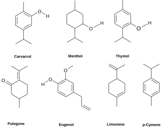
Figure 1 – Chemical structure of several metabolites: carvacrol, menthol and thymol, pulegone, eugenol, limonene and p-cymene. ... 6
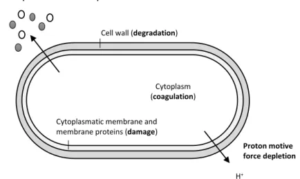
Figure 2 – Overview of locations and mode of EOs action: degradation of cell wall, damage of cytoplasmic membrane and of membrane proteins, leakage of cell contents, coagulation of cytoplasm and depletion of the proton motive force. Adapted from (Burt 2004). ... 7
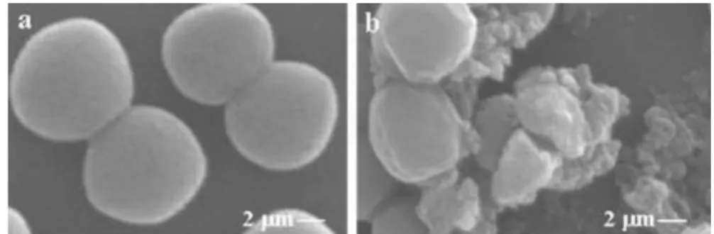
Figure 3 - Scanning electron micrographs of S. aureus. At the left (a) the bacteria were not exposed to EOs and at the right (b) exposition effects can be observed (Cleistocalys operculatus buds EO at its MIC concentration) Adapted from (Dung et al. 2008). ... 8
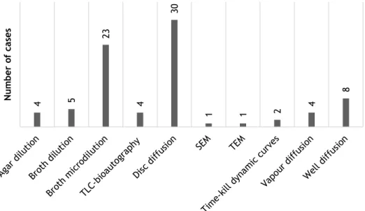
Figure 4 - Distribution of the different antimicrobial activity assays in the literature consulted. The survey was made from a total of 49 publications and some of them apply more than one method. Detailed information can be accessed in Appendix B. ... 14
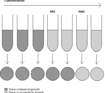
Figure 5 - Schematic representation of MIC and MBC. With the increasing of the concentration of antimicrobial solutions, the biomass present will decrease until the achievement of MIC and MBC. ... 16
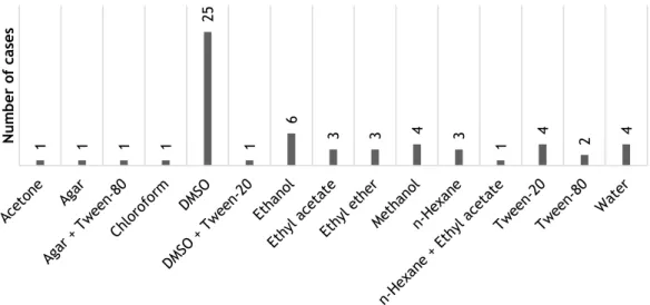
Figure 6 - Distribution of the different solvents used in the literature. The survey was made from a total of 49 publications and some of them apply more than one method. Detailed information can be accessed in Appendix B. ... 17
Figure 7- Representation of the Petri dishes used in vapour diffusion assays with 7 ml MHA in the bottom and 2 ml in the upper part used: a) example of a Petri dish used in the assays; b) representative scheme of vapour diffusion process in this type of systems..25
Figure C.1 - S. aureus calibration curve. . ... 61
Figure D.2 - X. campestris detailed results of of inhibition zone percentage in vapour phase assay: Clove Bud oil (CB-EO), Pennyroyal oil (PR-EO), eugenol and pulegone (n =3). 64
Figure F.1 - Appearance of beads control solution and maize treated with diluted bleach after 1 week (168 h) incubation at 30 °C. Only one plate per sample is shown (n = 3). ... 65
Figure F.2 - Appearance of beads control and maize treated with UV cycle after 1 week (168 h) incubation at 30 °C. Only one plate per sample is shown (n = 3). ... 66
Figure F.3 - Appearance of maize and bacteria seeded in medium with different fungicides after 48h incubation at 30 °C. Only one plate per sample is shown (n = 3). ... 67
Table 1 - Overview of the most usual definitions in the literature of minimum inhibitory
concentration (MIC) and minimum bactericidal concentration (MBC). ... 15
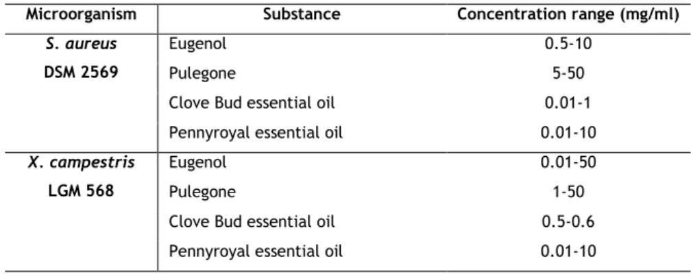
Table 2 – Microorganisms and concentration ranges of the antimicrobials used in MIC determination. ... 23
Table 3 - Summary of wells content in the broth microdilution assay. ... 23
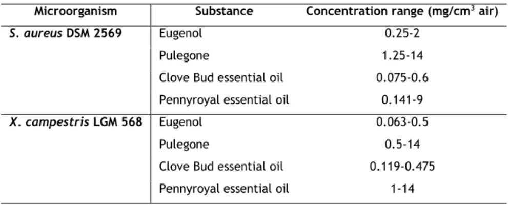
Table 4 - Antimicrobial concentration ranges used for MID determination. ... 25
Table 5 – Overview of the controls used in MID determination. ... 25
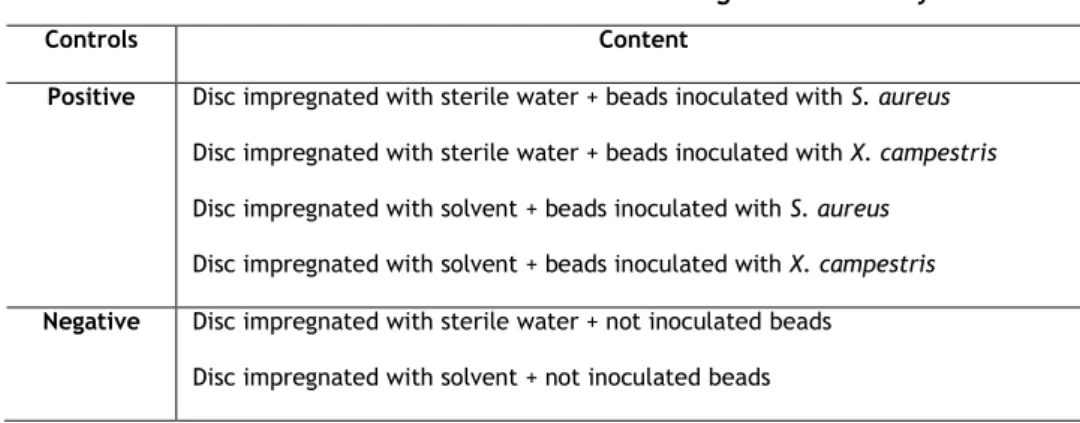
Table 6 – Overview of the controls used in glass beads assay. ... 26
Table 7 - Composition and average MIC value for the fungicides used. ... 27
Table 8 - Disc diffusion assay results (n = 2). ... 29
Table 9 – Minimum inhibitory (MIC) and bactericidal (MBC) concentrations. ... 32
Table 10 - Minimum inhibitory doses (MID) results (n = 3). ... 34
Table 11 - Estimated cell density in the beads resultant of cell counts (n = 3). ... 36
Table A.1 - Composition of S. aromaticum essential oil. ... 51
Table A.2 - Composition of M. pulegium essential oil according with different authors. . 53
Table B.1 – Survey from the methodologies applied in the literature in a total of 49 papers. ... 54
ATCC – American Type Culture Collection BHI – Brain Heart Infusion Medium CB-EO – Clove Bud Oil
CCUG – Culture Collection, University of Göteborg
CFU – Colony-Forming Unit
CLSI – Clinical and Laboratory Standards Institute
𝑑𝐼𝑛ℎ𝑖𝑏𝑖𝑡𝑖𝑜𝑛 - Inhibition Radius DMSO – Dimethyl Sulfoxide DNA – Deoxyribonucleic Acid 𝑑𝑃𝑒𝑡𝑟𝑖 𝑑𝑖𝑠ℎ - Petri Dish Radius
DSM – Culture Collection, DSMZ-Deutsche Sammlung von Mikroorganismen und Zellkulturen GmbH
EC – European Commission EO – Essential Oil
FDA - Food and Drug Administration (FDA)
GRAS – Generally Recognized as Safe HPTLC - High Performance Thin-Layer Chromatography
INT – p-iodonitotetrazolium violet LB – Luria Broth
LMG – Culture Collection, Laboratorium voor Microbiologie Universiteit Gent MBC – Minimum Bactericidal Concentration
MEA – Malte Extract Agar
MHA – Müller-Hinton Agar Medium MIC – Minimum Inhibitory Concentration MID - Minimum Inhibitory Dose
MTT – 3-(4,5-dimethylthiazol-2- yl)-2,5-diphenyltetrazolium bromide
OD – Optical Density
OPLC – Overpressured-Layer Chromatography
PEC – Planar Electrochromatography PCA – Plate Count Agar
PDA – Potato Dextrose Agar PR-EO – Pennyroyal Oil
REACH – Registration, Evaluation, Authorisation and Restriction of Chemicals System
SEM – Scanning Electron Microscope TEM – Transmission Electron Microscopy TLC – Thin-Layer Chromatography
1. Introduction
Essential oils (EOs) are formed as secondary metabolites by plants and they have always been used in traditional medicine due to their antibacterial, antiviral, antifungal and insecticidal activities. Such functions enable EOs to fulfil their primary role, which is plants´ protection. Moreover, herbs of those plants are used as spices in kitchens worldwide (Lang & Buchbauer 2012; Bakkali et al. 2008).
Nowadays EOs became once again popular since the available synthetic drugs are often related with unpleasant side effects and drug resistance may occur (Lang & Buchbauer 2012; Bakkali et al. 2008). Additionally, research has been made in order to apply these substances in the agricultural domain. In fact, diseases caused by plant pathogenic bacteria are an emergent concern of food safety. These bacteria can cause not only considerable losses in productivity and quality of harvests but also harm to the ones who ingest infected products. The management of plant disease is difficult and the complexity increases due to the large number of phytopathogenic bacteria and their easiness to spread along large distances through infected seeds. Despite such problems in disease control, there is a lack of antimicrobial agents likely to be applied in agriculture. The bactericides currently available in the market have high toxicity and are not biodegradable. The two main groups of bactericides existent are antibiotics and cooper compounds. Antibiotics are forbidden in most countries due to possible production of resistant strains. On the other hand, cooper agents are strictly controlled by the European Union due to their toxicity and environmental impact (Bajpai et al. 2011; Lo Cantore et al. 2009). Also, the green consumerism trends impel the development of new food products, especially the ones derived from plants since consumers prefer natural substance rather than synthetic ones (Lang & Buchbauer 2012; Lo Cantore et al. 2009).
Hence, the development of a biopreservation system capable to act against plant disease is a nowadays’ concern. Developing countries have particular interest to use this technology in order to protect minimal processed foods. Non-perishable food products preservation is a major interest since most of them are generally used not only as food but also as crop seeds. Thereby, spoilage of this product would prejudice populations both in the present and in the future.
The high volatility and low water solubility of EOs are, probably, the main barriers to the implementation of these natural antimicrobials in food preservation (Lang & Buchbauer
2012). To overcome these problems, special storage conditions are required to avoid volatilization and solubility can be enhanced using solvents or emulsifiers (which should not decrease antibacterial activity and must be suitable for food systems) (Burt 2004).
1.1. Objectives
In this Thesis the antimicrobial activity of pennyroyal (Mentha pulegium) and clove bud (Syzygium aromaticum) essential oils and their major components pulegone and eugenol was studied in order to analyse whether this substances would be suitable as biopreservatives for stored products. Contrary to the major part of the investigations made in the literature, both standard microorganisms and phytopathogenic ones were herein studied. The core of this work is directed to the study of Staphylococcus aureus and Xanthomonas campestris. Not only direct contact tests (as disc diffusion and broth microdilution) but also vapour phase assays were performed. A simulation of seed environment and an investigation using food matrix (maize) was attempted.
2. State of the art
2.1. Essential oils composition
Essential Oils (EOs) are liquid, volatile and rarely coloured lipid soluble substances with density normally lower than the one of water. They are synthetized in all plant organs such as flowers, herbs, buds, leaves, fruits, roots and others. Further, these compounds can be obtained by different methods: extraction, fermentation, steam distillation or expression (Solórzano-Santos & Miranda-Novales 2012; Bakkali et al. 2008). Nevertheless, concerning food and pharmaceutical applications, extraction by steam distillation or by expression are preferred (Bakkali et al. 2008).
Several works report antimicrobial activity of EOs dependence on different factors. Some of them can be the producing-part of plant under analysis, the stage of plant development, harvesting time, climatic and ecological conditions, and extraction methods used (Burt 2004; Fisher & Phillips 2008; Solórzano-Santos & Miranda-Novales 2012; Bakkali et al. 2008; Cosge et al. 2009; Lang & Buchbauer 2012; Ennajar et al. 2009; Ennajar et al. 2010; Hayouni et al. 2008; Bourgou et al. 2012).
Additionally, EOs are complex mixtures and may comprise from twenty to sixty individual components. Normally they have two major components in higher concentrations (20-70%) and several others in trace amounts. Even if the major constituents would determine the biological properties, typically the whole EO has greater antimicrobial activity than the pure constituents by themselves. Thereby the minor components may play a significant role in antibacterial activity suggesting that synergism must occur (Ahmadi et al. 2010; Bosnić et al. 2006; Bouhdid et al. 2009; Cosge et al. 2009; Dung et al. 2008; Mkaddem et al. 2009; Ennajar et al. 2010; Lopes-Lutz et al. 2008; Betoni et al. 2006; Burt 2004; Bakkali et al. 2008; Bourgou et al. 2012).
In the present work the EOs from the plants Syzygium aromaticum and Mentha pulegium and their major components (pulegone and eugenol, respectively) are of special interest. These substances are known to have antifungal, insecticidal and/or antibacterial activity, which would allow a satisfactory performance in the protection of stored products.
Additionally, such EOs are already used in food as flavourings, each means that approval for food application as biopreservatives would be easier to obtain (Hyldgaard et al. 2012).
In the Appendix A, the composition of Syzygium aromaticum and Mentha pulegium EOs are displayed. Several differences among samples can be perceived due to the several factors interfering in the EOs composition such as producing-part of plant under analysis, the stage of plant development and others already enumerated (Mahboubi & Haghi 2008).
2.1.1. Clove bud essential oil
Syzygium aromaticum is an evergreen tree belonging to the family Myrtaceae (Srivastava et al. 2005; Aneja & Joshi 2010). S. aromaticum oil (also known as clove bud oil, CB-EO) has been used for a long time in dentistry due to its analgesic, antiseptic and anti-inflammatory properties. Further, CB-EO has antibacterial, anthelmintic, antifungal, antiviral and anticarcinogenic properties (Srivastava et al. 2005; Chaieb et al. 2007; Lee et al. 2009; Aneja & Joshi 2010).
Several authors reported CB-EO composition (Srivastava et al. 2005; Aneja & Joshi 2010; Chaieb et al. 2007; Burt 2004; Lee et al. 2009; Edris & Malone 2012). The major constituent is eugenol which can be present in a concentration of 70 to 89% (see Table A.1 in the Appendix). Besides, the presence of other phenylpropanoids such as carvacrol, thymol, and cinnamaldehyde is usually reported (Srivastava et al. 2005; Aneja & Joshi 2010; Chaieb et al. 2007). Eugenol by itself reveals antioxidant and insecticidal properties (Chaieb et al. 2007) and it is strongly active in spite its low capacity to dissolve in water (Dorman & Deans 2000). Moreover, eugenol was shown to have antimicrobial activity also in the vapour phase (Goñi et al. 2009). Despite the recognized activity of eugenol by itself, there is some known synergism of the several components constituting the CB-EO, contributing to its antimicrobial activity (Lee et al. 2009). CB-EO is known to be effective against various types of bacteria, exhibiting a board spectrum of action (Lee et al. 2009; Aneja & Joshi 2010; Betoni et al. 2006), being effective against Gram-positive and Gram-negative bacteria (Dorman & Deans 2000) and as well against food spoilage bacteria (Hamed et al. 2012). In particular, high activity against S. aureus was demonstrated (Chaieb et al. 2007), whereas some resistance regarding Pseudomonas aeruginosa is reported (Lee et al. 2009; Hamed et al. 2012).
2.1.2. Pennyroyal essential oil
Mentha pulegium is also referred to as European pennyroyal and belongs to the family Lamiaceae. Since a long time M. pulegium EO (also known as pennyroyal oil, PR-EO) has been used in traditional medicine and aromatherapy. Moreover, it can also be found in foodstuff and cosmetics (Teixeira et al. 2012; Mahboubi & Haghi 2008). Nevertheless, only few papers (Teixeira et al. 2012; Franzios et al. 1997) reviewed M. pulegium EO composition, in opposition to the other EO herein addressed. The main constituent of M. pulegium EO is
known to be pulegone achieving concentrations from 23 to 76% as shown in Table A.2 from Appendix (Teixeira et al. 2012; Franzios et al. 1997).
The antimicrobial activity of this essential oil is normally associated to the presence of pulegone and other compounds as menthone and neo-menthol (Teixeira et al. 2012; Mkaddem et al. 2009). Additionally, the presence of high concentrations of piperitone is also recognized to contribute to the EO activity (Mahboubi & Haghi 2008). PR-EO is normally associated to its insecticidal activity and pulegone is known to have the same effectiveness or even higher (Franzios et al. 1997; Duru et al. 2004).
Concerning the spectrum of action, Mkaddem and coworkers (2009) claimed that EOs from mint species do not usually exhibit antimicrobial activity based on cell wall differences. Low susceptibility of Gram-negative bacteria has been reported as well (Teixeira et al. 2012; Mahboubi & Haghi 2008; Lang & Buchbauer 2012).
2.2. Chemical structure of essential oils
The chemical structure of EOs components is also a crucial characteristic since it affects the precise mode of action. There are two main chemical groups responsible for antimicrobial activity of EOs: terpenes and terpenoids, and aromatic and aliphatic constituents (Bakkali et al. 2008; Burt 2004). It should be noticed that the biosynthetic route of terpenes is independent of that of aromatic compounds. Even though, these two types of substances might coexist (Bakkali et al. 2008).
2.2.1. Terpenes and terpenoids
Terpenes are the most usual constituents of EOs, produced by a wide range of plants and they consist in combination of isoprene units (five-carbon-base units, C5) (Bakkali et al. 2008; Laciar et al. 2009). The two main terpenes types prevailing in EOs are monoterpenes (C10) and sesquiterpenes (C15), coupling two and three isoprene units, respectively (Bakkali et al. 2008). Terpenoids, also known as isoprenoids, can be described as terpenes with oxygen atoms as substituents. The nomenclature of terpenoids is similar to that of terpenes (Burt 2004).
Terpenoids and terpenes act as function of their lipophilic properties and thereby phospholipid bilayer appears to be the main target. Thus the central effects are related with inhibition of electron transport, protein translocation and enzyme-dependent reactions, as for instance phosphorylation (Dorman & Deans 2000; Laciar et al. 2009). Due to the outer membrane of Gram-negative bacteria, which comprises hydrophilic lipopolysaccharides, a barrier is created offering additional tolerance to hydrophobic antimicrobial compounds. Thereby, Gram-positive, which do not possess such barrier, would be more susceptible to
terpenoids-containing EOs (Dung et al. 2008; Mkaddem et al. 2009; Laciar et al. 2009; Ennajar et al. 2009).
2.2.2. Aromatic and aliphatic compounds
Aromatic compounds are derived from the phenylpropane pathway and EOs containing phenolic compounds are the strongest antimicrobials (Burt 2004; Bajpai et al. 2009; Cosge et al. 2009). For these EOs, the mechanism of action is similar to the one reported for other phenolic compounds and consequently the most notorious effects are in cellular membranes, similarly to terpenes and terpenoids. Phenolics not only attack membrane integrity, increasing permeability and release of intracellular molecules, but additionally they affect membrane function (as electron transportation, nutrient uptake, enzyme activity) and also protein and nucleic acid synthesis (Burt 2004; Bajpai et al. 2009).
Among the phenolic compounds, the presence of hydroxyl groups is determinant and can be proven by comparison of the activity of the phenolic carvacrol and the lack of activity of the non-phenolic menthol (see Figure 2). Additionally, the relative positions of these groups appear to influence the EO effectiveness. This is proven by carvacrol and thymol (see Figure 2) activity against Gram
-p
ositive and Gram-negative. Thymol, with a –OH group at the adjacent carbon to isopropyl group, (–CH(CH3)2), seems to be preferentially active against Gram-positive bacteria (Burt 2004; Dorman & Deans 2000), showing, for instance, low inhibition of Gram-negative bacteria such as Pseudomonas aeruginosa (Dorman & Deans 2000). O H O HO
HCarvacrol Menthol Thymol
O O H O
Pulegone Eugenol Limonene p-Cymene
Figure 2 – Chemical structure of several metabolites: carvacrol, menthol and thymol, pulegone, eugenol, limonene and p-cymene.
Concerning non-phenolic compounds, the structure of alkyl groups seems to affect antimicrobial activity. The existence of double bonds seems to favour activity since compounds having alkenyl groups exhibit enhanced activity when compared to others possessing alkyl groups. The presence of alkyl groups interferes in the partition coefficient, reducing the surface tension and modifying selectivity. The lack of activity of p-cymene (with alkyl group) versus the activity of limonene (with alkenyl group) confirms this condition (Burt 2004; Dorman & Deans 2000).
Regarding the two substances of special interest (Figure 2), pulegone can be classified as a cycle monoterpene with ketone function and eugenol as a phenolic aromatic compound (Bakkali et al. 2008).
2.3. Mode of antibacterial action of essential
oils
Regarding the antibacterial mode of action of EOs, additional research is needed since some doubts remain about the mechanisms involved. Nevertheless, several mechanisms have been proposed (Figure 3) and it is settled that EOs antibacterial activity is most-likely resultant from a combination of actions with several targets in the cell (Burt 2004).
Figure 3 – Overview of locations and mode of EOs action: degradation of cell wall, damage of cytoplasmic membrane and of membrane proteins, leakage of cell contents, coagulation of
cytoplasm and depletion of the proton motive force. Adapted from (Burt 2004).
Different authors used scanning electron microscopy (SEM) (Khan & Ahmad 2012; Dung et al. 2008; Bajpai et al. 2009) and transmission electron microscopy (TEM) (Fisher & Phillips 2008; Bouhdid et al. 2009) to observe morphological changes in the cell when contacting with different EOs. In Figure 4 electron micrographs clearly show that in the absence of EO the cell
Cell wall (degradation)
Cytoplasmatic membrane and membrane proteins (damage)
Proton motive force depletion
H+ Leakage of cytoplasmatic
constituents (metabolites and ions)
Cytoplasm (coagulation)
surface is smooth and regular contrary to cells in the presence of Cleistocalys operculatus essential oil. Hereby, EOs appear to deteriorate the morphology of cell membrane which starts to display several clefts, ending in disruption and lysis (Dung et al. 2008; Bakkali et al. 2008).
Figure 4 - Scanning electron micrographs of S. aureus. At the left (a) the bacteria were not exposed to EOs and at the right (b) exposition effects can be observed (Cleistocalys operculatus buds EO at
its MIC concentration). Adapted from (Dung et al. 2008).
The most important characteristic that enables the EOs action is their hydrophobicity. As hydrophobic compounds, EOs are able to partition into the lipids of cell membranes (e.g. bacterial cell membrane and mitochondria). Such action disturbs these structures which become more permeable, allowing the leakage of ions and metabolites. Even if up to a certain point leakage does not imply loss of cell viability, further leakage can be lethal (Nedorostova et al. 2009; Burt 2004; Solórzano-Santos & Miranda-Novales 2012; Fisher & Phillips 2008). In addition, a decrease of pH due to cell membrane disruption has been reported. This fact suggests that the control of cellular processes as for instance DNA transcription, protein synthesis or enzyme activity is lost (Fisher & Phillips 2008). Lipids and protein are damaged and cytoplasm might coagulate (Bakkali et al. 2008). Moreover respiration processes is known to be affected (Bouhdid et al. 2009; Pavithra et al. 2009).
Further, EOs appear to act on cell proteins from the cytoplasmatic membrane. There are two main possible mechanisms. The EO lipophilic molecules might accumulate in the lipid bilayer and disturb lipid-protein interactions or the lipidic compounds can directly interact with the protein hydrophobic parts (Burt 2004).
Even though the mechanisms of action are poorly understood, there are some evidences indicating that EOs mode of action is different from that of common antibiotics since, for example, methicillin-resistant strains were susceptible to EOs action (Mulyaningsih et al. 2010; Aneja & Joshi 2010; Chaieb et al. 2007).
2.3.1. Gram positive versus Gram negative susceptibility
Concerning susceptibility, it is often reported in the literature that Gram-positive bacteria seem to be slightly more sensitive to EOs action than Gram-negative ones (Laouer et al. 2009; Al-Reza et al. 2009; Bosnić et al. 2006; Bouhdid et al. 2009; Busatta et al. 2008;
Cosge et al. 2009; Ennajar et al. 2009; Ennajar et al. 2010; Hamed et al. 2012; Magina et al. 2009; Mahboubi & Haghi 2008; Mulyaningsih et al. 2010; Oyedeji et al. 2009; Shahat et al. 2008; Zarai et al. 2012; Nedorostova et al. 2009; Wang et al. 2012; Burt 2004; Fisher & Phillips 2008; Solórzano-Santos & Miranda-Novales 2012; Laciar et al. 2009; Chaieb et al. 2007). Usual explanations are related with the fact that Gram-negative are owners of an outer membrane surrounding the cell wall in opposition to Gram-positive. Thereby they offer higher resistance to EOs action, not allowing an easy diffusion through the lipopolysaccharide membrane and avoiding the accumulation of EOs in the cell (Burt 2004; Solórzano-Santos & Miranda-Novales 2012; Fisher & Phillips 2008; Bajpai et al. 2009; Busatta et al. 2008; Ennajar et al. 2009; Laciar et al. 2009; Magina et al. 2009; Mahboubi & Haghi 2008; Mulyaningsih et al. 2010; Teixeira et al. 2012).
However a reduced number of papers claim that there is no difference among Gram-positive and Gram-negative bacteria (Dorman & Deans 2000; Dung et al. 2008; Goñi et al. 2009; Mkaddem et al. 2009). Additionally, some authors (Burt 2004; Lang & Buchbauer 2012) report only a delay in EOs action concerning Gram-negative organisms. On the other hand, concerning antimicrobial vapour activity of EOs, the opinions seem to be consistent and it is reported that there is no difference among Gram-positive and Gram-negative bacteria (Bajpai et al. 2011; Lopez et al. 2005).
In the last decade, it was proposed that the degree of activity against organisms with different cell wall structure is due to individual components of EOs. This would explain the preferential activity of some EOs against certain type of bacteria. Given there are several factors affecting EOs composition, the susceptibility would differ in that extent (Burt 2004; Lang & Buchbauer 2012; Bajpai et al. 2009). Different studies (Ahmadi et al. 2010; Bosnić et al. 2006; Bouhdid et al. 2009; Cosge et al. 2009; Dung et al. 2008; Mkaddem et al. 2009; Ennajar et al. 2010; Lopes-Lutz et al. 2008; Betoni et al. 2006; Bourgou et al. 2012) support that the activity of the EO is due to additive, synergetic or antagonistic effects of individual components in specific quantities and thereby the same EO activity cannot be easily replicated due to the dependence on its composition and on different external factors. Further, the antimicrobial activity would be affected by EOs plant origin, composition and concentration (Lang & Buchbauer 2012; Bakkali et al. 2008; Cosge et al. 2009; Ennajar et al. 2009; Ennajar et al. 2010; Hayouni et al. 2008).
In the present study, Staphylococcus aureus and Xanthomonas campestris were used to assess the antimicrobial activity of CB-EO and PRO-EO and of their major constituents. S. aureus is a standard microorganism widely used in antibacterial assays and its popularity started due to their propensity to develop multi-drug-resistance (Solórzano-Santos & Miranda-Novales 2012). Today it is also used in antibacterial assays involving EO, allowing some degree of comparison with the results in literature. X. campestris is a phytopathogenic bacteria. Given it is intended to proceed to assays in food matrix using non-perishable products as maize, it is interesting to study a bacteria that normally spoilages products like this when stored.
2.3.1.1. Staphylococcus aureus
Staphylococcus aureus comprise Gram-positive bacteria commonly found in the human skin (Lang & Buchbauer 2012). These bacteria are often in the origin of food-borne diseases and skin infections (Solórzano-Santos & Miranda-Novales 2012; Ahmadi et al. 2010). Hence, elimination and control of S. aureus are main objectives of different industries, specially food-related ones (Ahmadi et al. 2010).
Staphylococcus genus, and specially S. aureus, is constantly reported in the literature as one of the most susceptible bacteria to EOs action (Laouer et al. 2009; Aneja & Joshi 2010; Bajpai et al. 2009; Betoni et al. 2006; Bosnić et al. 2006; Çetin et al. 2009; Ennajar et al. 2009; Goñi et al. 2009; Hamed et al. 2012; Mahboubi & Haghi 2008; Shahat et al. 2008; Zarai et al. 2012; Saeed et al. 2013; Nedorostova et al. 2009; Fatope et al. 2008; Bourgou et al. 2012; Chaieb et al. 2007). Actually, from the survey made, only one work out of forty nine (Hayouni et al. 2008) reports lack of antibacterial activity of Salvia officinalis L. and Schinus molle L. EOs against S. aureus.
2.3.1.2. Xanthomonas campestris
The genus Xanthomonas, comprising Gram-negative rods, causes diseases in several plants including cereals, affecting a wide range of plant parts. Even in developed countries, this fact make Xanthomonas one of the major problems in agriculture domain. The diseases control is extremely difficult and expensive as they can be easily transmitted from infected plants through different mechanisms as for instance rain waters, wind, birds or insects (Bajpai et al. 2011). Despite very few publications report the activity of EOs against Xanthomonas, some applications comprising EOs have been effective in inhibiting Xanthomonas growth (Bajpai et al. 2009; Inouye 2003). Additionally, along with other bacterial species (as for instance Pseudomonas syringae or Clavibacter michiganensis) Xanthomonas campestris is one of the major phytopathogenic bacteria type (Bajpai et al. 2011).
2.4. Overview of the methodologies
A wide range of assays to in vitro assessment of antimicrobial activity can be used. Such methods are usually classified as diffusion, dilution or bioautographic methods, and the most used are the first two (Burt 2004; Rios et al. 1988; Horváth et al. 2010; Lang & Buchbauer 2012).
The agar disc diffusion method is often employed and is frequently used as screening assay before further studies. Its application is useful for selection of antimicrobial substances
to be used. However, the comparison of different data concerning this method is difficult due to differences in operation conditions applied by different authors (Burt 2004).
On the other hand, antimicrobial activity assays are traditionally established and standardized for the utilization of antibiotics as active substances. As referred to above, essential oils and their components are volatile and complex viscous substance. Further, these substances are hydrophobic in opposition to antibiotics, which are generally hydrophilic. Hence the above mentioned methods were modified in order to be adapted to the utilization of EOs as active substance (Burt 2004; Horváth et al. 2010).
Concerning EOs antimicrobial activity study, different factors might bias the results. One of the major problems is their volatility, since substances with higher volatility evaporate faster and the EO activity would not be properly assessed. To avoid this problem, dilution tests are sometimes preferred. Nevertheless, hydrophobicity, which demands the use of surfactants or solvents, can distort the result. Each solvent has different properties which consequently may influence the EO activity in a different way (Lang & Buchbauer 2012). Also extraction methods of the EO can be a variable since the difference in organoleptic profile indicates differences in the composition of oils obtained by different extraction methods, which might influence the antimicrobial properties of the EO (Burt 2004). Further, there is a high difficulty in the comparison of published data, since individual modifications in the procedure conditions (even simple as volume of inoculum) can extremely influence the final result (Lang & Buchbauer 2012; Burt 2004).
Therefore, even if the different methods are described in the literature, no standardized method to evaluate antimicrobial activity of EOs against food-related microorganisms can be found in the literature (Burt 2004) even though several authors (Ebrahimabadi et al. 2010; Dung et al. 2008; Mahboubi & Haghi 2008; Hayouni et al. 2008; Saeed et al. 2013; Laciar et al. 2009; Hammer et al. 1999; Bajpai et al. 2009; El-Baroty et al. 2010; Mulyaningsih et al. 2010; Teixeira et al. 2012; Betoni et al. 2006; Wang & Liu 2010; Zarai et al. 2012) have been adapting Clinical and Laboratory Standards Institute (CLSI) antimicrobial susceptibility tests, designed for antibiotics, to EOs testing.
2.4.1. Diffusion methods
Agar diffusion is often used either as well diffusion, disc diffusion, cylinder diffusion or vapour diffusion. In the first case small holes are punched in the inoculated agar plate using simple devices such as a sterile Pasteur pipettes. The well is then filled with the EO which would diffuse into the agar.
Disc diffusion requires the placement of small paper discs impregnated with the EO onto the inoculated agar surface. The disc can be directly placed onto the agar after impregnation or EO can be allowed to dry in the disc before placement (Lang & Buchbauer 2012; Burt 2004).
Cylinder diffusion is a technique not so often used, as remarked by the lack of reference to it in several reviews about antimicrobial assays methodologies. This method is quite similar to well diffusion. Here, stainless steel or porcelain cylinders are placed on a previous inoculated Petri dish. The cylinders are filled with the antimicrobial substances and the Petri dishes are incubated. After that period the cylinders are removed and results evaluated (Choma & Grzelak 2011; Rios et al. 1988).
All these techniques result in an inhibition zone (halo) surrounding the area where the EO was placed. The size of this halo gives the strength of the active substance in a relative manner since enables the comparison between different substance activities (Lang & Buchbauer 2012; Burt 2004). It has been proposed in the literature (Fu et al. 2007) that inhibition diameters lower than 10 mm should be considered as weak antimicrobial activity and higher than 10 mm should be considered as satisfactory. As stated above, the main problem of this type of assay is the EOs low solubility in water which sometimes can lead to a false low antimicrobial activity. For instance, some EO present low activity in the agar diffusion but higher antimicrobial activity in dilution assays (Lang & Buchbauer 2012).
Still regarding diffusion assays, vapour phase methods are also applied, even though, less often (Lang & Buchbauer 2012; Inouye 2003). There are two variations of this technique nominated slow and fast evaporation. The most usual is fast evaporation (or inverted Petri dish method), in which a seeded agar plate is placed upside down onto a reservoir. A sterile filter paper impregnated with the volatile oil is placed bellow the seeded plate in order to allow vapour diffusion. Slow evaporation consists in a agar plate inoculated and a filter paper or glass dish containing EO placed inside a sealed container, which would allow the simulation of a closed system where the EO will slowly evaporate and exert its antibacterial action (Inouye 2003; Goñi et al. 2009; Fisher & Phillips 2008). In both cases, inhibition zone is measured and determination of minimum inhibitory doses (MID) is possible. The principal handicap of this technique relays also on the inexistence of a standardized method. Nevertheless, this type of assay takes advantage of EOs high volatility, overcoming the lack of solubility of these substances which is notorious in other type of assays (Lang & Buchbauer 2012; Inouye 2003).
2.4.2. Dilution tests
Dilution methods can use as support solid medium (agar) or liquid medium (broth). Both cases consider a concentration gradient of the substance and therefore a dilution series is made. The creation of a saturated moistened atmosphere is significantly useful to adjust the volatility (Lang & Buchbauer 2012). If agar is used, after dilution it is allowed to solidify onto the Petri plates (Rios et al. 1988).
Within broth dilution type, broth microdilution is often applied. This variant may comprise the growth of the microorganisms in a microwells plate and uses lower volumes (Choma & Grzelak 2011).
As mentioned above, the major advantage of dilution methods is the possibility to apply it to the study of water-soluble or insoluble compounds, thus being especially useful to EOs (Rios et al. 1988; Lang & Buchbauer 2012). Nevertheless it takes laborious handling and high costs (Lang & Buchbauer 2012).
Dilution methods allow the determination of the minimal inhibitory concentration (MIC) and, consequently, the determination of the EO strength against certain microorganism (Lang & Buchbauer 2012; Burt 2004; Fisher & Phillips 2008). In order to generalize the classification of antimicrobial activity of these substances it has been proposed the following classification: strong in the range 0.05-0.5 mg/ml, moderate for 0.6-1.5 mg/ml and weak if MIC is higher than 1.5 mg/ml (Sartoratto et al. 2004; Aligiannis et al. 2001; Magina et al. 2009).
2.4.3. Other methods
As stated, other often used method is bioautography which relays on the diffusion methods principle although the diffusion occurs from the chromatographic layer to the agar medium (Choma & Grzelak 2011). Therefore, it requires the execution of a previous EO characterization using chromatography and it can be efficaciously combined with different layer liquid chromatography methods (Rios et al. 1988; Horváth et al. 2010; Choma & Grzelak 2011). Moreover, bioautography can be subdivided in contact, direct or immersion types.
Besides, there are less typical methods. The determination of quickness and duration of antibacterial activity can be made using time-kill analysis by plotting the survival curves (number of viable cells in the broth after EO addition) versus time. Thereby, the measurement of optical density and plating out onto solid medium is required (Burt 2004; Lang & Buchbauer 2012). Other technique is the air-washer coupled with air-sampler which allows the study of the effect of EOs in air-born microorganisms (Lang & Buchbauer 2012). Further, the physical effects of antibacterial activity can be as well made using scanning electron microscopy (SEM) and transmission electron microscopy (TEM). These methods allow the assessment of damage of the bacterial cell wall and cytoplasm such as swelling, vacuolations or leakage (Burt 2004; Bouhdid et al. 2009; Fisher & Phillips 2008; Bakkali et al. 2008).
2.4.4. Incidence in the literature
According with a review of several publications, the most used method to assess EO antimicrobial activity is the disc diffusion assay soon followed by broth microdilution. The distribution of the several methods found can be seen in Figure 5. Moreover, concerning bioautography only the direct type was found. Within the literature assessed cylinder diffusion technique was not found.
Figure 5 - Distribution of the different antimicrobial activity assays in the literature consulted. The survey was made from a total of 49 publications and some of them apply more than one method.
Detailed information can be accessed in Appendix B.
2.4.5. Growth indicators
Antibacterial assays are occasionally coupled with bacterial growth indicators in order to easily analyze the effect of EOs growth inhibition. Examples are 2,3,5-triphenyltetrazolium chloride (TTC) (Sartoratto et al. 2004; O’Bryan et al. 2008; Laciar et al. 2009), 3-(4,5-dimethylthiazol-2- yl)-2,5-diphenyltetrazolium bromide (MTT) (Zarai et al. 2012; Horváth et al. 2010), p-iodonitotetrazolium violet (INT) (Oyedeji et al. 2009; El-Baroty et al. 2010) or 7-Hydroxy-3H-phenoxazin-3-one 10-oxide (Alamar Blue or resazurin) (Bouhdid et al. 2009; Valgas & Souza 2007).
Additionally, growth indicators capable of detect dehydrogenase activity (such as tetrazolium salts) are quite often in bioautography methods (Choma & Grzelak 2011).
2.4.6. Minimum inhibitory and bactericidal concentrations
The minimum inhibitory concentration (MIC) is normally used as a measure of the antibacterial performance of EOs. Nevertheless, MIC definition diverges between publications, which is an additional problem concerning data comparison. Some studies report as well, the minimum bactericidal concentration (MBC) whose definition can also diverge. An overview of the most usual definitions for both MIC and MBC is presented in Table 1. Figure 6 displays a schematic definition of both MIC and MBC. As stated, MIC and MBC are determined using dilution methods.
It should be noticed that despite the variations among the protocols and considerations taken by different authors, MICs determined by agar dilution are generally in the same range of magnitude (Burt 2004; Busatta et al. 2008) whereas broth dilution methods are not. This
4 5 23 4 30 1 1 2 4 8 Nu m b er of c as es
can be explained by the utilization of different techniques to determine the dilution assay end-point. The most frequent methods to do so are optical density (turbidity) and enumeration of colonies by viable count (after incubation in solid medium). Other methods can be listed such as visible growth (macroscopic evaluation), absorbance, colourimetry or conductivity (Burt 2004).
Table 1 - Overview of the most usual definitions in the literature of minimum inhibitory concentration (MIC) and minimum bactericidal concentration (MBC).
Term Definition Reference
MIC The lowest concentration inhibiting visible growth…
(Wang et al. 2012; Goñi et al. 2009; Adiguzel et al. 2009; Hammer et al. 1999; Kloucek et al. 2012; Betoni et al. 2006; Ahmadi et al. 2010; Oyedeji et al. 2009; Ebrahimabadi et al. 2010; Teixeira et al. 2012; CLSI 2012a)
…after macroscopic evaluation. (Al-Reza et al. 2009; Dung et al. 2008; Magina et al. 2009; Wang & Liu 2010)
…after 24h incubation period. (Pavithra et al. 2009) …after 24h incubation period at 37 °C. (El-Baroty et al. 2010) …24h for bacteria and 48h for fungi. (Mahboubi & Haghi 2008)
…indicated by the staining agent. (Sartoratto et al. 2004; Zarai et al. 2012; Bouhdid et al. 2009; Laciar et al. 2009; O’Bryan et al. 2008)
…indicated by no visible turbidity. (Joshi et al. 2008) …indicated by no visible turbidity after
24h incubation period.
(Lee et al. 2009) …indicated by the presence of a white
“pellet” on the well bottom.
(Hayouni et al. 2008) …resulting in a clearly visible inhibition
zone.
(Nedorostova et al. 2009; Aneja & Joshi 2010; Shahat et al. 2008)
…resulting in 80% reduction in visible growth when compared with that for substance-free sample.
(Havlik et al. 2009)
MBC The lowest concentration resulting in 99% absence of growth.
(Magina et al. 2009) The lowest concentration resulting in
absence of growth…
(Pavithra et al. 2009)
…on the solid media surface. (Dung et al. 2008; Mahboubi & Haghi 2008) …on the solid media surface,
determined by seeding 10 μL from each well on a plate which was then incubated for further 24 h at 37 °C.
(Wang et al. 2012)
Moreover, it is also possible to determine the minimum inhibitory dose (MID), generally defined as the minimum dose of the gaseous state to inhibit growth. Even so, definition can vary. The determination of MID is similar to that for MIC but instead of dilution methods it demands vapour diffusion techniques. There are two main problems related with MID determination. First, the incubation temperature can have an effect in the inhibition once
MID is dependent on the evaporation of the EO volatile components. Second, loss of vapour can occur due to absorption into the media (Fisher & Phillips 2008; Goñi et al. 2009).
Figure 6 - Schematic representation of MIC and MBC. With the increasing of the concentration of antimicrobial solutions, the biomass present will decrease until the achievement of MIC and MBC.
2.4.7. Usual solvents
In the literature it is often reported the utilization of different solvents to allow a better incorporation of the EO into the medium when using bacterial systems (Burt 2004). The importance of it rises when applying diffusion methods due to the water insolubility problems previously mentioned. However, after literature review several are the authors who do not report the utilization of any solvent when using diffusion techniques (Park et al. 2010; Pavithra et al. 2009; Hayouni et al. 2008; O’Bryan et al. 2008; Çetin et al. 2009; Mulyaningsih et al. 2010; Goñi et al. 2009; Busatta et al. 2008; Mkaddem et al. 2009; Joshi et al. 2008; Fatope et al. 2008; Nedorostova et al. 2009; Saeed et al. 2013; Bourgou et al. 2012; Ennajar et al. 2010; El-Baroty et al. 2010; Fatope et al. 2008).
It is notorious a preference in the application of DMSO as solvent in different method types (El-Baroty et al. 2010; Pavithra et al. 2009; Aneja & Joshi 2010; Mahboubi & Haghi 2008; Hayouni et al. 2008; Joshi et al. 2010; Laciar et al. 2009; Çetin et al. 2009; Ahmadi et al. 2010; Lopes-Lutz et al. 2008; Mulyaningsih et al. 2010; Dung et al. 2008; Adiguzel et al. 2009; Laouer et al. 2009; Magina et al. 2009; Shahat et al. 2008; Oyedeji et al. 2009; Wang & Liu 2010; Ebrahimabadi et al. 2010; Teixeira et al. 2012). In addition, other solvents reported are: acetone (Maggi et al. 2010), agar (O’Bryan et al. 2008; Bouhdid et al. 2009), Tween-80 (Ennajar et al. 2010; Wang & Liu 2010; Fu et al. 2007), Tween-20 (Wang et al. 2012; Hammer et al. 1999), ethanol (Zarai et al. 2012; Horváth et al. 2010; Teixeira et al. 2012), methanol (Lee et al. 2009; Adiguzel et al. 2009; Bajpai et al. 2009; Saeed et al. 2013), ethyl acetate
(Sartoratto et al. 2004; Kloucek et al. 2012; Bajpai et al. 2009), ethyl ether (Goñi et al. 2009; Lopez et al. 2005), n-hexane (Bajpai et al. 2009; Ahmadi et al. 2010), water (Teixeira et al. 2012; Cosge et al. 2009; Saeed et al. 2013) and chloroform (Bajpai et al. 2009). Tween-20 was reported as more suitable for solubilization than Tween-80. Both compounds are normally attractive due to their low toxicity and safety regarding food and pharmaceutical applications (Edris & Malone 2012). In some cases, combination of two solvents are found. From the consulted publications, Aneja and Joshi (2010) used a combination of DMSO and Tween-20, Laciar et al. (2009) used n-hexane with ethyl acetate and Havlik et al. (2009) used agar and Tween-80. Figure 7 illustrates the incidence of different solvents used.
Figure 7 - Distribution of the different solvents used in the literature. The survey was made from a total of 49 publications and some of them apply more than one solvent. Detailed information can be
accessed in Appendix B.
Additionally, it was previously reported that the utilization of substances to solubilize the EO can decrease its antibacterial effect. An evidence of that is for instance the utilization of Tween-80 as neutralizer of some disinfectants (Burt 2004). On the opposite, it was shown that agar at 0.05% (w/v) improves the antibacterial effect of some types of EOs by enhancement of the dispersion in water (Burt et al. 2005). In addition, 0.2% (w/v) agar was proven to enable an homogeneous dispersion of the EOs with better performance than the one obtained with Tween-80 or ethanol (Burt et al. 2005; Burt 2004).
Also regarding Tween, some studies reported the utilization of this substances as carbon source especially for P. aeruginosa, since the members of genus Pseudomonas are typically capable of use several exogenous substrates as carbon source (Howe & Ward 1976). Tween-80 is most frequently used as carbon source than Tween-20. Both compounds derive from fatty acids which justify their utilization as carbon source for some application such as lipase or medium-chain-length polyhydroxyalkanoates production (Zouaoui & Bouziane 2011; Chan et al. 2006). Therefore, despite its advantages for food and pharmaceutical applications, Tween do not seem to be the best choice as solvent, in particular Tween-80.
1 1 1 1 25 1 6 3 3 4 3 1 4 2 4 Nu m b er of c as es
Even if in one work DMSO is reported as prejudicial to antimicrobial activity (Hili et al. 1997), in the works where DMSO is used as negative control, no inhibition zone for DMSO is shown, and thus, no activity can be seen (Bosnić et al. 2006; Aneja & Joshi 2010). Thereby, DMSO seems to be the most advantageous solvent to be applied.
2.5. Applications of essential oils as
preservatives in food systems
Since food systems are environments prone to microorganisms’ proliferation, food spoilage is usual. Food-borne diseases are caused due to ingestion of food contaminated with pathogenic microorganisms and/or their toxins (Solórzano-Santos & Miranda-Novales 2012).
Thereby, EOs have potential to be applied in package coating containing the antimicrobial compound or even by development of biodegradable coating films (Hamed et al. 2012; Havlik et al. 2009). Besides, EOs are able to reduce bacterial population when applied directly in the soil and this will, consequently, avoid spoilage of fresh organic products cultivated in that soil (Solórzano-Santos & Miranda-Novales 2012).
It should be highlighted that, when handling with antibacterial compounds in food systems, an improved incorporation is a main concern. Often, lack of water solubility of EOs reduces availability for antimicrobial action. Moreover, comparing food systems tests with in vitro tests, food systems have greater availability of nutrients which may allow bacteria to repair damaged cells faster and thus, be less susceptible. This leads to the use of higher concentrations than the ones used in in vitro. Such fact may be a problem, since exceeding the acceptable flavour and/or odour thresholds would imply a stronger herbal aroma and thus consumer’s rejection once the original food aroma is changed (Shah et al. 2013; Velázquez-Nuñez et al. 2013).
Moreover, different parameters of the foodstuff are of great concern. Both intrinsic (as fat, protein, water content; antioxidants; preservatives; pH; salt and additives) and extrinsic properties (temperature, packing in vacuum, gas or air; characteristics of the microorganism) are indeed important and can affect antimicrobial activity of EOs (Burt 2004; Havlik et al. 2009; Hayouni et al. 2008). Actually, it is known that the affinity of food components (as proteins and fat) can bind and solubilize phenolic compounds reducing their availability to act against bacteria (Hayouni et al. 2008; Shah et al. 2013; O’Bryan et al. 2008). Moreover, in food systems, MIC must be achieved in all points of the food to ensure protection and thereby concentration would be higher (Havlik et al. 2009).
As stated before, different studies concerning application of EOs as biopresevatives have been accomplished. The EOs act by reducing or eliminating the pathogens. Food quality
can, thus, be improved and to do so EOs effectiveness was already assessed for some food products (Solórzano-Santos & Miranda-Novales 2012). Indeed, eugenol was already tested as seed disinfectant, showing promising results (Lang & Buchbauer 2012; Lo Cantore et al. 2009). Actually, Lo Cantore research group (2009) was able to lower the X. campestris bacterial population of infected been seeds from 2.6x10⁶ to 7.0x10² CFU/ml using eugenol emulsions. Despite the major components of the EO could be used isolated, it is more sustainable to use the whole EO. Furthermore there is the additional advantage of the synergetic effects of the minor components which normally increase antimicrobial activity (Burt 2004).
Several topics must to be taken in consideration and in the next sub-section different methods of application of this technology are summarized (Solórzano-Santos & Miranda-Novales 2012; Fisher & Phillips 2008).
2.5.1. Available technologies for implementation of EOs as
biopreservatives
Notwithstanding lack of investigation about implementation of EOs as biopreservatives in foods, there are some works reporting a few solutions.
Several works have demonstrated the potential of microencapsulation using either proteins or polysaccharides (Shah et al. 2013; Hayouni et al. 2008). Recently, some other studies (Shah et al. 2013; Edris & Malone 2012; Hamed et al. 2012) describe microencapsulation by the utilization of synthetic surfactants which would assemble in oil-water (O/W) micelles at the critical micelle concentration (CMC), forming micro or nanoemulsions. In the interior of the micelles, a hydrophobic environment is provided allowing EOs solubilization (Rodrigues et al. 2013). These methods aim to reduce toxicity, to protect the active substances from deterioration and to avoid the alteration of organoleptic characteristics of the foodstuff (Hayouni et al. 2008).
The use of EOs vapour diffusion can be a safer alternative by eliminating organoleptic issues since EOs would not contact directly with foodstuff (Dorman & Deans 2000; Ennajar et al. 2010; Fisher & Phillips 2008). Other greater advantage of this approach is the vehicle. When in solution, EOs tend to form micelles which would suppress the complete attachment of the EO to the microorganism. On the other hand, vapour phase dispersion allows free attachment (Fisher & Phillips 2008). Actually, when comparing MIDs and MICs, the first ones seem to be lower. This represents an advantage since foodstuff properties would be protected by the utilization of lower doses. Even so, dispersion of the oils as vapours instead of the natural evaporation must be analysed, as heating the oils to increase evaporation can affect the oils antimicrobial properties (Solórzano-Santos & Miranda-Novales 2012; Fisher & Phillips 2008).
2.5.2. Legal aspects
Regarding legal aspects, European Commission (EC) allows the utilization of some EOs in foods as flavourings (Burt 2004). United States Food and Drug Administration (FDA) has classified these compounds as generally recognized as safe (GRAS) or as approved food additives (Burt 2004; Fisher & Phillips 2008). Nevertheless, some EO components can cause irritation and toxicity (Burt 2004). Even so, pulegone and eugenol (principal components of the EOs studied in the present work) are approved by the above mentioned lists (Burt 2004; FDA 2012; EC 2002; Kollanoor Johny et al. 2010). Besides, the organic solvents commonly used to enhance solubility involve dosing restrictions (Edris & Malone 2012) and some cautions must be taken since high doses of EOs (>0.05% v/v) are proven to have cytotoxic effects (Fisher & Phillips 2008). Despite these facts, most of these oils are available for purchase as a whole or contained in pharmaceutical or cosmetic products, which indicates that possible toxic properties do not prohibit their commercialization and utilization (Hammer et al. 1999).
The use of EOs in food is regulated by other directive concerning to flavourings for use in foodstuffs, the directive 88/388/CEE. This regulation allows the utilization with restriction in foodstuffs of some substances forbidden for direct application (among them there is pulegone). Further, recently the REACH system (registration, evaluation, authorization and restriction of chemicals, EC regulation 1907/2006) was implemented. The target substances are the ones produced in amounts of more than one ton per year. Therefore, until the present, EOs were not submitted to this regulation and probably would not be due to the lack of patents applicable to plant extracts (Vigan 2010).
3. Materials and methods
3.1. Reagents
Dimethyl-sulfoxide (DMSO) (≥ 99.5%), (R)-(+)-pulegone (≥ 85%), Triton x-100 and Tween-20 were obtained from Sigma-Aldrich. Eugenol (99%) and stabilized (+)-limonene (96%) were purchased from Acros Organics. Clove bud oil (CB-EO) and pennyroyal oil (PR-EO) were acquired from Inovia International. Müller-Hinton Agar (MHA) and Brain Heart Infusion Agar (BHI) were obtained from Oxoid and Müller-Hinton Broth (MHB) from HiMedia. Plate Count Agar (PCA), Potato Dextrose Agar (PDA) and Luria Broth (LB) were acquired from Liofilchem and Malte Extract Agar (MEA) from Merk. The fungicides Benlate, Previcur N and Baycor 300 were obtained from commercial brands.
3.2. Microorganisms, culture and stock
conditions
The standard bacteria Staphylococcus aureus DSM 2569 and Pseudomonas aeruginosa DSM 1117 and the phytopathogenic bacteria Xanthomonas campestris LGM 568, Clavibacter michiganensis DSM 46364, and Rathayibacter tritici DSM 7486 were used during the assays.
Bacteria stocks were made in LB with 50% glycerol and maintained at -80ºC. BHI was used to transfer the bacteria from the freezing medium since this is the recommended media by CLSI standards (CLSI 2012a; CLSI 2012b; Inouye 2003). The incubation time was 24 h for standard bacteria and 48 h for phytopathogenic bacteria.
MHA and MHB were used for the antimicrobial assays since these are the most suitable media to such purpose (CLSI 2012a; CLSI 2012b; Inouye 2003). Preliminary assays revealed difficulties when S. aureus and X. campestris were used in suspension since in the absence of dispersants aggregation of cells seemed to occur. Thus, the surfactant Triton x-100 was used
as dispersant and was added to the liquid medium (MHB) at a concentration of 0.001% (Manuel 2007).
3.3. Antimicrobial stock solutions
Due to the low solubility in water of the compounds to be tested, preliminary tests using 10% DMSO or 5% Tween-20 in sterile water were made. These concentrations were chosen since they are the most subscribed in the literature (Hammer et al. 1999; Adiguzel et al. 2009; Mahboubi & Haghi 2008; Ebrahimabadi et al. 2010; Valgas & Souza 2007; Ahmadi et al. 2010). DMSO was found to be the best solvent and thereby it was used to disperse the antimicrobials in all the assays carried out.
3.4. Disc diffusion assay
Disc diffusion method was used as screening assay in order to evaluate the relative antimicrobial activity of eugenol, limonene, pulegone, PR-EO and CB-EO at the concentrations listed in Table 8. The antimicrobial activity was tested against bacteria also listed in Table 8 (Results and Discussion section). The protocol employed was an adaptation of CLSI (CLSI 2012b) normally used for antibiotics antimicrobial activity tests since there is a lack of established and standardized protocols for EOs testing (Burt 2004). CLSI methods were chosen to be used as guideline since the more standardized the method, the more reproducible it is (Hammer et al. 1999; CLSI 2012a; CLSI 2012b).
For each organism, a suspension was prepared in MHB with 0.001% Triton x-100 and its optical density (OD) was adjusted to 0.5 McFarland standard (absorbance around 0.08-0.1 at 600 nm). MHA was used to spread on plate the suspension with a sterile swab and 6 mm sterile filter paper discs were place over the inoculated plate. Then, 3 µl of each antimicrobial solution were used to impregnate the paper discs (20-25 µm porosity). This volume was chosen in order to prevent leakage of the compounds to the area around the disc. Phytopathogenic bacteria were allowed to incubate during 48 h at 30 ºC. S. aureus and P. aeruginosa incubated during 24 h at 37 ºC. Inhibition halos were then measured.
The antimicrobial solutions were constituted by the non-diluted substances or by dispersion of those using sterile water with 10% DMSO. The concentrations tested were chosen
based in the range of the expected minimum inhibitory concentration determined by preliminary results of broth microdilution assays. Sterile water and solvent were used as negative controls.
3.5. Minimum inhibitory concentration (MIC)
determination
MIC was determined by broth microdilution assay. Once again, CLSI (CLSI 2012b) methods were chosen to be used as guideline. According with the CLSI standards, broth microdilution assays should start from a OD representing around 105 CFU/ml (CLSI 2012a; CLSI 2012b). Such requirement led to the need of calibration curves figuring OD vs colony forming units per ml (CFU/ml) for the microorganisms to be used in broth microdilution assays. The calibration curve for X. campestris and S. aureus can be seen in Appendix C.
The inoculum was prepared in MHB with 0.001% Triton x-100 using individual colonies previously grown in BHI. OD was adjusted in order to obtain 1x10⁷ CFU/ml. A 96-well plate was used. At each well, 10 µL of the suspension was mixed with 100 µL of MHB, and with 100 µL of antimicrobial solution. Thus, it is possible to achieve an initial cell density of about 105 CFU/ml. The range of concentration of each antimicrobial tested is displayed in Table 2. In negative controls, sterile water and solvent were used instead of inoculum. Positive controls were made using sterile water and solvent instead of the antimicrobials solutions (Table 3). Each assay was performed in triplicate.
Table 2 – Microorganisms and concentration ranges of the antimicrobials used in MIC determination.
Microorganism Substance Concentration range (mg/ml)
S. aureus DSM 2569
Eugenol 0.5-10
Pulegone 5-50
Clove Bud essential oil 0.01-1 Pennyroyal essential oil 0.01-10
X. campestris LGM 568
Eugenol 0.01-50
Pulegone 1-50
Clove Bud essential oil 0.5-0.6 Pennyroyal essential oil 0.01-10
Table 3 - Summary of wells content in the broth microdilution assay.
Sample Content
Antimicrobial solutions
10 µL of bacterial suspension + 100 µL of MHB + 100 µL of antimicrobial solution
Positive controls
10 µL of bacterial suspension + 100 µL of MHB + 100 µL of sterile water 10 µL of bacterial suspension + 100 µL of MHB + 100 µL of solvent
Negative controls
10 µL of sterile water+ 100 µL of MHB + 100 µL of sterile water 10 µL of solvent + 100 µL of MHB + 100 µL of solvent