Faculdade de Ciências
Departamento de Química e Bioquímica
Effect of palmitoylated synaptic proteins and
biologically relevant calcium concentrations on
the compartmentalization of PI(4,5)P
2in
membrane model systems and in the plasma
membrane of PC12 cells
Maria João Martins Sarmento
Mestrado em Bioquímica
(ramo Bioquímica Médica)
Dissertação de Tese de Mestrado orientada por
Dr. Fábio Fernandes e co-orientada por Dra. Ana Coutinho
v
Acknowledgments/ Agradecimentos
After concluding this work, I wish to acknowledge everyone that somehow helped and supported its realization.
In the first place, I want to thank to Professor José Manuel Gaspar Martinho for allowing me to execute this project in the Centro de Química-Física Molecular (CQFM) of the Instituto Superior Técnico (IST). I specially want to express my gratitude to Professor Manuel Prieto for receiving me in his group (Molecular Biophysics Group) and for all the support and willingness demonstrated. I also want to acknowledge Professor Joaquim M. S. Cabral for allowing me to perform all the cellular work at the Institute for Biotechnology and Bioengineering (IBB/ IST) and Dr. Catarina Madeira for all the technical support and useful help kindly provided.
My most thankful acknowledgment is for Dr. Fábio Fernandes, my supervisor in this work. I really want to thank all the support and dedication, for being available at all times and, above all, for the huge patience he had with me. Also, I want to direct a special word to Dr. Ana Coutinho that encouraged and supported me during all the year.
For the great work environment, I want to thank all the laboratory colleagues and friends. They were an exceptional help and supported me in all the possible ways. I particularly want to express my gratitude to Sandra Pinto for helping me with the cell biology studies.
Financial support from FCT must be acknowledged, for the concession of an investigator grant in the context of the project “Relationship between PI(4,5)P2 compartmentalization and palmitoylated synaptic proteins in membrane model systems and in the plasma membrane of neuron and non-neuron like cells.” (PTDC/QUI-BIQ/112067/2009).
To all my friends, I warmly want to express my sincere gratitude for supporting and encouraging me all the way along. Particularly, I want to thank Joana and Samuel for their help, not only this year but also in previous ones, for their kind support, their confidence in me, for their gigantic patience and most of all, for making laugh when I needed to. Also, I want to direct a really special word to Francisco, for putting up with me during difficult times, for his care and patience, for his encouragement and kind support. The simple presence of these 3 persons really meant something to me during this year.
vi
Finalmente, e como não podia deixar de ser, quero agradecer todo o apoio da minha família. Aos meus pais, João e Aida, pelo apoio e confiança incondicionais que sempre demonstraram, pelo respeito por todas as minhas opções, por aturarem com o mau feitio nos piores momentos e com a parvoíce nos melhores, por tudo o que sempre fizeram, e continuam a fazer, por mim e por todo o amor: muito obrigado. Não tinha chegado até aqui sem eles. Para terminar, quero agradecer a uma pessoa muito especial que esteve ao meu lado desde que me lembro: o meu irmão Pedro. Obrigado por todo o apoio e compreensão, pelas gargalhadas frequentes e pela confiança incondicional.
vii
Table of Contents
Acknowledgments/ Agradecimentos v
Table of Contents vii
Abstract xi Resumo xiii Abbreviations xix 1. Introduction 1 1.1. Phosphatidylinositol 4,5-bisphosphate ... 1 1.1.1 Structural characteristics ... 1 1.1.2 Metabolism ... 2 1.1.3 Cellular functions ... 3
1.1.4 Imaging PI(4,5)P2 in vivo – Pleckstrin Homology (PH) domains... 5
1.2. Lateral organization of PI(4,5)P2 in vitro ... 7
1.2.1 Membrane model systems ... 7
1.2.2 PI(4,5)P2 clustering... 8
1.3. Neuronal exocytosis ... 10
1.3.1 Studying synapses in vivo – PC12 cells ... 11
1.3.2 Synaptic machinery – the SNARE complex ... 12
1.3.3 SNAP-25 palmitoylation ... 15
1.4. Membrane distribution of t-SNAREs ... 15
1.4.1 Role of cholesterol ... 16
1.4.2 Relationship between PI(4,5)P2 location and exocytic sites ... 16
viii
2. Materials and Methods 19
2.1. Materials and chemical reagents ... 19
2.2. Cell lines ... 20
2.3. DNA constructs ... 20
2.4. Biochemical methods ... 20
2.4.1 Cell culture and maintenance ... 20
2.4.2 DNA amplification and purification ... 21
2.4.3 Transfection using cationic liposomes ... 21
2.4.4 Differentiation of PC12 cells ... 21
2.4.5 Membrane labelling ... 22
2.4.6 Palmitoylation inhibition ... 22
2.5. FRET microscopy in living cells ... 22
2.5.1 FRET fundamental concepts ... 22
2.5.2 FRET pairs ... 24
2.5.3 FRET microscopy methodologies ... 25
2.5.3.1 Intensity based FRET microscopy 26 2.5.3.2 G factor determination through a FRET-FLIM approach 28 2.5.3.3 Experimental conditions 29 2.6. Studies using membrane model systems ... 30
2.6.1 Preparation of giant unilamellar vesicles ... 30
2.6.2 Immobilization of GUVs using the avidin-biotin method ... 31
2.6.3 Preparation of large unilamellar vesicles ... 32
2.6.4 Confocal fluorescence microscopy ... 32
2.6.5 Steady-state fluorescence spectroscopy ... 35
2.6.6 Time-resolved fluorescence measurements ... 36
ix
2.6.6.2 Experimental conditions 38
2.7. Image analysis ... 38
3. Results and discussion 39 3.1. Clustering of PI(4,5)P2 in the plasma membrane of non-neuronal (HEK293) and neuron-like (PC12) cells ... 39
3.1.1 Confirmation of PLCδ1-PH-GFP sorting to the plasma membrane of PC12 cells ... 39
3.1.2 Determination of the G factor for FRET analysis through the filter cube method ... 40
3.1.3 HEK293 cells ... 42
3.1.4 PC12 cells ... 45
3.2. Protein palmitoylation and PI(4,5)P2 distribution in HEK293 and PC12 cells ... 48
3.2.1 Influence of palmitoylation on the localization of SNAP-25 in non-differentiated PC12 cells ... 49
3.3. Studies in membrane model systems ... 53
3.3.1 Optimization of an immobilization procedure for GUVs ... 53
3.3.1.1 POPC:Chol:PSM (1:1:1) – Liquid ordered/liquid disordered phase coexistence GUVs 55 3.3.1.2 DOPC:DPPC (1:1) – Gel/fluid phase coexistence GUVs 66 3.3.2 Effect of calcium concentration on the membrane distribution of PI(4,5)P2 in model membranes ... 70
4. Conclusions and Perspectives 77
xi
Abstract
Phosphatidylinositol 4,5-bisphosphate (PI(4,5)P2) is a minor component of the
plasma membrane of eukaryotic cells that is essential for several cellular mechanisms, including organization of the actin cytoskeleton, membrane trafficking, endocytosis and exocytosis. The regulation of these processes is thought to involve the local enrichment of PI(4,5)P2 in the plasma membrane at particular timings.
In the first part of this work, we aimed to better characterize the mechanisms responsible for PI(4,5)P2 clustering in the plasma membrane of neuronal-like (PC12)
and non-neuronal (HEK293) cells. Using FRET microscopy carried out with PH-domains labelled with fluorescent proteins (PH-CFP and PG-YFP), we detected a highly clustered distribution of PI(4,5)P2 in PC12 cells and some indication of clustering
below the confocal resolution for HEK293 cells. Moreover, FRET efficiency decreased after inhibition of protein palmitoylation, emphasizing the importance of the interaction between palmitoylated proteins and PI(4,5)P2 for its lateral membrane organization.
Additionally, since we were also interested in studying PI(4,5)P2 distribution in
giant unilamellar vesicles (GUVs), we optimized a GUV immobilization method based on the interaction of biotinylated lipids with an avidin-coated surface. Three different biotinylated lipids were studied: DOPE-Cap-biotin, DPPE-Cap-biotin and DPPE-biotin. Since PI(4,5)P2 has been shown to partition into lipid rafts, studies with
POPC:Chol:PSM (1:1:1) GUVs presenting a coexistence of liquid ordered/liquid disordered phases were performed. Our results showed that the immobilization conditions and the type of immobilizing lipid significantly influence the distribution of lipid domains within GUVs, potentially generating artefacts in lipid phase coexistence studies.
By imaging GUVs with a fluorescent analogue of PI(4,5)P2, we observed a Ca2+
induced clustering of the fluorescent lipid at concentrations (>100 μM) known to occur in the proximity of calcium channels after synaptic stimulation. Then, PI(4,5)P2 can
potentially act as a lipidic calcium sensor, regulating not only local plasma membrane charge and curvature but also synaptic protein organization.
Keywords: PI(4,5)P2 distribution, exocytosis, protein palmitoylation, calcium, GUV
xiii
Resumo
O 4,5-bisfostato de fosfatidilinositol (PI(4,5)P2) é um componente minoritário do
folheto externo da membrana plasmática das células eucariotas. Este lípido constitui cerca de 1% da quantidade total de fosfolípidos presentes na membrana, quantidade esta que é mantida em estado estacionário através de contínuas fosforilações e desfosforilações deste fosfoinositol.
O PI(4,5)P2 está envolvido em diversos processos fisiológicos que são essenciais
à manutenção celular. Por exemplo, é o precursor de 3 segundos mensageiros (1,4,5-trisfosfato de inositol (IP3), diacilglicerol (DAG) e 3,4,5-trisfosfato de fosfatidilinositol
(PI(3,4,5)P3)) e funciona ainda ele próprio como segundo mensageiro, controlando a
função de diversas proteínas de membrana e activando diversos enzimas, como os envolvidos no metabolismo dos fosfoinositóis. Por outro lado, existem na célula várias proteínas que apresentam na sua estrutura domínios com grande afinidade para moléculas de PI(4,5)P2, como por exemplo os domínios homólogos da pleckstrina
(domínios PH). As interacções destes domínios com o fosfoinositol desempenham um importante papel na regulação de diferentes processos a nível celular, como sejam a organização do citosqueleto de actina, o tráfego membranar, a endocitose e a exocitose.
Em neurónios, pensa-se que o PI(4,5)P2 seja um factor chave na regulação da
sinapse. A fusão de vesículas com a membrana plasmática é mediada por um conjunto de proteínas chamadas soluble NSF (N-ethylmaleimide-sensitive factor)
attachment protein receptors (SNAREs) que partilham o mesmo motivo SNARE na sua
estrutura. Em neurónios e células de tipo neuronal, o complexo SNARE responsável pela fusão é constituído por 3 proteínas SNARE, duas presentes na membrana plasmática (sintaxina-1 e a synaptosomal-associated protein of 25 kDa (SNAP-25)) e uma presente na membrana das vesículas (sinaptobrevina or vesicle-associated
membrane protein 2 (VAMP2)). Tanto a sintaxina como a sinaptobrevina apresentam
um segmento transmembranar e, por isso, estão inseridas na membrana. Por outro lado, a SNAP-25, embora não tenha qualquer segmento transmembranar, está ancorada na membrana plasmática através de múltiplas palmitoilações. Em células PC12, tanto a sintaxina-1 como a SNAP-25 co-localizam-se parcialmente com microdomínios de PI(4,5)P2.
Apesar da estrutura do PI(4,5)P2 não favorecer a curvatura negativa da
xiv
vesículas à membrana é essencial ao início da fusão dependente de cálcio. Adicionalmente, o PI(4,5)P2 inibe a fusão de lipossomas mediada por SNAREs,
inibição essa que pode ser ultrapassada através da inclusão da proteína CAPS (Ca2+ -dependent activator protein for secretion) nas vesículas. Este resultado sugere que o
PI(4,5)P2 regula a fusão de vesículas sinápticas inibindo-a directamente, ao mesmo
tempo que recruta proteínas necessárias à transmissão mediada por SNAREs possivelmente através de uma mecanismo dependente da concentração de cálcio intracelular.
Neste projecto pretendia-se elucidar o papel da palmitoilação das proteínas e de diferentes concentrações de cálcio na distribuição do PI(4,5)P2 na membrana
plasmática. Para isso, foram seguidas duas estratégias distintas e complementares. Na primeira parte deste trabalho estudou-se a distribuição de PI(4,5)P2 na
membrana plasmática de células neuronais (PC12) e não neuronais (HEK293). Para isso foram realizados estudos de microscopia confocal de fluorescência e FRET por imagem, recorrendo a construções de domínios PH ligados a proteínas fluorescentes (o dador PH-CFP e o aceitante PH-YFP) como sensores da densidade lateral de PI(4,5)P2 nas membranas plasmáticas. Em células HEK293, embora não tenham sido
observadas heterogeneidades na distribuição lateral do PI(4,5)P2 na membrana acima
da resolução confocal, as eficiências de FRET entre PH-CFP e PH-YFP eram quase independentes da concentração de aceitante. Desta forma, conclui-se que deverá existir uma organização das moléculas de PI(4,5)P2 abaixo do nível de resolução do
microscópio (< 250 nm). Quando as células HEK293 foram expostas ao inibidor de palmitoilação 2-bromo-palmitato (2BP), observou-se uma ligeira diminuição na eficiência de FRET, o que revela a importância da interacção do PI(4,5)P2 com
proteínas palmitoiladas na sua distribuição intracelular, mesmo em células não neuronais. No caso das células PC12 diferenciadas através da exposição (3 dias) ao factor de crescimento neural (NGF), foi possível observar a segregação de moléculas de PI(4,5)P2 em agregados que apresentavam eficiência de FRET elevada. Contudo,
os agregados não tinham todos a mesma densidade, sendo que os clusters com maior intensidade de fluorescência apresentavam menor eficiência de FRET. A fonte desta discrepância é desconhecida e deverá ser estudada em maior detalhe no futuro.
A influência da palmitoilação na localização da proteína SNAP-25 em células PC12 foi também estudada. Expondo as células PC12 diferenciadas a 2BP, foi possível observar a re-localização da SNAP-25-GFP da membrana para o citosol. Contudo, possivelmente devido à sua interacção com a sintaxina-1 na membrana
xv plasmática, só cerca de metade da proteína SNAP-25-GFP deixou de se ligar à membrana plasmática. No futuro, espera-se que esta ferramenta seja útil no estudo dos efeitos da palmitoilação da SNAP-25 na distribuição de PI(4,5)P2 em células
PC12.
Na segunda parte deste projecto, estudou-se a distribuição lateral de PI(4,5)P2 na
membrana de vesículas unilamelares gigantes (GUVs), na presença de diferentes concentrações de cálcio. Inicialmente, e com o intuito de obter dados de microscopia confocal de elevada qualidade, foi necessário optimizar um método de imobilização das vesículas durante o tempo de aquisição dos dados. O método escolhido tem por base a interacção de lípidos biotinilados com uma superfície revestida com avidina. Duas misturas lipídicas foram estudadas, POPC:Chol:PSM (1:1:1), que apresenta coexistência de fases líquido ordenado (lo)/líquido desordenado (ld), e DOPC:DPPC (1:1) que apresenta coexistência fases gel/fluido. Três lípidos biotinilados diferentes (DOPE-Cap-biotin, DPPE-Cap-biotin e DPPE-biotin) foram também utilizados. Note-se que sistemas modelo que apresentam coexistência de fases lo/ld têm grande relevância para estudos sobre a distribuição de PI(4,5)P2 na membrana plasmática,
uma vez que já foi observada a partição de PI(4,5)P2 para jangadas lipídicas ricas em
colesterol e esfingomielina.
Através da utilização de microscopia confocal de fluorescência foi possível estudar a influência do método de imobilização na forma das vesículas gigantes e na distribuição dos domínios lipídicos. Quando se utilizaram baixas concentrações de lípido biotinilado, a forma das GUVs não era afectada significativamente. Para concentrações mais elevadas de lípido biotinilado, a taxa de colapso das vesículas aumentava significativamente e as vesículas apresentavam frequentemente deformações em relação à sua forma esférica.
A interacção das vesículas com a superfície sólida da câmara, mesmo na ausência de lípidos biotinilados e de avidina, induzia uma redistribuição de domínios lipídicos, havendo uma preferência de interacção das vesículas gigantes com a superfície através da fase mais organizada (fase gel ou lo). Quanto aos lípidos biotinilados estudados, estes apresentavam claramente uma preferência para diferentes fases lipídicas dependente da sua estrutura química (tal como seria de esperar), o que também influencia a distribuição lateral dos domínios lipídicos. Foi também desenvolvido um método para quantificar a partição de lípidos biotinilados entre 2 fases lipídicas coexistentes, cuja aplicação do método ao lípido DOPE-Cap-biotina permitiu a recuperação de um coeficiente de partição líquido ordernado/líquido
xvi
disordenado de 0,26. Desta forma, concluiu-se que os estudos de separação de fases em GUVs que fazem uso da popular estratégia de medir as áreas de cada fase apenas no hemisfério superior da vesícula (mais afastado da lamela), estão susceptíveis a artefactos gerados pelo tipo de lípido imobilizador usado ou pela interacção da própria vesícula com a superfície imobilizadora. Conclui-se então que para minimizar estes artefactos nestes estudos, será útil estudar cada vesícula gigante na sua totalidade. Adicionalmente, uma vez que o lípido biotinilado que induziu menores diferenças entre a composição de cada hemisfério foi o DPPE-biotina, deverá ser o fosfolípido preferencialmente utilizado em estudos de GUVs imobilizados que apresentem coexistência de fases lo/ld.
Finalmente, é conhecido de estudos anteriores que concentrações elevadas de Ca2+ (1 mM) afectam a distribuição lateral do PI(4,5)P2. Contudo, o efeito de
concentrações mais fisiológicas deste catião (100 nM a 100 μM) é ainda desconhecido. Actualmente, considera-se que após um estímulo, a concentração de cálcio nas sinapses na vizinhança imediata dos canais de cálcio activados pode aumentar até aos 100 μM, a partir de um estado basal de 100 nM. Neste trabalho, estudámos o efeito de concentrações fisiológicas de cálcio na distribuição de uma sonda fluorescente análoga de PI(4,5)P2 (TopFluor-PI(4,5)P2). Através do estudo das
propriedades de emissão de fluorescência da sonda em membranas fluidas, foi possível observar que a baixas concentrações (<1%), a sonda aparentava difundir-se livremente na membrana, provavelmente sob a forma monomérica. A maiores concentrações do análogo fluorescente, foi detectada a formação de complexos de TopFluor-PI(4,5)P2 na membrana, através da realização de um estudo de
self-quenching de fluorescência. Contudo, estes complexos dever-se-ão à presença do
fluoróforo TopFluor no fosfolípido derivatizado, uma vez que não foram observados com outro análogo fluorescente de PI(4,5)P2. Desta forma, para os estudos com o
cálcio, incorporaram-se baixas quantidades (razão molar 1:500) de TopFluor-PI(4,5)P2
nas GUVs para garantir que estávamos a observar unicamente interacções mediadas pelo grupo inositol. Através das imagens de microscopia confocal, foi verificada a formação de grandes agregados de PI(4,5)P2 na presença de 100 μM e 1 mM de
cálcio.
Estes resultados são importantes para o estudo da função do PI(4,5)P2,
especialmente nos mecanismos de exocitose. Após o estímulo sináptico, a entrada de cálcio deverá levar a uma reorganização massiva da distribuição do PI(4,5)P2. Este
fenómeno terá o potencial de não só alterar as propriedades de carga e curvatura locais da membrana plasmática nas zonas activas de sinapses, como de modificar a
xvii distribuição e actividade das proteínas responsáveis pela maquinaria sináptica. O PI(4,5)P2 poderá ser, desta forma, um sensor lipídico de cálcio, permitindo à célula um
xix
Abbreviations
2BP, 2-bromopalmitate
A431 cells, human epithelial carcinoma cells
AF594-WGA, alexa fluor 594-wheat germ agglutinin ANTH domain, AP180 N-terminal homology domain AP, adaptor proteins
Arp2/3, actin-related protein 2/3 BFP, blue fluorescent protein
CAPS, Ca2+-dependent activator protein for secretion
CFP, cyan fluorescent protein Chol, cholesterol
DAG, diacylglycerol
DHHC motif, aspartate-histidine-histidine-cystein motif DMEM, Dulbecco’s modified Eagle’s medium
DOPC, 1,2-dioleoyl-sn-glycero-3-phosphocholine
DOPE-Cap-biotin, 1,2-dioleoyl-sn-glycero-3-phosphoethanolamine-N-(cap biotinyl) DPPC, 1,2-dipalmitoyl- sn-glycero-3-phosphocholine
DPPE-biotin, 1,2-dipalmitoyl-sn-glycero-3-phosphoethanolamine-N-(biotinyl)
DPPE-Cap-biotin, 1,2-dipalmitoyl-sn-glycero-3-phosphoethanolamine-N-(cap biotinyl) ENTH domain, epsin N-terminal homology domain
ERM proteins, ezrin/radixin/moesin proteins FBS, fetal calf serum
FCS, fluorescence correlation spectroscopy
xx
FITC, fluorescein isothiocyanate
FLIM, fluorescence lifetime imaging microscopy FRET, Förster resonance energy transfer GAP-23, growth associated protein of 43 kDa GFP, green fluorescent protein
GUV, giant unilamellar vesicle
HEK293, human embryonic kidney 293 IP3, inositol trisphosphate
IPP 5-Ptase, inositol polyphosphate 5-phosphatase IRF, instrument response function
ld, liquid disordered
LDCV, large dense core vesicle lo, liquid ordered
LUV, large unilamellar vesicle
MARCKS, myristyolated alanine-rich C kinase substrate
NBD-DPPE,
1,2-dipalmitoyl-sn-glycero-3-phosphoethanolamine-N-(7-nitro-2-1,3-benzoxa-diazol-4-yl)
NGF, nerve growth factor PA, phosphatidic acid PAT, palmitoyl transferase PBS, phosphate buffered saline
PC12 cell, adrenal pheochromocytoma cell PH domain, pleckstrin homology domain
PI(3,4,5)P3, phosphatidylinositol 3,4,5-trisphosphate
xxi
PI(4,5)P2 4-Ptase, phosphatidylinositol 4,5-bisphosphate 4-phosphatases
PI(4,5)P2, phosphatidylinositol 4,5-bisphosphate
PI, phosphoinosite
PI3KI, type I phosphatidylinositol 3-kinase
PI4K, phosphatidylinositol-4-monophosphate kinases PIKfyve, phosphatidylinositol-3-phosphate 5-kinase
PIP4KII, type II phosphatidylinositol-5-phosphate 4-kinase PIP5KI, type I phosphatidylinositol-4-phosphate 5-kinase PKC, protein kinase C
PLC, phospholipase C PLD, phospholipase D
POPC, palmitoyl-2-oleoyl-sn-glycero-3-phosphocholine PSM, N-palmitoyl-D-erythro-sphingosylphosphorylcholine PTEN, phosphatase and tensin homologue on chromosome 10 PX domain, phox homology domain
Rho-DOPE, 1,2-dioleoyl-sn-glycero-3-phosphoethanolamine-N-(lissamine rhodamine B
sulfonyl)
ROI, region of interest SBT, spectral bleedthrough SLB, supported lipid bilayer
SNAP-25, synaptosomal-associated protein of 25 kDa
SNARE, NSF (N-ethylmaleimide-sensitive factor) attachment protein receptor TCSPC, time-correlated single photon counting
TCSPT, time-correlated single-photon timing TMR, transmembrane region
xxii
TopFluor-PI(4,5)P2, 1-oleoyl-2-{6-[4-(dipyrrometheneboron difluoride) butanoyl] amino} hexanoyl-sn-glycero-3-phosphoinositol-4,5-bisphosphate
t-PnA, trans-parinaric acid
t-SNARE, target membrane SNARE
VAMP2, vesicle-associated membrane protein 2 (or synaptobrevin) v-SNARE, vesicle membrane SNARE
WASP, Wiskott–Aldrich syndrome protein
1
1. Introduction
In the 1950s, Hokin and co-workers discovered that pancreatic secretion upon cell stimulation was linked to the phosphorylation of plasma membrane lipids (Hokin and Hokin, 1953). Five years later, they found that acetylcholinergic stimulation of cortical sympathetic ganglions also leads to this kind of lipid modification, particularly on phosphoinositides (PIs) and phosphatidic acid (PA) (Hokin and Hokin, 1958). These discoveries quickly drew in the interest of the scientific community around the PI family of phospholipids, since it became clear that PIs could play a central role in the regulation of membrane trafficking processes.
In the past decades, an enormous quantity of studies focused on PIs and their functions in cells (Haucke, 2005;Dumas et al., 2010). One of the most studied PI is phosphatidylinositol 4,5-bisphosphate (PI(4,5)P2). This lipid has been associated to a
great variety of vital processes in cells, and is a good example of PIs functional multiplicity and biological relevance.
1.1. Phosphatidylinositol 4,5-bisphosphate
1.1.1 Structural characteristics
The structure of the most common PI(4,5)P2 form present in mammalian cell
membranes is presented in figure 1.
Figure 1. Predominant structure of PI(4,5)P2 in mammalian cell membranes. Stearic acid is found at the sn1 position and arachidonic acid is found at sn2. Adapted from (McLaughlin et al., 2002).
2
From figure 1, we can see that PI(4,5)P2 has a large head group and typically one
highly unsaturated carbon chain. These two characteristics are expected to naturally favour the lipid localization to less rigidified areas of the membrane (McLaughlin et al., 2002).
Another important property is that the net charge of PI(4,5)P2 can be altered by
numerous factors, such as pH, ionic force or lipid-protein interactions. For example, at pH 7.0, the phosphates in the 4 and 5 positions, due to their pK values, should be protonated and unprotonated, respectively (van Paridon et al., 1986). Although we would expect a net charge of -4 at pH 7.0, at high ionic strenghts it is -3, as a result of the interaction of the lipid with cations in solution (Toner et al., 1988) or proteins (Wang et al., 2001). Thus, depending on the local environment, PI(4,5)P2 can exhibit a net
charge of -3, -4 or -5 (McLaughlin et al., 2002).
1.1.2 Metabolism
PI(4,5)P2 is a minor component of the inner leaflet of the plasma membrane
(Kwiatkowska, 2010). It constitutes around 1% of the total membrane phospholipids (Ferrell and Huestis, 1984). The total quantity of this PI in the plasma membrane remains at a steady-state as a result of continuous and sequential phosphorylation and dephosphorylation (figure 2).
Figure 2. General eukaryotic metabolism of PI(4,5)P2. Adapted from (Kwiatkowska, 2010).
As shown in figure 2, eukariotic cells have three pathways for PI(4,5)P2 synthesis:
3 (PIP5KI) and type II phosphatidylinositol-5-phosphate 4-kinase (PIP4KII), and a dephosphorylation catalyzed by phosphatase and tensin homologue on chromosome 10 (PTEN). However, they do not equally contribute to PI(4,5)P2 concentration. In fact,
the major route for its synthesis is the phosphorylation of phosphatidylinositol 4-monophosphate (PI(4)P) by the PIP5KI (Toker, 1998), since the substrate is present in the plasma membrane in larger quantities (Kwiatkowska, 2010).
The balance between synthesis and degradation is maintained by the hydrolysis catalyzed by phospholipase C (PLC) and the type I phosphatidylinositol 3-kinase (PI3KI)-driven phosphorylation, besides the contribution of different phosphatases (Cockcroft and De Matteis, 2001). Along with the control of PI(4,5)P2 levels, these two
reactions generate three important second messengers, inositol triphosphate (IP3),
diacylglycerol (DAG) and phosphatidylinositol 3,4,5-trisphosphate (PI(3,4,5)P3)
(Berridge and Irvine, 1984).
The cellular location of all these reactions depends on the location of the correspondent enzymes. The great majority of these processes occur in the cytosolic face of the plasma membrane and are tightly regulated. At a smaller scale, some of them can take place along the secretory pathway, mostly in the Golgi apparatus (Payrastre et al., 2001).
1.1.3 Cellular functions
PI(4,5)P2 is implicated in many physiological processes that are crucial to the
cells. As already mentioned, it is the precursor of three second messengers, IP3, DAG
and PI(3,4,5)P3, each of them with distinct and important functions. IP3 stimulates the
release of Ca2+ stored in the endoplasmic reticulum (Berridge, 2009), whereas DAG is related with membrane anchoring and activation of the protein kinase C (PKC) (Hurley and Misra, 2000). PI(3,4,5)P3 regulates the localization and function of several proteins,
including the phagocytosis-implicated protein ARF6 (Czech, 2000).
On the other hand, PI(4,5)P2 can act as a second messenger itself, affecting a
great variety of processes. It can control the activity of integral membrane proteins, such as transporters and ion channels (Kwiatkowska, 2010), and activate several enzymes including the ones involved in the metabolism of phosphoinositides. For example, the voltage-gated K+ channel (Zhang et al., 2003) and the phospholipase D
4
(PLD), which generates the phosphatidic acid necessary to the PIP5PK activation (Liscovitch et al., 1994), are both PI(4,5)P2-activated.
Notwithstanding, much of the PI(4,5)P2 functional multiplicity arises from its
capacity of binding several protein domains. In other words, it functions as an anchor that targets proteins to the plasma membrane, controlling their activity in time and space. Over the past twenty years, several distinct domains were shown to bind PI(4,5)P2: the pleckstrin homology (PH) domain of PLCδ1, the band
4.1-ezrin-radixin-moesin (FERM), the AP180 N-terminal homology (ANTH), the epsin N-terminal homology (ENTH) and the phox homology (PX) domains (Lemmon, 2003). Beyond these, electrostatic interactions can also occur between the PI(4,5)P2 negative
head-group and the unstructured polybasic region presented in some proteins (Lemmon, 2003). Together, these lipid-protein interactions play a key role in living cells, regulating the actin cytoskeleton attachment and reorganization, membrane trafficking, endocytosis and exocytosis (McLaughlin et al., 2002).
It was already shown that a decrease in PI(4,5)P2 levels result in a
depolymerization and release of the actin cytoskeleton from the membrane (Raucher et al., 2000). As show in the figure below, the remodelling and maintenance of the cytoskeleton is regulated by the presence of this phosphoinositide. In fact, it can for example bind the Wiskott–Aldrich syndrome protein (WASP) that, through the activation of the actin-related protein 2/3 (Arp2/3) complex, is responsible for the remodeling and branching of the actin filaments (Sechi and Wehland, 2000).
Figure 3. PI(4,5)P2-WASP interaction and its effect on actin polymerization. Adapted from (Sechi and
5 A great number of other proteins interact directly with PI(4,5)P2 to regulate the
cellular cytoskeleton. Ezrin/radixin/moesin (ERM) proteins, vinculin and talin also anchor the actin filaments to the membrane (Logan and Mandato, 2006), while pleckstrin and the myristyolated alanine-rich C kinase substrate (MARCKS) is related to lamellipodia and membrane ruffles formation (Honda et al., 1999). In addition, profilin, cofilin, gelsolin, destrin, α-actinin and filamin take action in actin capping and severing.
As pointed out before, PI(4,5)P2 is also associated to membrane trafficking. Its
functions are not well clarified, but it has already been related to several steps of clathrin-mediated endocytosis and exocytosis. In the first case, PI(4,5)P2 was shown to
bind several endocytic proteins along the process. Initially, it interacts with clathrin adaptor proteins such as AP-1, AP-2 and AP180, during the recruitment of the clathrin coat (Ford et al., 2001). Latter in the mechanism, it was shown to be related with the fission of endocytic pits and clathrin uncoating, through the GTPase dynamin (Jost et al., 1998) and the phosphatase synaptojanin-1 (Cremona et al., 1999), respectively.
As well as in endocytosis, PI(4,5)P2 plays different roles throughout exocytosis. It
interacts with specific proteins during each phase of the process: vesicle docking, priming and fusion (Lang et al., 2008). Although PI(4,5)P2 regulation of exocytosis is
poorly understood, it is already known that, for example, PI(4,5)P2 binds
synaptotagmin, a protein associated with the Ca2+-triggered exocytosis, rabphilin3, the effector of rab3 proteins responsible for controlling the SNARE complex formation (Chung et al., 1998) and the SNARE protein syntaxin-1, as is described further down in this work (Aoyagi et al., 2005).
1.1.4 Imaging PI(4,5)P2 in vivo – Pleckstrin Homology (PH) domains
Pleckstrin homology domains represent the major family of PI binding domains, with more than 250 known members. They present around 120 aminoacid residues and were first described in 1993 in pleckstrin, the major substrate of PKC in platelets (Halet, 2005).
Despite the extremely low sequence homology (7-23%), PH domains exhibit a much conserved tertiary structure based on seven β-strands that shape two orthogonal β-sheets and one C-terminal α-helix, forming an electrostatically polarized binding pocket. The binding to phosphoinositides involves basic residues located in the
β-6
strands and inter-strands loops, establishing multiple hydrogen bonds with the PIs phosphate groups. The great variability in the length and sequence of the loops is responsible for the diversity of specificity and affinity of PIs recognition by PH domains (Lemmon and Ferguson, 2000;Halet, 2005)
To report PI(4,5)P2 distribution in vivo, the most commonly used domain is the
already mentioned PH domain of PLCδ1 (PLCδ1-PH, see figure 4) fused with a
fluorescent protein (Halet, 2005). PLCδ1-PH was shown to be necessary and sufficient
for the interaction of PLC with the plasma membrane, i.e., it represents a non-catalytic binding site that recruits the enzyme to PI(4,5)P2-enriched membrane regions
(Lemmon et al., 1996). However, since it binds PI(4,5)P2 through its IP3 headgroup,
PLCδ1-PH shows 10 to 20-fold higher affinity to IP3 than to PI(4,5)P2 (Gamper and
Shapiro, 2007). Notwithstanding, due mostly to PI(4,5)P2 higher concentration in cells,
in most cell lines PLCδ1-PH-GFP is a good PI(4,5)P2 sensor, better than an IP3 reporter
(Balla, 2007).
Figure 4. PLCδ1-PH as the binding pocket for PI(4,5)P2 molecules. Adapted from (Lemmon, 2003).
In addition, the interaction between PLCδ1-PH-GFP and PI(4,5)P2 was observed to
be highly dynamic. In this way, PI(4,5)P2 is believed to remain accessible to
endogenous proteins, non-interfering significantly with the physiology of the cell if the protein is expressed at a reasonable level (Halet, 2005).
7 1.2. Lateral organization of PI(4,5)P2 in vitro
The most intriguing question about PI(4,5)P2 is how can it regulate so many
mechanisms having such a low and constant concentration in cells. In fact, PI(4,5)P2
regulation of biological processes generally seems to depend on localized membrane variations of its concentration. Here, the lateral organization of PI(4,5)P2 in fluid
membranes is discussed, based on studies carried out with membrane model systems.
1.2.1 Membrane model systems
In living cells there are many factors that contribute to modify the biophysical properties of membranes and their interaction with proteins. In this context, it is very helpful to use simplified artificial models of cell membranes, whose conditions can be closely controlled. Despite all the variants that can be altered or introduced depending on the experiment, there are two main types of model systems typically used to mimic cellular membranes in microscopy studies: giant unilamellar vesicles (GUVs) and supported lipid bilayers (SLBs), as shown in figure 5.
Figure 5. Model systems used to mimic cellular membranes. A) supported lipid bilayer; B) GUV (images
are not represented to scale; GUV diameter is typically around 10-100 μm). Adapted from (Peetla et al., 2009).
In the supported lipid bilayer model, the membrane lays down on a solid surface, which is usually a polar substrate such as silicon, mica or even glass (Peetla et al., 2009). The lipid molecules are not in direct contact with the surface, since they have between them an aqueous sheet with a length of 1-2 nm that ensures the lateral and rotational motility of the lipids. However, it remains some friction with the solid surface, and lipid diffusion in SLBs is more than two times slower than in vesicles. Moreover,
8
the water layer is not always sufficiently thick to accommodate the big extramembrane domains of transmembrane proteins, what can result in movement restrictions, immobilization or even denaturation of the protein (Loose and Schwille, 2009). For this reason, hydrated polymer cushions (Wagner and Tamm, 2000) or hydrogels (Sackmann, 1996) are often used between the SLB and the substrate, adding complexity to the system. Nevertheless, the use of a solid surface guarantees that the membrane maintains its well-defined geometry and does not deform even in case of protein binding. Other advantages are facilitated microscopic observation and the possibility of performing real time measurements of lipid dynamics with single molecule techniques (Loose and Schwille, 2009).
GUVs are lipid vesicles with an internal aqueous compartment that can float freely in aqueous solutions (Peetla et al., 2009). As a result of their cell-like size (10-100 µm) and spherical closed bilayer geometry, they are the most biomimetic membrane model in use. In addition, they do not present the lipid movement restrictions observed in SLBs (Kahya, 2010). However, contrary to SLBs, they can suffer deformations and their movement increases the difficulty of performing accurate microscopic measurements.
To overcome the disadvantages of both models it was necessary to combine the structural ideal characteristics of GUVs with the technical approaches that could easily be performed in SLBs. The most common way to reach this purpose is to immobilize GUVs in a solid surface, e.g. through DNA hybridization or taking advantage of the biotin-avidin/streptavidin interaction properties (Chan and Boxer, 2007).
1.2.2 PI(4,5)P2 clustering
As pointed out before, the total cellular PI(4,5)P2 concentration does not change
much over time. On the other hand, the cellular levels of PI(4,5)P2 ligands are greater
than the PI(4,5)P2 concentration itself (Levental et al., 2009). Notwithstanding, it
regulates several important signaling pathways in time and space through local variations of its concentration. In other words, the regulation of these processes must be achieved by localized enrichment of PI(4,5)P2 in the plasma membrane at particular
sites and timings. Here, we will describe these local deviations of homogeneity in membrane composition as clusters.
9 One of the mechanisms suggested to explain PI(4,5)P2 membrane clustering is
the formation of hydrogen bonds between the head-groups of PI(4,5)P2 molecules. This
implies a spontaneous aggregation of the molecules dependent of pH, which should occur regardless the membrane composition (Redfern and Gericke, 2005). However, it was clearly demonstrated that PI(4,5)P2 does not form domains in fluid-phase large
unilamellar vesicles (LUVs) composed of 1-palmitoyl-2-oleoyl-sn-glycero-3-phosphocholine (POPC) in a pH range from 4,8 to 8,4 (Fernandes et al., 2006). This means that, although hydrogen bonds could thermodynamically favor the clustering, they are not the major factor leading to PI(4,5)P2 localized enrichment.
Nevertheless, clustering of PI(4,5)P2 molecules was already observed in human
epithelial carcinoma cells (A431). PI(4,5)P2 seemed to partition to
cholesterol/sphingomyelin-enriched membrane rafts (Pike and Casey, 1996) and, in the presence of a cholesterol removing agent (methyl-β-cyclodextrin), PI(4,5)P2
compartmentalization was abolished (Pike and Miller, 1998). In this way, cholesterol appears to play a crucial role in PI domains formation. On the other hand, the sn-2 acyl chain of PI(4,5)P2 is highly polyunsaturated and is not expected to partition
spontaneously to lipid rafts. In fact, in lipid monolayers it is segregated from cholesterol-enriched rafts, partitioning preferentially to the fluid phase (Levental et al., 2009). This suggests that in living cells there must be other underlying mechanisms driving PI(4,5)P2 molecules to cluster in more ordered areas of the membrane.
One hypothesis is the localized synthesis of PI(4,5)P2 that could increase its
concentration in microscopic regions of the plasma membrane (Payrastre et al., 2001). This is supported by the discovery of the co-localization of phosphatidylinositol-4-monophosphate kinases (PI4K) with rafts (Botelho et al., 2000). However, due to lateral diffusion, the molecules are expected to diffuse away much faster than they can be produced (McLaughlin et al., 2002). Once more, this process alone should not be able to drive localized enrichment of PI(4,5)P2.
Another factor to be considered is the presence of several proteins with PI(4,5)P2
affinity, which are able to electrostatically sequester PI(4,5)P2 (Laux et al., 2000) even
in model systems (Rauch et al., 2002). That is the case of the MARCKS protein or the growth associated protein of 43 kDa (GAP-23). To be able to concentrate the lipid in a membrane domain, proteins have to be themselves concentrated in the domain and show high affinity to PI(4,5)P2. When these proteins bind PI(4,5)P2 in a membrane
domain, they promote a decrease in its local diffusion coefficient up to 10-fold, causing the lipid to accumulate till ten times more than the free molecules (McLaughlin et al.,
10
2002). Combining the protein effect with the localized synthesis, it is possible to efficiently compartmentalize PI(4,5)P2 in membranes, allowing cells to control several
processes in a fast way, without changing PI(4,5)P2 global concentration.
1.3. Neuronal exocytosis
Exocytosis is the process by which an intracellular vesicle merges with the plasma membrane. It occurs in eukaryotic cells and plays two main functions: the addition of membrane components to the plasma membrane and the release of the vesicle contents to the extracellular space.
There are two main types of exocytosis. Constitutive exocytosis occurs in every eukaryotic cell in the absence of any stimulus. Its principal function is to maintain the lipid and protein levels in the plasma membrane and extracellular environment. The second type is the regulated exocytosis and is limited to cells with specific functions, only taking place in response to a stimulus (Lin and Scheller, 2000).
Ca2+-triggered exocytosis, e.g. neurotransmitter release in neurons (figure 6), is a good example of regulated exocytosis.
11 Cells spend a lot of energy to keep the intracellular calcium levels at a low range (~100 nM, while outside the cells is 2 mM), pumping Ca2+ ions to internal storage organelles as the endoplasmic reticulum or to the extracellular space. A low cytosolic Ca2+ concentration is essential for neurons as this allows for a fast and effective response to the influx of Ca2+ after stimulation (Clapham, 1995;Greer and Greenberg, 2008).
In pre-synaptic terminals, the process of synaptic vesicle fusion begins with a transient event in which the plasma membrane quickly depolarizes (membrane potential is changed from -70 mV to +40 mV), followed by an also fast repolarization – the action potential. Membrane depolarization leads to calcium influx by opening of voltage-gated Ca2+ channels, increasing the local intracellular concentration of this ion, which in turn results in a downstream synchronized vesicle fusion with the plasma membrane and the consequent release of neurotransmitters (Clapham, 1995;Neher and Sakaba, 2008).
1.3.1 Studying synapses in vivo – PC12 cells
Adrenal pheochromocytoma cells (PC12) were first cloned from a solid pheochromocytoma tumor of a white rat (Greene and Tischler, 1976). They are small (6-14 µm diameter), round and present a doubling time of 48-96 hours (Fujita et al., 1989). PC12 is the most commonly used cell line to study regulated exocytosis, since they can be easily cultured, pharmacologically manipulated, and acquire a sympathetic ganglion neuron-like phenotype after exposure to the nerve growth factor (NGF) (Westerink and Ewing, 2008). When treated with NGF, PC12 cells stop division and start to extend ramifications called neurites, as shown in figure 7. Removal of NGF leads to degeneration of neurites and re-establishment of cell cycle and division, indicating the reversible character of this differentiation.
12
Figure 7. Differentiation of PC12 cells after exposure to NGF during A) 0h, B) 24h, C) 48h and D) 96h.
Adapted from (Vancha et al., 2004).
PC12 cells actually present some other interesting and useful characteristics beyond the neuronal morphology. They synthesize and store dopamine and sometimes noradrenalin that are released upon Ca2+-dependent depolarization. They present the protein machinery responsible for the synapse in neurons and also develop electrical excitability, closely mimicking the behaviour of neurons (Westerink and Ewing, 2008). These features establish undifferentiated and differentiated PC12 cells as good models to study neuronal chemical transmission and other synapse-related processes.
1.3.2 Synaptic machinery – the SNARE complex
Given that for fusion of two distinct membranes to occur a high energy barrier must be overcomed, it soon became clear that this process do not occur spontaneously and that there must be other factors facilitating the process. On the other hand, concerning synaptic exocytosis, the high degree of specificity suggests the intervention of several proteins controlling fusion. In fact, clostridial toxins were shown to strongly inhibit neurotransmitters release and the interest in their substrates has been increasing over the past 20 years. These are called soluble NSF (N-ethylmaleimide-sensitive factor) attachment protein receptors (SNAREs) and play a crucial role in exocytosis (Schiavo et al., 1992).
SNAREs are a superfamily of small membrane proteins that share a segment in the cytosolic domain with 60-70 aminoacid residues, called SNARE motif. In neurons
13 and neuron-like cells, three SNARE proteins constitute the basic machinery catalyzing the fusion of synaptic vesicles with the plasma membrane: syntaxin-1 and the synaptosomal-associated protein of 25 kDa (SNAP-25) present in the target/plasma membrane (also called t-SNAREs) and synaptobrevin or vesicle-associated membrane protein 2 (VAMP2) in the vesicle membrane (v-SNARE) (Halemani et al., 2010).
Figure 8. Representation of proteins from the core SNARE complex, their location and sequential motifs.
Adapted from (Lang, 2007).
As shown in figure 8, both syntaxin-1 and VAMP2 are attached to the membrane by a transmembrane region (TMR) and have only one SNARE motif. In contrast, SNAP-25 presents two SNARE motifs and no TMR, being the anchoring to the membrane guaranteed by several post-translational palmitoylations.
The fusion reaction requires the formation of a stable complex composed by the three SNARE motifs from the t-SNAREs and the one from synaptobrevin (Sutton et al., 1998). This complex is called the core SNARE complex, is resistant to clostridial toxins and its assembly is completely sequential. Initially, as synaptobrevin does not interact with any of the t-SNAREs individually, it is necessary that syntaxin-1 and SNAP-25 interact to form a binary complex capable of functioning as a receptor for synaptobrevin. In the ternary complex, the four helices form a parallel and twisted bundle stabilized by 16 layers of interaction (figure 9a). All these layers are hydrophobic, with the exception of the central ionic one, highly conserved and composed by three glutamines from the t-SNAREs (one from each SNARE motif) and one arginine from the SNARE motif of synaptobrevin (Lin and Scheller, 2000;Halemani
14
et al., 2010). This discovery gave rise to an alternative classification for SNARE proteins: t-SNAREs are considered Q-SNAREs because they contribute with glutamines to the central layer of the complex, and v-SNAREs are said to be R-SNAREs as they contribute with an arginine (Fasshauer et al., 1998). At this point, as the SNARE motifs of syntaxin-1 and synaptobrevin are adjacent to the respective TMR, the parallel arrangement of the helices imply that the SNARE complex must put the two membranes in great proximity as shown in figure 9b, suggesting that the assembly of the complex could provide the energy needed for fusion (Rizo and Rosenmund, 2008).
SNARE proteins are the basic machinery for fusion, since they are able to promote membrane fusion when reconstituted in lipid vesicles (Weber et al., 1998). However, in living cells, a multitude of other proteins are expressed to guarantee the tight control and high efficiency that is required. Munc13, Munc18-1, complexin and synaptotagmin-1 are some of these proteins (Rizo and Rosenmund, 2008), as presented in figure 9c.
Figure 9. SNARE complex. A) Representation of the 16 layers of interaction between the SNARE motifs.
Adapted from (Fasshauer et al., 1998); B) Assembled complex and membranes proximity. Adapted from (Dun et al., 2010) ; C) Positions of other proteins involved in the fusion reaction. Adapted from (Rizo and Rosenmund, 2008).
15
1.3.3 SNAP-25 palmitoylation
As pointed out before, SNAP-25 anchoring to the plasma membrane is achieved by multiple palmitoylations in the linker region between the two SNARE motifs. In this manner, this post-translational modification is essential to SNAP-25’s function.
SNAP-25 has a cystein (Cys)-enriched domain between residues 84-92 that contain four closely spaced Cys residues. The thioester linkage of palmitate to these Cys residues (S-palmitoylation) is carried out by enzymes from the palmitoyl transferase (PAT) family, which share the same aspartate-histidine-histidine-cystein (DHHC) motif in a Cys-rich region. PATs are membrane proteins with several known localizations within the cell, including the endoplasmic reticulum, the Golgi apparatus and the plasma membrane (Prescott et al., 2009).
Palmitoylation increases SNAP-25 lipid affinity, playing an important role in the sorting of the protein to the cytosolic face of the plasma membrane (Greaves and Chamberlain, 2007) and, consequently, in the synaptic process. In fact, SNAP-25 mutants that lack the Cys-enriched domain or in which Cys were replaced by alanines failed to form SNARE complexes and do not promote regulated exocytosis. This seems to indicate that palmitoylation is fundamental to locally concentrate SNAP-25 molecules in the membrane, leading to the formation of enough SNARE complexes to support Ca2+-dependent exocytosis (Koticha et al., 2002).
1.4. Membrane distribution of t-SNAREs
t-SNAREs were found to be highly organized in the plasma membrane of PC12
cells. Syntaxin-1a and SNAP-25 are segregated in clusters with high local concentration, and both proteins seem to overlap in more that 86% of the clusters. These clusters have around 50 nm and are usually composed of 30 to 40 syntaxin molecules (Rickman et al., 2010). Since these clusters are preferential sites for vesicle docking and membrane fusion (Lang et al., 2001), it is important to understand what regulates their formation and composition. Although the SNARE protein machinery is essential to membrane fusion, it is now becoming clear that the lipid environment around SNARE proteins plays an important role concerning synapse vesicle release efficiency and regulation.
16
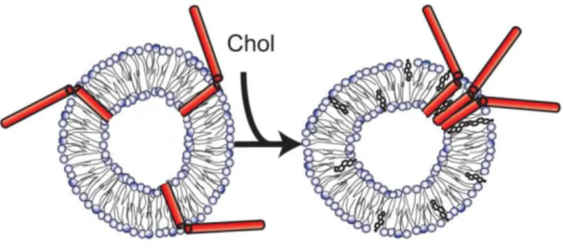
1.4.1 Role of cholesterol
In membrane model systems, the inclusion of cholesterol was shown to promote clustering of syntaxin-1 even in lipid bilayers that do not favour the formation of liquid ordered domains (Murray and Tamm, 2009) (figure 10).
Figure 10. Model for cholesterol-induced cluster formation of syntaxin. Adapted from (Murray and Tamm,
2009).
In PC12 cells, t-SNARE clusters also seem to depend on the presence of cholesterol-enriched rafts. When cholesterol is removed from the plasma membrane, clusters disintegrate and exocytosis decreases, suggesting that cholesterol guarantees the high concentration of SNAREs needed to efficient fusion to occur (Lang et al., 2001).
On the other hand, cholesterol appears to have an effect in SNAP-25 targeting to the plasma membrane. Due to the presence of several palmitates in SNAP-25 structure, cholesterol may modulate its activity through a differential insertion of the palmitate groups in the membrane (Rickman et al., 2010).
1.4.2 Relationship between PI(4,5)P2 location and exocytic sites
Because syntaxin-1 has a juxtamembrane region with basic aminoacid residues, in living cells it interacts with acidic phospholipids as PA and several PIs, including PI(4,5)P2. Protein-lipid interactions were proposed to structurally and electrostatically
contribute to reduce the energy barrier for fusion in the specific sites of exocytosis (Lam et al., 2008).
17 In the plasma membrane of PC12 cells, PI(4,5)P2 molecules seem to be organized
in microdomains and these domains partially co-localize with syntaxin-1 clusters and docked large dense core vesicles (LDCVs) (Aoyagi et al., 2005), as shown in figure 11.
Figure 11. Part of the docked LDCVs is co-localized with PI(4,5)P2 microdomains and syntaxin clusters.
Immunostaining for A) chromogranin B present in LDCVs of PC12 cells, B) PI(4,5)P2 and C) syntaxin-1; D)
Overlap image. Bar, 2 µm. Adapted from (Aoyagi et al., 2005).
The biophysical properties of PI(4,5)P2 do not favour the membrane negative
curvature that is necessary for fusion. However, it is highly concentrated in the vesicle docking sites, at around 6 mol%, and it is essential for the priming step of Ca2+ -dependent fusion, in which synaptic vesicles are converted to a ready-releasable state. Moreover, PI(4,5)P2 was observed to inhibit SNARE-dependent liposome fusion, but
this inhibition was abolished with the inclusion a priming protein like the Ca2+ -dependent activator protein for secretion (CAPS). This suggests that PI(4,5)P2
regulates fusion by directly inhibiting it and, at the same time, recruiting the priming proteins that facilitate the SNARE-dependent synaptic transmission upon Ca2+ stimulation (James et al., 2008).
18
1.5. Main objectives and work organization
The main focus of this project was to elucidate the role of synaptic protein palmitoylation and different calcium concentrations in the distribution of PI(4,5)P2 in the
plasma membrane. To address these questions, two distinct and complementary strategies were implemented.
In the first part of this work, in vivo experiments were carried out with neuron-like (PC12) and non-neuronal (HEK293) cells. Making use of PH-domains specificity to PI(4,5)P2 molecules, FRET microscopy experiments between CFP (donor) and
PH-YFP (acceptor) were performed to evaluate PI(4,5)P2’s distribution in the plasma
membrane in the absence and in the presence of the palmitoylation inhibitor 2BP. The efficiency of this inhibitor at the cellular level was assessed by following the alterations in SNAP-25 localization at the plasma membrane, using confocal fluorescence microscopy.
The second set of experiments aimed to understand what is the basic effect of physiological concentrations of calcium ions in the distribution of PI(4,5)P2 in fluid lipid
membranes. A self-quenching fluorescence study of the fluorescent analogue TopFluor-PI(4,5)P2 was first performed in POPC LUVs to characterize the behaviour of
this fluorescent lipid probe. Then, POPC GUVs incorporating the fluorescent phosphoinositide were imaged by confocal microscopy in the presence of different calcium concentrations to assess its influence on PI(4,5)P2 lateral distribution.
In addition, a GUV immobilization method based on the interaction of biotinylated lipids with an avidin coated surface was also optimized. This was necessary to allow the acquisition of confocal data with higher quality and the future use of single molecule methods.
19
2. Materials and Methods
2.1. Materials and chemical reagents
1-Palmitoyl-2-oleoyl-sn-glycero-3-phosphocholine (POPC), 1,2-di-oleoyl-sn-glycero-3-phosphocholine (DOPC), 1,2-dipalmitoyl-sn-glycero-3-phosphocholine (DPPC), N-palmitoyl-D-erythro-sphingosylphosphorylcholine (PSM), 1,2-dioleoyl-sn-glycero-3-phosphoethanolamine-N-(cap biotinyl) (DOPE-Cap-biotin),
1,2-dipalmitoyl-sn-glycero-3-phosphoethanolamine-N-(biotinyl) (DPPE-biotin), 1,2-dipalmitoyl-sn-glycero-3-phosphoethanolamine-N-(cap biotinyl) (DPPE-Cap-biotin), 1,2-dioleoyl-sn-glycero-3-phosphoethanolamine-N-(lissamine rhodamine B sulfonyl) (Rho-DOPE, ε(559 nm, chloroform) = 95x103
M-1cm-1, (Pinto et al., 2008)), 1,2-dipalmitoyl-sn-glycero-3-phosphoethanolamine-N-(7-nitro-2-1,3-benzoxa-diazol-4-yl) (NBD-DPPE, ε(458 nm, chloroform) = 21x103
M-1cm-1, (de Almeida et al., 2005)) and 1-oleoyl-2-{6-[4-(dipyrrometheneboron difluoride) butanoyl] amino} hexanoyl-sn-glycero-3-phosphoinositol-4,5-bisphosphate (TopFluor-PI(4,5)P2, ε(495 nm, methanol) = 80x103
M-1cm-1 (Invitrogen)) were obtained from Avanti Polar Lipids (Alabaster, AL). Avidin from egg white, extrAvidin-FITC conjugate (ε(494 nm) = 84x103 M-1cm-1 (Invitrogen))
and 2-bromopalmitate (2BP) were from Sigma Chemical Co. (St. Louis, MO). Trans-parinaric acid (t-PnA, ε(299 nm, ethanol) = 89x103 M-1cm-1 (de Almeida et al., 2005)),
the membrane probe Alexa Fluor 594-wheat germ agglutinin (λex= 590 nm; λem= 617
nm) and cell culture reagents were obtained from Invitrogen (Breda, The Netherlands). All organic solvents were UVASOL grade from Merck (Darmstadt, Germany).
Lipid stock solutions were all prepared in chloroform, with the exception of TopFluor-PI(4,5)P2 and t-PnA that were prepared in chloroform/methanol (2:1) and
20
2.2. Cell lines
PC12 cells were used as neuron models. As a non-neuronal control, HEK293 (Human Embryonic Kidney 293) cells were utilized, since they do not present neuron-like properties such as all the machinery of synapse.
2.3. DNA constructs
All pcDNA3 plasmid constructs containing PH domains were obtained by material transfer agreement from the laboratory of Dr. K. Jalink (Netherlands Cancer Institute). CMV-SNAP-25-GFP was a kind gift of Dr. R. Jahn (Max Planck for Biophysical Chemistry). pEGFP-N1 vector was from Clontech (Mountain View, CA). Venus-N1 (Nagai et al., 2002) and monomeric Cerulean C1 (Rizzo et al., 2004) were PCR amplified and inserted into the ptagRFP-N vector (Evrogen, Moscow) via AgeI/NotI, and NheI/NotI respectively. CMV-Cerulean-Venus construct (C11V) was prepared by using a primer for inserting downstream of Cerulean a EcoRI site, and cloning Cerulean into CMV-Venus via NheI/EcoRI.
2.4. Biochemical methods
2.4.1 Cell culture and maintenance
PC12 and HEK293 cells were grown in adherence in flasks coated with poly-L-lysine (Sigma Chemical Co., St. Louis, MO) and maintained in Dulbecco’s modified
Eagle’s medium (DMEM) with 10% fetal calf serum (FBS) and 1% penicillin- streptomycin (Sigma Chemical Co., St. Louis, MO), at 37 ºC in an humidified
21
2.4.2 DNA amplification and purification
To transform DH5α bacteria, 40 µL of competent cells were first incubated with ~0.5 µg of DNA, on ice for 30 minutes. Heat-shock (45 seconds at 42 ºC) was performed, followed by further incubation on ice during 1 minute. Bacteria were then grown in antibiotic-free LB medium for 1h at 37 ºC at 250 rpm, allowing them to express antibiotic resistance. Subsequently, cells were plated into LB-agar supplemented with 100 µg/mL of the proper antibiotic, and left at 37 ºC overnight. A colony of transformed bacteria was grown in LB medium supplemented with the appropriate antibiotic at 37 ºC (250 rpm) overnight and used to purify the DNA.
Purification was carried out using the ZyppyTM Plasmid Miniprep Kit from Zymo Research (Irvine, CA). DNA concentration was determined by absorbance measurements at 260 nm (Ԑ=0.02 µg/mL) in a Shimadzu UVPC-3100 spectrophotometer (Shimadzu, Kyoto, Japan). 1x1 cm quartz cuvettes from Hellma Analytics (Müllheim, Germany) were used.
2.4.3 Transfection using cationic liposomes
Cells were seeded in eight-well µ-Slides from Ibidi (Munich, Germany) coated with poly-L-lysine at 30 000 cells per well to achieve around 80% of confluence. Transfection was performed using Lipofectamine 2000 (Invitrogen, Carlsbad, CA) for 5h at 0.5 µg plasmid DNA per well. Following transfection, cells were incubated in DMEM without phenol-red for 2-5 days.
2.4.4 Differentiation of PC12 cells
After transfection, PC12 cells were differentiated replacing the growth medium by NGF 100 ng/mL in DMEM with 2% FBS. Cells were exposed to NGF during at least 3 days and the growth medium supplemented with NGF was renewed each 2 days.
22
2.4.5 Membrane labelling
To label the plasma membrane, Alexa Fluor 594-wheat germ agglutinin (Invitrogen, Carlsbad, CA) was used at a concentration of 5 μg/mL (in PBS). Before measurements, cells were incubated with the probe at 37 ºC during 10 min and then washed with PBS.
2.4.6 Palmitoylation inhibition
Inhibition of palmitoylation was performed 24h after transfection using the palmitate analogue 2-bromopalmitate (2BP). Cells were exposed to a concentration of 50 µM (or 150 μM before FRET experiments) of 2BP and the time of exposure depended on the experiment.
2.5. FRET microscopy in living cells
2.5.1 FRET fundamental concepts
Förster Resonance Energy Transfer (FRET) occurs when a fluorophore in the excited state (donor molecule) displays non-radiative transfer of energy to a molecule in the ground state (the acceptor), through a long range dipole-dipole coupling mechanism (Sun et al., 2011). For FRET to occur, there are three essential conditions that must be achieved: i) donor and acceptor have to be in proximity (1-10 nm); ii) there must be spectral overlap between the spectra of donor emission and acceptor absorption, as shown in figure 12; iii) the absorption transition dipole moment of the acceptor cannot be perpendicular to the emission transition moment of the donor (Pietraszewska-Bogiel and Gadella, 2010).
23
Figure 12. Required conditions for occurrence of FRET. Adapted from (Vogel et al., 2006).
The FRET efficiency ( ) is the fraction of donor molecules that transfer energy to acceptor molecules and, for transfer to a single acceptor, is described by the following equation:
Eq. 1
where is the distance between donor and acceptor molecules and is the distance at which FRET efficiency is 50% (named Förster distance). The value of is characteristic of each donor-acceptor pair. Note that FRET efficiency is dependent on the inverse of the sixth power of the distance that separates the two fluorophores.
As pointed out before, in addition to fluorophores proximity, a spectral overlap and a good orientation of the dipole moments are also required:
Eq. 2
where is a constant (8,79x10⁻¹¹ M cm nm2). The Förster radius depends on the refraction index ( ), the quantum yield of the donor ( ), the relative orientation of the donor emission and acceptor absorption dipoles ( ), and the degree of overlap between the donor emission and acceptor absorption spectra ( ).