UNIVERSIDADE DA BEIRA INTERIOR
Ciências da SaúdePurification of supercoiled HPV-16 E6/E7 plasmid
using a modified monolithic support
Lúcia Filipa Alves Amorim
Dissertação para a obtenção do Grau de Mestre em
Ciências Biomédicas
(2º Ciclo de estudos)
Orientadora: Profª. Doutora Ângela Sousa
Coorientadora: Profª. Doutora Carla Cruz
Acknowledgments
Firstly, I would like to thank to my supervisors Doctor Ângela Sousa and Doctor Carla Cruz, for all their guidance, advice, availability throughout this year, and continuous help in the development of this work. It was a privilege to work and learn with them.
I would also like to acknowledge to University of Beira Interior, in particular the Health Sciences Research Centre, where all the work was developed.
I am also grateful to all my colleagues in the Biotechnology and Biomolecular Sciences group for all the help, advice and support.
Thanks to my friends, for all the laughter and pranks, that always made a difficult day easier, for all the coffees and the patience.
Finally, I am deeply thankful to my parents, for everything they taught me, for all the sacrifices they have made, for all the support, the advices, for believing in me and for their love.
Resumo
Cerca de 15% dos cancros em humanos são causados por vírus. Por exemplo, o Vírus do Papiloma Humano encontra-se associado a mais de 99% dos casos de cancro do colo do útero. As vacinas preventivas contra o Vírus do Papiloma Humano existentes no mercado apenas induzem imunidade mediada por anticorpos e são completamente ineficazes na presença de infeção. Deste modo, os problemas associados a infecções pelo Vírus do Papiloma Humano e progressões tumorais continuam a aumentar. A necessidade de atenuar as lesões associadas ao Vírus do Papiloma Humano levou ao desenvolvimento de vacinas de DNA.
As vacinas de DNA surgiram como uma estratégia versátil para induzir respostas imunes, quer celulares, quer humorais. Para além disso, o DNA plasmídico (pDNA) surge como um transportador promissor para entrega de genes, uma vez que é produzido de forma simples, com elevado grau de pureza e baixo custo, e apresenta capacidade de transfectar células eucarióticas com níveis de expressão satisfatórios.
Assim sendo, nos últimos anos, os níveis de plasmídeo necessário para aplicações farmacêuticas levaram ao desenvolvimento de novos suportes cromatográficos, com elevada capacidade de ligação e seletividade pela isoforma superenrolada do plasmídeo. As matrizes monolíticas são consideradas ideais para purificar biomoléculas como o pDNA. Estes suportes apresentam estruturas tridimensionais de poros interconectados que permitem a transferência de massa por convecção e fornecem elevadas capacidades de ligação, o que torna estas matrizes inovadoras. Por outro lado, a cromatografia de afinidade com aminoácidos revelou-se uma abordagem promissora devido ao bioreconhecimento seletivo da isoforma superenrolada do pDNA, uma vez que esta estratégia se baseia na ocorrência natural de várias interações entre proteínas e ácidos nucleicos, em organismos biológicos, que maioritariamente envolvem aminoácidos básicos como a L-histidina.
As interações entre aminoácidos imobilizados e as diferentes isoformas de plasmideos podem ser estudadas por ressonância de plasma de superfície (RPS). O conhecimento prévio da afinidade aminoácido/pDNA pode posteriormente ser explorado na purificação através da cromatografia de afinidade.
Desta forma, um dos objetivos deste trabalho é utilizar a técnica de RPS para selecionar o aminoácido L-histidina ou um dos seus derivados, benzil-L-histidina e metil-L-histidina, que tem maior afinidade com o pDNA, para posteriormente imobilizar o ligando mais promissor numa matriz monolítica.
Inicialmente foram realizadas várias experiências de RPS utilizando três plasmídeos de tamanhos diferentes (6,05, 8,70 e 14 quilo pares de bases) e com as isoformas previamente separadas (circular aberta, superenrolada e linear). Os resultados revelaram que, no geral, a afinidade dos plasmídeos para o ligando de L-histidina e seus derivados, era elevada (KD >10-8
3,34 x 10⁻¹⁰ ± 0,0209 M. Desta forma, a L-histidina foi o aminoácido selecionado para ser
imobilizado na matriz monolítica.
Após preparação do suporte monolítico de L-histidina, foram realizados vários estudos cromatográficos com amostras mencionadas anteriormente. No geral, a isoforma superenrolada promoveu interações fortes com o monolito de L-histidina e a separação das isoformas foi conseguida. A separação das isoformas dos plasmídeos não se alterou com variação da taxa de fluxo. Estudos de capacidade de ligação dinâmica do monolito L-histidina revelaram que a sua capacidade de ligação máxima foi 11,03 mg/mL, com uma taxa de fluxo de 0,5 mL/min e uma solução de plasmídeo de 0,05 mg/mL. Estes resultados foram comparados com os valores de capacidade do suporte convencional de L-histidina-agarose, de um monolito não modificado e um monolito modificado com outro aminoácido, a aginina. A maior diferença nos resultados foi verificada com a matriz convencional de L-histidina-agarose, em que a capacidade do monolito de L-histidina foi cerca de vinte e nove vezes maior do que a capacidade da matriz convencional, usando as mesmas condições de saturação. Em relação ao monolito não modificado e à matriz monolítica modificada com o aminoácido arginina, os valores de capacidade do monolito de L-histidina revelaram ser ligeiramente superiores, em ambos os casos.
De um modo geral, a cromatografia de afinidade pode beneficiar das análises de afinidade exploradas por RPS. A combinação do ligando de L-histidina com o suporte monolítico permitiu a separação da isoforma superenrolada do plasmídeo HPV-16 E6/E7.
Palavras-chave
Capacidade dinâmica de ligação, DNA plasmídico superenrolado, Ligandos de afinidade, Monolito de L-histidina, Ressonância de plasma de superficíe, Vírus do Papiloma Humano.
Abstract
About 15% of human cancers are caused by viruses. For instance, the particular case of the high-risk human papillomavirus (HPV) is associated with more than 99% of cervical carcinomas. The preventive vaccines for HPV infection available in the market only induce the antibody immunity and are completely ineffective when the infection is already present. Therefore, the problematic associated to HPV infections and tumor progressions continue to be unsolved. Thus, the urge to attenuate the HPV associated lesions led to the development of DNA vaccines.
DNA vaccines emerged as a versatile strategy to induce both humoural and cellular immune immune responses. In addition, plasmid DNA (pDNA) arose as a promising vehicle for gene delivery, due to its simple manufacturing process with high purity degree and low cost, as well as its ability to transfect eukaryotic cells with satisfactory expression levels.
Therefore, in the last years, the growing demand of pharmaceutical-grade pDNA fostered the development of new chromatographic supports, allowing high capacity and selectivity by the supercoiled (sc) pDNA. The innovative monolithic matrices are considered advantageous supports to purify large biomolecules, such as pDNA, due to their tridimensional characteristics of interconnected pores, which allows good mass transfer properties and binding capacity. Amino acid-affinity chromatography has revealed to be a promise approach that selectively recognizes the sc pDNA, since this strategy is based on natural occurrence of multiple interactions between proteins and nucleic acids in biological organisms, which mainly involve basic amino acids such as L-histidine.
Surface Plasmon Resonance (SPR) Biosensor can be used to exploit the interactions between immobilized amino acids and different plasmid topologies to provide further structural information for affinity chromatography purification.
Thus, the aim of this work was to perform a screening of L-histidine amino acid and their derivatives, Im-benzyl-L-histidine and L-methyl-L-histidine, employing the SPR technique in order to modify a monolithic support with the selected ligand, to purify pDNA. Several experiments were performed with three plasmids of different sizes (6.05, 8.70 and 14 kilo base pairs) and different isoforms (open circular, sc and linear), separately. The results revealed that the overall affinity of plasmids to L-histidine ligand and their derivatives was high (KD >10-8 M) and the highest affinity was found for HPV-16 E6/E7/L-histidine interaction,
3.34 x 10⁻¹⁰ ± 0.0209 M. Therefore, L-histidine was selected for immobilization on a
monolithic matrix.
After preparation of the histidine monolithic support, chromatographic studies were also accomplished with the aforementioned samples. In general, the sc isoform developed strong interactions with the support and the separation of plasmid isoforms was achieved by decreasing ammonium sulfate concentration. The separation of plasmid isoforms remained
unchanged by flow rate variations. The breakthrough experiments of L-histidine monolith revealed satisfactory dynamic binding capacity when compared to other matrices.
Overall, affinity chromatography can benefit from affinity analysis experiments provided by SPR biosensor, and the combination of L-histidine ligand with the monolithic support can be a promising strategy to purify the sc pDNA with the desirable purity degree for pharmaceutical applications, such as the DNA vaccines directed against the HPVs.
Keywords
Affinity ligands, Dynamic binding capacity, Human papillomavirus, L-Histidine monolith, Supercoiled plasmid DNA, Surface Plasmon Resonance.
Table of Contents
Chapter I- Introduction 1
1.Gene therapy and DNA vaccines 3 1.1.Delivery systems 4 1.1.1.Viral and non-viral delivery systems: advantages and limitations 6
1.1.1.1.Viral delivery systems 6
1.1.1.2.Non-viral delivery systems 7
2.Therapeutic applications 9
2.1.Human papillomavirus 10
3.Plasmid DNA technology 12
3.1.Plasmid DNA biosynthesis 12
3.2.Downstream processing 12
3.2.1.Primary isolation 13
3.3.Plasmid DNA purification 13
3.3.1.Chromatographic techniques 14 3.3.1.1.Size exclusion chromatography 14 3.3.1.2.Ion exchange chromatography 14 3.3.1.3.Hydrophobic interaction chromatography 15 3.3.1.4.Affinity chromatography 15
3.3.2.Ligands selection 17
3.3.2.1.L-Histidine and derivatives 17 3.3.2.2.Surface Plasmon Resonance 18
3.3.3.Stationary phase 19
3.3.3.1.Monoliths 19
4.Aims 20
Chapter II – Materials and methods 23
2.1.Materials 25 2.1.1.Reagents 25 2.1.2.Plasmids 25 2.1.3.Instrumentation 25 2.1.4.Specifications 26 2.2.Methods 26
2.2.1.Plasmid amplification by bacterial production 26 2.2.2.Alkaline lysis and pre-purification of pDNA samples with NZYTech Kit 27 2.2.3.Plasmid isoforms preparation 27
2.2.3.1.Supercoiled isoform 27
2.2.3.3.Open circular isoform 27 2.2.4.Agarose gel electrophoresis 28
2.2.5.SPR studies 28
2.2.5.1.Amino acids immobilization 28
2.2.5.2. Affinity analysis 29
2.2.6.1H NMR experiments 29
2.2.7.Cromatographic studies 29
2.2.7.1. Amino acid immobilization 29
2.2.7.2.Linear gradients 29
2.2.7.3.Stepwise gradients 30
2.2.7.4.Dynamic binding capacity 30
2.2.7.5.Monolith regeneration 31
Chapter III – Results and discussion 33
3.Ligands screening 35
3.1. Amino acid immobilization 35
3.2.SPR binding experiments 35
3.2.1.Running buffer selection for affinity experiments 35
3.2.2.Affinity data analysis 37
4.Cromatographic experiments 40
4.1.L-Histidine immobilization 40
4.2.Evaluation of elution conditions on pDNA retention 41 4.2.1.Elution buffer composition 41
4.2.2.Ionic strength influence 42
4.2.3.pH influence 42
4.3.Chromatographic profile of different plasmid isoforms 43
4.3.1.pVAX1-LacZ (6.05 kbp) 44
4.3.1.1.pVAX1-LacZ linear isoform 44 4.3.1.2.pVAX1-LacZ open circular isoform 44 4.3.1.3.pVAX1-LacZ supercoiled isoform 45
4.3.2.HPV-16 E6/E7 (8.70 kbp) 46
4.3.2.1. HPV-16 E6/E7 linear isoform 46 4.3.2.2. HPV-16 E6/E7 open circular isoform 47 4.3.2.3. HPV-16 E6/E7 supercoiled isoform 48 4.3.3.pcDNA3-based plasmid (14 kbp) 48 4.3.3.1.pcDNA3-based plasmid linear isoform 48 4.3.3.2.pcDNA3-based plasmid open circular isoform 49 4.3.3.3.pcDNA3-based plasmid supercoiled isoform 50 4.4.Plasmid isoforms separation 51
4.4.3.pcDNA3-based plasmid 52 4.5.Effect of flow rates on plasmid isoforms separation 54
4.6.Dynamic binding capacity 56
Chapter IV – Conclusions and future perspectives 61
Chapter V – Bibliography 67
List of Figures
Chapter I- Introduction
Figure 1. Gene therapy and DNA vaccines. 3 Figure 2. Barriers to delivery systems. 5 Figure 3. Distribution of delivery systems used in clinical trials. 6 Figure 4. Indications addressed by gene therapy clinical trials. 9 Figure 5. HPV genome: structure and organization. 10 Figure 6. Graphical representation of the main goals in the present work. 20
Chapter III- Results and discussion
Figure 7. Examples of sensorgrams in which no signal was detected. 36 Figure 8. Examples of sensorgrams and equilibrium binding analyses. 38 Figure 9. Decreasing linear gradients performed with non-immobilized epoxy
disk and L-histidine modified monolith. 40 Figure 10. Decreasing linear gradient performed with HEPES buffer. 41 Figure 11. Increasing linear gradient performed with NaCl. 42 Figure 12. Decreasing linear gradient performed with elution buffer at pH
6.0. 43
Figure 13. Decreasing linear gradient performed with pVAX1-LacZ linear
isoform sample. 44
Figure 14. Decreasing linear gradient performed with pVAX1-LacZ open
circular isoform sample. 45
Figure 15. Decreasing linear gradient performed with pVAX1-LacZ supercoiled
isoform sample. 46
Figure 16. Decreasing linear gradient performed with HPV-16 E6/E7 linear
isoform sample. 47
Figure 17. Decreasing linear gradient performed with HPV-16 E6/E7 open
circular isoform sample. 47
Figure 18. Decreasing linear gradient performed with HPV-16 E6/E7
supercoiled isoform sample. 48
Figure 19. Decreasing linear gradient performed with pcDNA3-based plasmid
linear isoform sample. 49
Figure 20. Decreasing linear gradient performed with pcDNA3-based plasmid
open circular isoform sample. 49
Figure 21. Decreasing linear gradient performed with pcDNA3-based plasmid
supercoiled isoform sample. 50
Figure 22. Stepwise gradient used in pVAX1-LacZ plasmid isoforms separation
Figure 23. Stepwise gradient used in HPV-16 E6/E7 plasmid isoforms
separation (2.94 M and 0M ammonium sulfate). 52 Figure 24. Stepwise gradient used in pcDNA3-based plasmid isoforms
separation (2.91 M and 0M ammonium sulfate). 53 Figure 25. Flow rate effect on pVAX1-LacZ isoforms separation. 55
List of Tables
Chapter I- Introduction
Table 1. Main limitations and advantages of the most widely used viral delivery systems.
7 Table 2. Regulatory agencies specifications. 12 Table 3. Affinity chromatography methodologies for nucleic acids
purification. 16
Chapter III- Results and discussion
Table 4. Equilibrium data analysis of plasmids in 10 mM of HEPES pH 7.4. 39 Table 5. Dynamic binding capacity results of L-histidine monolith at 0.5 and 1
List of Acronyms
BCA Bicinchoninic acid
ºC Celsius degree
CDI CarbonylDiImidazole
DBC Dynamic binding capacity
D2H Deuterated water
cm Centimeter
DNA Desoxirribonucleic acid
E. coli Escherichia coli
EDC 1-ethyl-3-(3- dimethylaminopropyl) carbodiimide
EDTA Ethylene-diamine tetraacetic acid
EU Endotoxin Units
g gram
gDNA Genomic DNA
HBS-EP HEPES, NaCl, P20 surfactant, EDTA
HIV Human Immunodeficiency Virus
1H NMR Proton Nuclear Magnetic Resonance
HPV Human Pappilomavirus
kbp kilo base pairs
KH2PO4 Monopotassium phosphate
K2HPO4 Dipotassium phosphate
KD Dissociation constant
L liter
LAL Limulus amebocyte lysate
LB Luria-Broth ln Linear M Molar mM milimolar MHz Megahertz min minutes mL milliliter
mRNA Messenger RNA
NaCl Sodium chloride
NaOH Sodium hydroxide
NHS N-hydroxysuccinimide
(NH4)2SO4 Ammonium sulfate
oc Open circular
OD600 Optical density at 600 nm
PCR Polymerase chain reaction
pDNA Plasmid DNA
RNA Ribonucleic acid
RU Ressonance Units
s Seconds
sc Supercoiled
SDS Sodium dodecylsulfate
siRNA Small interfering RNA
SPR Surface Plasmon Resonace
TAE Tris, acetic acid, EDTA
Tris Tris(hydroxymethyl) aminomethane
URR Upstream regulatory region
UV Ultraviolet
V volt
1. Gene therapy and DNA vaccines
Gene therapy may be defined as the cure, treatment or prevention of human diseases using nucleic acids. This can be achieved by gene addition, gene correction and gene knockdown, depending on the type of disease, being the gene addition the most attempted strategy, since the other aforementioned strategies are technically more difficult (Kauffman et al., 2013). Gene therapy can be categorized into germ line and somatic gene therapy. In the somatic gene therapy approach, the changes resulting from insertion of genetic material in some target cells will not pass along to the next generation, whereas in germ line gene therapy approach, the therapeutic or modified gene will be passed onto the next generation, but regulatory agencies do not allow such approach due to technical and ethical reasons (Haritha
et al, 2012).
Somatic gene therapy can be performed using two different strategies to vector administration, in vivo and ex vivo (Kauffman et al., 2013; Haritha et al, 2012). The in vitro strategy involves the direct vector delivery into the target cells of the patient, as shown in figure 1. The therapy achievement relies on efficient uptake of the therapeutic gene by the target cells, and its prior uptake by nucleus, intracellular degradation and its expression capability. The ex vivo strategy involves the target cells isolation from the patient, genetic modification outside the body and autologous transplant of the modified cells into the patient (figure1), being less likely to trigger immune responses (Naldini, 2011).
(A)
(B)
Figure 1. Gene transfer strategies. (A) in vivo strategy: Direct vector delivery to patient cells. (B)
ex vivo strategy: Isolation, genetic modification and transplantation of modified cells into the patient (Kauffman et al., 2013).
On the other hand, DNA vaccines are based on the conventional vaccines principle, since they are used to produce immunological responses against infectious agents. Nevertheless, conventional protein based vaccines only induce an humoral response from the immune system and vaccines derived from live attenuated organisms induce cellular immune response, while DNA vaccines are able to stimulate both antibodies and cell-mediated components (Liu, 2011).
DNA vaccines take advantage of genetic information that is delivered by a system able to induce an immune response against a given antigen. Upon inoculation, the individual shall produce a strong and enduring immune response against the encoded protein antigen, associated to the pathology (Liu, 2010). Therefore, DNA vaccines may be advantageous in terms of safety when compared with conventional vaccines, such as conventional live virus vaccines, since can reverse to a pathogenic form. Besides, DNA vaccines can be manufactured in a relatively cost-effective manner and easily stored. Other advantages are the versatility and rapid design of DNA vaccines, since multiple antigenic genes can be given in one formulation (Liu, 2010; Saade et al., 2012).
1.1. Delivery systems
Several delivery systems have been developed over the years to transport the therapeutic gene to the target cells. Therapeutic genes can be categorized into gene inhibitors or gene inductors.
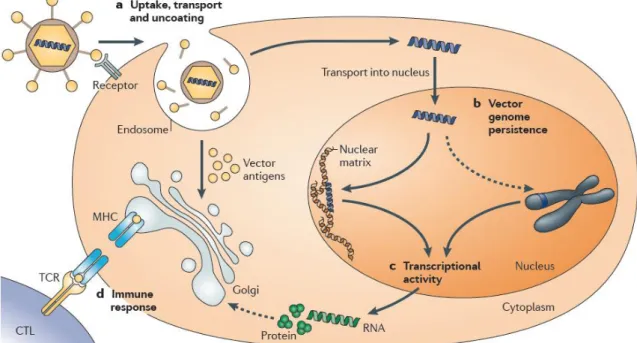
The gene inhibitors category include: oligonucleotides, ribozymes, DNAzymes, aptamers and small interfering RNAs (siRNAs). Oligonucleotides are short single-stranded segments of DNA able to selectively inhibit the expression of a single protein; ribozymes interact at the mRNA level, the RNA molecules are capable of sequence specific cleaving of mRNA; DNAzymes present strong catalytic activity and have high potential as gene suppression agents; aptamers are small single-stranded or double-stranded nucleic acid segments that can directly interact with proteins; siRNAs are short double-stranded RNA segments which nucleotide bases are complementary to the mRNA sequence of the protein whose transcription is blocked. Gene inductors, such as plasmids containing transgenes, are high molecular weight, double-stranded DNA constructs, with transgenes that encode specific proteins (Patil et al., 2005). Although some limitations and problems have been solved or minimized, some barriers to deliver the naked therapeutic nucleic acids still remain unsolved (Kay, 2011). Those barriers are the vector uptake, transport and uncoating, vector genome persistence, sustained transcriptional expression and the host immune response, as it is represented in figure 2.
After vector administration, several parameters influence successful transgene expression of therapeutic levels, namely the vascular supply, endothelial barriers, vector size and interactions between host cell receptors and the vector ligand. The cell membrane, endosomal escape and the nuclear membrane are the three barriers that non-viral delivery systems have to overcome until to reach the nucleus, while virus have efficient mechanisms to enter the cell and localize the nucleus (Thomas et al., 2003; Kay, 2011).
The vector genome persistence depends whether it integrates into host cellular chromatin or predominantly persists in the cell nucleus as extrachromosomal episomes. Non-integrating vectors can mediate persistent transgene expression in non-proliferating cells, but integrating vectors are more widely used when the genetic alteration should remain stable in dividing cells (Thomas et al., 2003), although they present the risk of insertional mutagenesis (Biasco
et al., 2012).
Sustained transcriptional expression refers to the fact that the transgene expression may be desired during lifetime or limited periods, depending on the specific target disease (Thomas
et al., 2003; Kay, 2011).
The transgene product or the vector may be recognized by the host immune system as foreigner and therefore, an immune response can be triggered, limiting the transgene product expression (Thomas et al., 2003; Kay, 2011).
These aforementioned limitations illustrate the importance of correct selection of a suitable delivery system to carry the therapeutic nucleic acid for a specific purpose. Nevertheless, the delivery systems can be categorized under viral delivery systems and non-viral delivery systems and each category present advantages and limitations.
Figure 2. Barriers to delivery systems: a) vector uptake, transport and uncoating, vectors are internalized by
various processes and they must reach escape the endosome to reach the nucleus without being degraded. b) Vector genome persistence, the DNA can exist as an episomal molecule or be integrated into the host chromosome. c) Sustained transcriptional expression that should match the time required to treat the target disease. d) The host immune response can limit the gene therapy success (Kay, 2011).
1.1.1. Viral and non-viral delivery systems: advantages and limitations
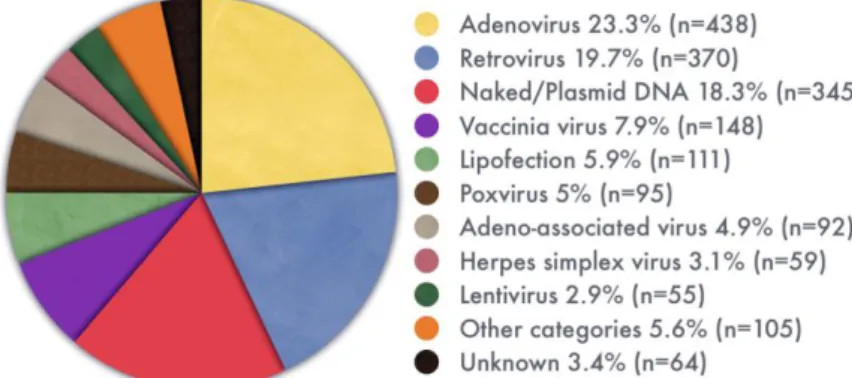
The most widely used vectors are the viral delivery systems, adenovirus and retrovirus, followed by the non-viral delivery system, plasmid DNA (figure 3) that has received significant attention over the last years, becoming increasingly common (Ginn et al., 2013).
The plasmid DNA (pDNA) emerges as a promising vehicle for gene delivery because it is a double-stranded biomolecule with high molecular weight, easily constructed with any trangenes of interest, obtained in large scale under satisfactory purity degree by a simple manufacturing process with low cost, and able to transfect eukaryotic cells with satisfactory expression levels. A more detailed description of pDNA technology is presented in section 3. The obstacle of naked pDNA clinical application is the efficiency to cross the extra- and intracellular barriers, but this problem is being addressed with delivery techniques, addition of adjuvants, and various prime-boost strategies (Saade and Petrovsky, 2012; Coban et al., 2008).
1.1.1.1. Viral delivery systems
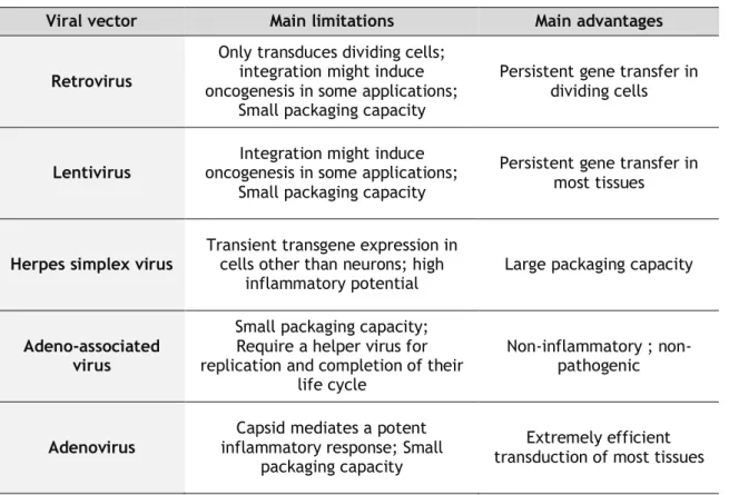
The virus mediated gene transfer is highly efficient because gain easily access to host cells and make use of their machinery to facilitate the replication. Viral delivery systems such as retrovirus, lentivirus, adenovirus, adeno-associated virus, and herpes simplex virus have been widely used in clinical trials (Walter and Stein, 2000) and their main advantages and limitations are summarized in table 1.
Table 1. Main limitations and advantages of the most widely used viral delivery systems (adapted from
Thomas et al., 2003).
Other limitations associated with viral delivery systems include hampered production, repeated administrations due to host inflammatory responses and potential insertional mutagenesis of some viral delivery systems (Haritha et al., 2012).
1.1.1.2. Non-viral delivery systems
The use of non-viral delivery systems became popular to overcome the bottlenecks associated with the use of viral delivery. Non-viral delivery consists on synthetic or natural compounds or physical forces to deliver the therapeutic nucleic acid into the cell. They are considered less toxic and immunogenic, since they do not contain viral contaminants. The non-viral gene delivery offers other advantages, once there is no size limit on the amount of genes that they can deliver. They are also easily produced and allow repeated administration, without triggering inflammatory responses (Al-Dosari and Gao, 2009).
The major drawbacks associated with non-viral delivery systems, such as the low efficacy of cell invasion when compared with viral vectors and the short-lived gene expression, are being addressed with constant developments in the field (Kamimura et al., 2011).
Viral vector Main limitations Main advantages
Retrovirus
Only transduces dividing cells; integration might induce oncogenesis in some applications;
Small packaging capacity
Persistent gene transfer in dividing cells
Lentivirus
Integration might induce oncogenesis in some applications;
Small packaging capacity
Persistent gene transfer in most tissues
Herpes simplex virus Transient transgene expression in cells other than neurons; high
inflammatory potential Large packaging capacity
Adeno-associated virus
Small packaging capacity; Require a helper virus for replication and completion of their
life cycle
Non-inflammatory ; non-pathogenic
Adenovirus
Capsid mediates a potent inflammatory response; Small
packaging capacity
Extremely efficient transduction of most tissues
The viral gene delivery methods can be divided into physical and chemical-based non-viral delivery methods.
The use of physical forces to improve the gene transfer can be performed by jet injection, hydrodynamic gene transfer, gene gun, electroporation and sonoporation. Jet injection is accomplished through a high-speed stream of therapeutic nucleic acid solution driven by a pressurized gas to penetrate the skin and the underlying tissues. This injection methodology does not induce tissue damage or significant inflammatory reactions at jet-injection sites (Ren et al., 2002). The hydrodynamic gene transfer is based on the injection of a large therapeutic nucleic acid volume in short period of time, which leads to a reversible permeability change in the endothelial lining and the generation of transient pores. This invasive procedure has been modified in order to achieve its clinical applicability (Fabre et
al., 2008). The gene gun delivery method depends on the impact of heavy metal particles
coated with the therapeutic nucleic acid on target tissues (Uchida et al., 2002). These particles are accelerated by highly pressurized inert gas and can easily cross the cell and nuclear membranes, releasing the nucleic acid adsorbed on their surface into the nucleus. Electroporation uses an electric field to alter the cell permeability. The therapeutic nucleic acid is injected to the target tissues and then electric pulses are applied (Marti et al., 2004). On the other hand, sonoporation uses ultrasound waves to create plasma membrane defects by acoustic cavitation (Nomikou et al., 2013).
Examples of chemical-based delivery systems are cationic lipids and cationic polymers. These vectors form condensed complexes with therapeutic nucleic acids, negatively charged, through electrostatic interactions. The complexes facilitate cell uptake and intracellular delivery and protect the therapeutic nucleic acid (Morille et al., 2008).
Overall, an ideal carrier has low toxicity and immunogenicity and higher efficiency. However it has not yet been achieved; despite several efforts continue to be made to develop hybrid carriers. The large array of novel carriers is in continuous investigation to improve safety and enhanced therapeutic efficacy. In these carriers, new abilities are added or certain undesirable elements are replaced (Thomas et al., 2003; Huang and Kamihira, 2013).
2. Therapeutic applications
From the beginning, gene therapy substantial progresses have been made in this field. Important milestones were achieved, as for example, China became the first country to approve a gene therapy based product for clinical use, Gendicine, an adenoviral vector for the treatment of head- and neck squamous cell carcinoma (Zhaohui, 2005). In Europe, gene therapy has also taken the next step with approval of Glybera, an adeno-associated viral vector for the treatment of familial lipoprotein lipase deficiency (Wirth and Yla-Herttuala, 2013). These achievements open perspectives of more gene-based therapies in near future. Cancer, cardiovascular disease and inherited monogenic diseases lead the ranking of diseases addressed in clinical trials by gene therapy (Ginn et al., 2013), as can be seen in figure 4.
However, due to the enormous therapeutic potential of gene therapy, several other conditions can be addressed.
A broad range of viral and non viral diseases are being addressed in DNA vaccine clinical trials. The majority of clinical trials are focused in human immunodeficiency virus (HIV) and cancers. Influenza, hepatitis B, hepatitis C, malaria and human papillomavirus (HPV) are also being exploited in clinical trials (Ferraro et al., 2011).
The preventive HPV vaccines in the market, Gardasil and Cervarix do not induce appreciable levels of cellular immune responses, and thus, the burden of HPV infections or HPV-associated lesions proceeds to increase (Ferraro et al., 2011). Therefore, strong cellular responses are required and DNA vaccines appear promising candidates to induce the desired immune response.
2.1. Human papillomavirus
About 15% of all human cancers are caused by viruses. Human papillomaviruses are responsible for at least 5% of tumors worldwide, although not sufficient cause of cervical cancer, the HPV infection represents the major etiologic factor in the neoplasia development (Tota et al., 2011). Other cancer subsets like oropharyngeal, penile, vaginal, vulvar and anal cancer are also attributed to HPV (Trottier and Burchell, 2009), which is currently one of the most common sexually transmitted infections worldwide (Dunne et al., 2007).
Human papillomavirus are small, circular and double-stranded DNA virus, members of the Papillomaviridae family. All HPV show a pronounced tropism for epithelial cells, and infections by these viruses are associated with hyperproliferative lesions of mucosa and skin (Howley and Lowry, 2007). These mucosal HPVs can be classified as low- or high-risk, according to the outcome of associated lesions. Low-risk HPVs, such as HPV6 or HPV11, cause genital warts, benign lesions, whereas high-risk HPVs, like HPV16 or HPV18, cause intraephitelial lesions with the propensity for malignant progression (Schlecht et al., 2001). High-risk HPVs are associated with greater than 99% of cervical carcinomas (Schiffman et al., 2007).
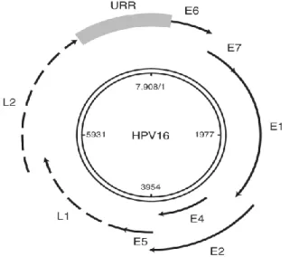
All HPV viruses share a common genome organization of about 7.9 kbp. The circular DNA can be divided into three functional regions: a non-coding regulatory region, known as the upstream regulatory region (URR), which modulates viral DNA replication and gene transcription; a region composed by HPV early genes (E1,E2, E3, E4, E5, E6,E7), which code for proteins related with viral genome transcription, replication, persistence and regulation of cell proliferation. And finally, a region that codes for HPV late genes (L1 and L2) and structural genes, which is responsible for the major and minor capsid proteins codification (as shown in figure 5) (Doorslaer and Burk, 2010).
HPV life cycle is associated with epithelial differentiation. The basal epithelial cells are initially infected by HPVs, where the viral episome is maintained extrachromosomally at low copy number (viral genome is maintained at about 100 copies per cell). A productive infection with genome amplification, capsid protein expression and virion mounting occurs in cells from the suprabasal layers. On one hand, cells above the basal layer are terminally differentiated, on the other HPV does not code for its own set of replication proteins. Thus, the HPV has to induce host cells re-entry in S-phase to take advantage of host replication machinery (Doorslaer and Burk, 2010).
The products of E6 and E7 early genes are essential in the process of HPV induced cellular immortalization and transformation (DeFilippis et al., 2003). The E7 expression, the major HPV oncoprotein, is sufficient to immortalize primary human epithelial cells at low frequency. E7 interacts with cellular regulatory protein complexes and alters or neutralizes their normal functions, leading to impaired cell cycle arrest responses and deregulation of cellular differentiation and apoptosis mechanisms. E7 interacts with retinoblastoma protein and mediates its degradation, affecting its growth-suppressive function. As consequence, transcriptional genes necessary for S-phase entry and progression are activated. The E6 oncoprotein targets the tumor suppressor p53, leading to its proteosomal degradation (Howie et al, 2009; McLaughlin-Drubin and Münger, 2009).
3. Plasmid DNA technology
3.1. Plasmid DNA biosynthesis
The demand for clinical grade pDNA application has led to technology development in the pDNA manufacturing. Efforts to achieve high upstream plasmid yields have been made in vector design (Huang and Kamihira, 2013), host strain (Carnes et al., 2006) and fermentation processes (Williams et al., 2009a).
The origin of replication and selection marker are the most important prokaryotic vector elements that are functionally necessary for host production and directly influence the outcome of manufacturing process. The origin of replication is essential to propagate the plasmid as an extrachromosomal DNA element in the host cells, and the selection marker is essential to select bacterial clones carrying the plasmid, through the plasmids antibiotic-resistance gene (Williams et al., 2009b; Tolmachov, 2009).
Plasmids, encoding the gene of interest, are generally biosynthesized by autonomous replication in Escherichia coli (E. coli) hosts, a bacterium widely used in recombinant proteins safe production. The fermentation process key parameters are cell density, plasmid copy number, homogeneity and yield that are strongly influenced by medium-composition and harvesting point (Carnes et al., 2011). Plasmid yield is maximized by the increase of cell density measured by optical density units (dry or wet cell mass per unit culture volume). The optimum harvesting time point is critical in order to obtain high sc isoform homogeneity at the end of the fermentation process, since other plasmid topologies can arise by the action of nucleases and they are difficult to separate from the sc isoform (Williams, 2013).
3.2. Downstream processing
The downstream process is intended to remove host impurities, such as RNA, gDNA and endotoxins to acceptable levels (table 2 summarizes impurity levels acceptable by regulatory agencies).
Table 2. Regulatory agencies specifications (Stadler et al., 2004; Ferreira et al., 2000).
Impurity FDA specifications
Appearance Plasmid homogeneity
Proteins RNA gDNA
Clear, colorless solution <97% supercoiled
Not detectable (by micro-BCA method) Not detectable (by 0.8% agarose gel)
The main challenge of this step remains in common characteristics shared by pDNA and host impurities, namely the negative charge (RNA, gDNA and endotoxins), molecular mass (gDNA and endotoxins) and hydrophobicity (endotoxins) (Stadler et al., 2004; Ferreira, 2005).
The presence of impurities may detrimentally affect the vaccine performance. For instance, impurities like endotoxins may reduce the transfection efficiency, produce cytotoxic effects on mammalian cells and, if present in large amounts in vivo, can produce symptoms of toxic shock syndrome (Davis et al., 1996).
3.2.1. Primary isolation
Following fermentation, the host cells are disintegrated and the plasmids are released for the extracellular medium. Several processes have been developed to disrupt the bacterial cells and release their content but the method of choice and more widely used is alkaline lysis, firstly described by Birnboim and Doly (Birnboim and Doly, 1979), or its variations (Holmes and Quigley, 1981).
In alkaline lysis, the cell disruption is achieved at high pH (pH 12) with sodium hydroxide (NaOH) and sodium dodecylsulfate (SDS) to disintegrate the cell walls. In this process the pH value and the residence time have to be accurately controlled to avoid plasmid degradation, since these alkaline conditions also promote sc pDNA unwind. If the lysis process is carried out above pH 12.5 or if in pH extremes, the anchor base pairs, that prevented the complete separation of complementary strains, may be lost, resulting in denatured pDNA. Potassium acetate is subsequent applied to neutralization, which precipitates SDS together with denatured gDNA and cellular debris. Thus, precipitated impurities can be removed by filtration or centrifugation and the majority of pDNA remains in the supernatant. During these steps, mixing procedures have to be done with care since shear forces can induce damages in the supercoiled plasmid isoform (Prather, 2003).
3.3. Plasmid DNA purification
The recovery and purification of pDNA from a clarified cell lysate may involve several techniques. The techniques most widely used in this process are precipitation and pDNA extraction by organic solvents, ultrafiltration and predominately different chromatographic processes (Prather, 2003; Prazeres and Ferreira, 2004). In general, the lysate sample can be first concentrated by an alcohol precipitation and then clarified by a chaotropic salt precipitation.
Chromatography techniques have been used, singly or combined, to achieve a final pDNA product that respects the regulatory agencies recommendations to be used as a biotherapeutic agent (Prazeres and Ferreira, 2004). Analytical chromatography is also a very useful tool to monitor the pDNA quality through the production process (Diogo et al., 2003).
3.3.1. Chromatographic techniques
In separation and purification of pDNA by chromatography, the analytes are distributed between the stationary and mobile phase. This technique exploits different types of interactions that the analyte can develop with the stationary or mobile phases. The analyte with relative affinity for both phases determines the interaction magnitude. The analyte that strongly interact with stationary phase will be retained longer in the chromatographic system. The successful chromatographic separation depends upon the selection of the most appropriate chromatographic process followed by the optimization of the binding and elution conditions associated with the separation (Urthaler et al., 2005).
3.3.1.1. Size exclusion chromatography
The principle of the size exclusion chromatography involves separation of molecules based on their molecular size and shape. The analytes are passed through the column particles with a narrow range of pore sizes. Larger analytes will be excluded from the pores and will pass through the interstitial spaces between the particles, being the firsts to appear in the eluate. Smaller analytes will be distributed between the mobile phase, inside and outside the particles, and will therefore pass the column at slower rate, being eluted in last. Size exclusion chromatography can be used to separate pDNA on basis of size and can be used separately or sequentially with other chromatographic methods. Since this method shows some drawbacks, such as limited capacity and selectivity for pDNA (Prazeres and Ferreira, 2004; Horn et al., 1995), it is not preferred as an initial purification step but as an ideal polishing step, enabling residual contaminants removal with simultaneous buffer exchange into an appropriate buffer for storage or formulation.
3.3.1.2. Ion exchange chromatography
Ion exchange chromatography is frequently chosen for the separation and purification of charged molecules (proteins, peptides, nucleic acids, polynucleotides), based on the attraction between oppositely charged ion exchanger, the stationary phase and analyte. There are two types of ion exchangers, cation and anion exchangers. The negatively charged groups will attract positively charged cations and the positively charged groups will attract negatively charged anions, respectively. Anion exchange chromatography can be applied for pDNA separation, the negatively charged phosphate groups on the pDNA backbone will
gradient, the different nucleic acids should elute according of the increase charge density, depending on their chain length and conformation (Quaak et al., 2009). The main challenge to achieve pDNA purification with a single anion exchange chromatography step is the lack of selectivity, owing to similar binding affinities between the pDNA and impurities (Lyddiat and O’Sullivan, 1998; Ongkudon and Dankuah, 2011).
3.3.1.3. Hydrophobic interaction chromatography
This technique takes advantage of the higher hydrophobicity of single stranded nucleic acids and endotoxins. The pDNA have the hydrophobic bases packed inside the double helix, and thus, the hydrophobic interaction of pDNA with the stationary phase is minimal, whereas single stranded nucleic acid impurities show a higher exposure of the hydrophobic groups (Ferreira, 2005; Urthaler et al., 2005). The major drawback of hydrophobic interaction chromatography is associated with high salt concentration used in the binding and elution strategies (Iuliano et al., 2002).
3.3.1.4. Affinity chromatography
Affinity chromatography is the most specific separation technique. As a classical definition, this technique is based on a selective association between the solute and a complementary molecule, the ligand. The association is reversible and therefore the solute can recovered in the active conformation.
The ligands, with affinity to target molecules on the solute, are covalently attached on a chromatographic matrix. The target molecules elution can be promoted by competitive agent addition or pH, ionic strength or polarity conditions change (Roque and Lowe, 2008).
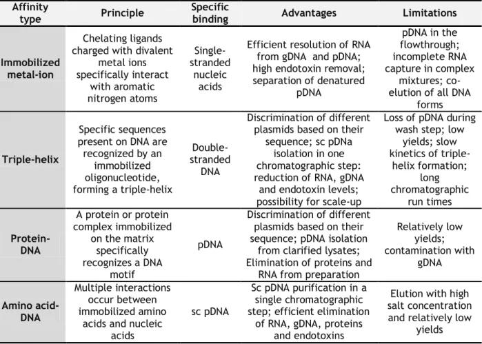
The natural biological processes, such as molecular recognition, are exploited by several affinity chromatographic modalities employed in pDNA purification. Immobilized metal-ion, triple-helix, protein-DNA and amino acid-DNA affinity chromatography have been employed with a specific ligand to separate pDNA on the basis of its biological function or chemical structure (Ghanem et al., 2013). Table 3 summarizes the principle, disadvantages and advantages of these four affinity chromatography methodologies.
Table 3. Affinity chromatography methodologies for nucleic acids purification (adapted from Sousa et al., 2008a).
Affinity
type Principle
Specific
binding Advantages Limitations
Immobilized metal-ion
Chelating ligands charged with divalent
metal ions specifically interact with aromatic nitrogen atoms Single-stranded nucleic acids
Efficient resolution of RNA from gDNA and pDNA; high endotoxin removal; separation of denatured pDNA pDNA in the flowthrough; incomplete RNA capture in complex mixtures; co-elution of all DNA
forms
Triple-helix
Specific sequences present on DNA are recognized by an immobilized oligonucleotide, forming a triple-helix Double-stranded DNA Discrimination of different plasmids based on their
sequence; sc pDNa isolation in one chromatographic step: reduction of RNA, gDNA
and endotoxin levels; possibility for scale-up
Loss of pDNA during wash step; low
yields; slow kinetics of triple-helix formation; long chromatographic run times Protein-DNA A protein or protein complex immobilized on the matrix specifically recognizes a DNA motif pDNA Discrimination of different plasmids based on their sequence; pDNA isolation
from clarified lysates; Elimination of proteins and
RNA from preparation
Relatively low yields; contamination with gDNA Amino acid-DNA Multiple interactions occur between immobilized amino
acids and nucleic acids
sc pDNA
Sc pDNA purification in a single chromatographic step; efficient elimination
of RNA, gDNA, proteins and endotoxins
Elution with high salt concentration and relatively low
yields
These methodologies have the power to eliminate additional steps while increasing yields and improving process economics.
The selection of the amino acids as affinity ligands was based on natural occurrence of many different interactions between proteins and nucleic acids in biological organisms, which mainly involve basic amino acids such as L-histidine or L-arginine (Sousa et al., 2010).
Due to the common characteristics shared by pDNA and host impurities, several chromatographic steps had to be applied in order to obtain the purified sc pDNA isoform. Therefore, amino acid-DNA affinity chromatography has revealed to be a promissory approach, since allows a specific binding with sc pDNA isoform in a single chromatographic step, through specific amino acids bound to the agarose matrix, such as L-histidine (Sousa et
al., 2006), L-arginine (Soares et al., 2008b) and L-lysine (Sousa et al., 2009), eliminating the
remaining pDNA isoforms and all the impurities present on sc pDNA-containing extract.
3.3.2. Ligands selection
characteristics of pDNA becomes relevant in order to select suitable ligands for purification strategies.
Briefly, plasmids are DNA molecules double stranded and covalently closed, forming a closed loop. Each strand of a pDNA molecule consists of a linear polymer of deoxyribonucleotides linked by phosphodiester bonds. When pH<4, the phosphate groups are negatively charged. The two anti-parallel strands are connected to each other by hydrogen bounds between complementary nucleotides in each strand and the two strands wind around a common axis originating the right handed double helix structure. This structure has highly hydrophobic grooves, due to the close packing of the aromatic bases, with accessible aromatic electrons and available sites for hydrogen bounding, which can be reached by solvent and ligand molecules. These structural and physicochemical characteristics can be exploited in purification strategies, namely to select appropriate ligands to interact with pDNA (Ferreira, 2005).
Synthetic and natural compounds such as dyes (Clonis et al., 2000), metal chelates (Yuchi et al., 2000) and amino acids can be used as affinity ligands. In addition, several atomic studies have described preferential interactions occurring between particular positively charged amino acids and nucleic acid bases (Hoffman et al., 2004). Although L-histidine, L-arginine and L-lysine belong to the positively charged amino acids group, the agarose matrices with these immobilized ligands applied on pDNA chromatography have showed different elution behavior.
3.3.2.1. L-histidine and derivatives
The use of L-histidine as an affinity ligand was first reported in a variety of peptides and proteins purification (Amourache and Vijayalakshmi, 1984) and more recently, as above mentioned, in pDNA purification using an agarose conventional matrix (Sousa et al., 2006). The versatility of L-histidine in molecular interactions arises from its unique structure, which distinguishes it among the twenty natural amino acids. The imidazole group in L-histidine side chain plays a major role in interactions. The molecular interactions of L-histidine can be classified into cation- π interactions (the imidazole motif is an aromatic ring that can interact with metallic cations or organic cations), π-π stacking interactions and hydrogen-π interactions (since the structure of L-histidine aromatic ring allow π-π stacking interactions and the polar hydrogen atom of L-histidine can form hydrogen-π bonds with other aromatic structures), coordinate bond interactions (due to the lone electron pair in the nitrogen atom of imidazole ring) and hydrogen bond interactions (since imidazole system is a hydrogen bond donor and acceptor) (Caramelo-Nunes et al., 2014; Liao et al., 2013).
These L-histidine features derived from the imidazole ring characteristics, which highlighted the potential of this amino acid as a suitable ligand for chromatographic purposes. The use of
L-histidine derivatives, such as Im-benzyl-L-histidine and L-methyl-L-histidine, composed by benzyl and methyl groups in position 1 of imidazole ring appear as promising ligands as well.
3.3.2.2. Surface plasmon resonance
The reversible biomolecular interactions between amino acids and pDNA molecules can be assessed by a label-free method, surface plasmon resonance (SPR). Thus, this method can be used to perform a screening of suitable ligands for affinity chromatography.
The SPR main components are the optical light source, a sensor chip and a detection system. The sensor chip, the biorecognition transducer, has a thin gold layer coupled to a glass layer; the glass layer has higher refractive index than gold. When a light beam propagates in a medium of high refractive index (glass) and encounters an interface at a medium of low refractive index (gold) is totally reflected at a specific angle, this physical phenomenon is called total internal reflection. Despite the light being fully reflected, the electromagnetic field component penetrates over a short distance into the medium that has the lower refractive index (gold). The leaked electromagnetic field component is referred as the evanescent field, and the wave amplitude attenuates exponentially from the interface. The evanescent wave excites electrons within the gold layer, yielding surface plasmons that propagate parallel to the interface. The ligands are immobilized on the sensor chip gold surface (coated chemically to enhance surface immobilization) and the analytes are injected into a continuous flow of running buffer. Once the analyte interacts non-covalently with the immobilized ligand, a change in molecular weight occurs and therefore the resonance angle shifts (Grupta and Verma, 2009; Ritzefeld and Sewald, 2011). A change in the SPR angle of 0.1 degrees corresponds to a change of 1000 RU (resonance units) in SPR signal (Thillaivinayagalingam et al., 2010).
Biacore (GE Healthcare) is the main supplier in the SPR market (Rich and Myszka, 2010) and every standard device is equipped with an integrated microfluidic cartridge that forms four flow cells on the sensor chip. Usually the first flow cell is used as blank, to subtract the responses obtained in the other three flow cells, which can be used to immobilize three different ligands (Majka and Speck, 2007).
There are several sensor chips available in market, all with the common gold surface but coated chemically different. The sensor chip CM5, from Biacore, is the most versatile chip with a matrix carboxymethylated dextran, covalently attached to the gold surface, in order to promote a hydrophilic environment for interaction with biomolecules, without interfering with the SPR signal. Advantageous for interactions involving small molecules with high surface stability, providing accuracy and precision and allowing repeated analysis on the same surface, accordingly to manufacturer’s instructions.
sensor chip CM5 surface, using amine coupling (Fischer, 2010), and the pDNA can be injected on the surface, in order to evaluate the affinity of interactions, label-free and with low sample consumption. SPR becomes a valuable tool to select suitable ligands for chromatographic experiments.
3.3.3. Stationary phase
A ligand with high selectivity will aid in the pDNA chromatography, however conventional matrices exhibit some limitations that will only be surpassed with a suitable chromatographic matrix.
Over the last years, several efforts have been made to overcome the chromatography bottlenecks associated with the stationary phases. An ideal matrix would be inexpensive, able to preserve the sensitive three-dimensional structure of large biomolecules but rigid enough, in order to avoid swelling or shrinking due to higher flow rates. More rigid stationary phases would resist to higher pressures and consequently higher linear velocities, enabling faster separations. At this point, the main barrier to fast chromatographic procedures would be the mass transfer between the mobile and stationary phases (Mihelic et al, 2000; Sousa et al., 2012). Thus, functionally distinct from porous particle-based columns, the monolithic columns appeared as attractive supports for chromatographic purification procedures, especially of large biomolecules (Endres et al., 2003).
3.3.3.1. Monoliths
The monolithic support is a rigid macroporous polymer column, made by in-situ polymerization within the confines of a chromatographic column, requiring no packing operations. The polymerization procedure allows optimization of porous properties of the monolith in terms of amount and pore size, depending on the polymerization temperature, which enable a plethora of applications (Merhar et al., 2003; Vlakh and Tennikova, 2007). Moreover, monoliths are highly porous (Svec, 2010) and the pores are interconnected, forming a network of channels with large binding area (Endres et al., 2003). The entire mobile phase is forced to flow through the monolith and convection becomes the dominant transport mechanism. Mass transport based on convection by laminar flow is an important feature on the separation of large biomolecules, such as pDNA. This results in flow-unaffected resolution and dynamic binding capacity that enables fast chromatographic procedures with low back pressure (Yamamoto and Kita, 2005). Time is a crucial factor in plasmid purification due to the possible pDNA degradation that may affect its structure and conformation. Besides the crucial role of these parameters on interaction, the desired conformation for clinical proposes (sc isoform) may be affected by longer chromatographic procedures.
Overall, the combination of the versatility and capacity of monolithic supports with the selectivity and specificity of amino acid ligands becomes a promising strategy to find the ideal chromatographic support, to recognize and purify the sc pDNA isoform from the non-effective pDNA topologies and host components, with high purity degree and productivity.
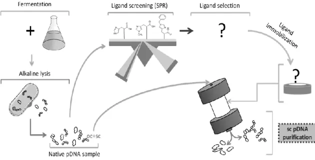
4. Aims
Given the importance of DNA vaccines and the use of pDNA vectors previously mentioned, the main goals of the present work are: the use of the SPR technique to perform a screening of binding affinity between three suitable ligands (Histidine, Im-benzyl-histidine and L-methyl-L-histidine) and pDNA samples; the immobilization of the selected ligand on a CIMTM
epoxy monolithic disk; Isoforms separation of three plasmids with different sizes; evaluation of the flow rate effect on isoforms separation; assessment of the dynamic binding capacity of the modified monolith. Figure 6 represents graphically the general aims of this work.
2.1. Materials
2.1.1. Reagents
The Hind III restriction enzyme, the GreenSafe Premium and the NZYTech Plasmid Maxi Columns were purchased from NZYTech (Lisbon, Portugal). Hyper Ladder I (Bioline, London, UK) was used as a DNA molecular weight marker.
Sodium chloride and ammonium sulfate were purchased from Panreac (Barcelona, Spain), tris(hydroxymethyl) aminomethane (Tris) from Merck (Darmstadt, Germany), 4-(2-hydroxyethyl)-1-piperazineethanesulfonic acid (HEPES) and borate buffer were from Sigma Aldrich (St.Louis, MO, USA).
2.1.2. Plasmids
The 6.05 kbp pVAX1-LacZ plasmid was provided by Invitrogen (Carlsband, CA, USA), the 8.702 kbp HPV-16 E6/E7 plasmid, Addgene plasmid 8641 (Münger et al., 1989), and the 14 kbp pcDNA3-myc-FLNa S2152A plasmid, Addgene plasmid 8983, (pcDNA3-based plasmid) (Woo et
al., 2004) were provided by Addgene (Cambrige, USA).
2.1.3. Instrumentation
Ultrospec 3000 UV/Visible Spectrophotometer (Pharmacia Biotech, Cambridge, England) was used to determine samples concentration of nucleic acids.
Agarose gels were revealed under UV light in a transilluminator system (ILC Lda, Lisbon, Portugal).
All SPR experiments were performed using a BIAcore T200 system and the BIAevaluation software was used for data analysis.
The 1H NMR spectra were recorded on a Bruker Avance III 600 MHz spectrometer equipped
with a four-channel Quadruple (QCI) resonance probe and all spectra were processed with the software topspin 3.1 (Bruker).
Chromatographic experiments were performed by using the AKTA Purifier system (GE Healthcare Biosciences Uppsala, Sweden) and the control system software was Unicorn version 5.11.
2.1.4. Specifications
All solutions, used in SPR and chromatographic experiments, were freshly prepared using deionized ultra-pure grade water, purified with a Mili-Q system from Millipore (Billerica, MA, USA) and analytical grade reagents were used. Elution buffers were filtered through a 0.20 µm pore size membrane (Schleicher Schuell, Dassel, Germany) and degassed ultrasonically. Chromatographic experiments were carried out with monolithic disks of 0.34 mL bed volume (average pore size of 1500 nm in diameter) modified with L-histidine amino acid and kindly provided by BIA Separations (Ajdovščina, Slovenia).
All experiments were conducted at room temperature unless otherwise stated.
2.2. Methods
2.2.1. Plasmids amplification by bacterial production
The pVAX1-LacZ, HPV-16 E6/E7 and pcDNA3-based plasmid amplification were performed by autonomous replication in Escherichia coli (E. coli) DH5α, after transformation. To ensure the exclusive growth of transformed cells, antibiotics were applied as selection markers. The medium was supplemented with 30 µg kanamycin/mL for cells transformed with pVAX1-LacZ, 100 µg ampicillin/mL for cells transformed with HPV-16 E6/E7 and 100 µg ampicillin/mL and 50 µg Neomycin/mL for cells transformed with pcDNA3-based plasmid.
The transformed cells harboring each plasmid were separately grown in plates at 37 ºC and the medium used was Luria-Broth (LB) agar, supplemented with the aforementioned antibiotics in accordance with the plasmid of transformed cells. In pre-fermentation, strides from plates of each plasmid were inoculated into 250 mL shake flasks with 62.5 mL of Terrific Broth (TB) medium (20 g/L tryptone, 24 g/L yeast extract, 4 mL/L glycerol, 0.017 M KH2PO4
and 0.072 M K2HPO4), with the respective selection marker for each plasmid. The
pre-fermentation was carried out at 37 ºC under 250 rpm shaking.
The optical density of the culture medium at a wavelength of 600 nm (OD600) was used to
evaluate the transformed cells growth. When OD600 reached approximately a value of 2.6, the
appropriate amount of pre-fermentation was inoculated in fresh TB medium, in order to start the fermentation with an OD600 of approximately 0.2. Growth was carried out under the same
conditions but in 1L shake flasks with 250 mL of fermentation medium in order to maintain the same oxygenation conditions of pre-fermentation. Growth was suspended at the mid-log phase (OD600~7) in order to achieve cells enriched with sc pDNA isoform.
2.2.2. Alkaline lysis and pre-purification of pDNA samples with
NZYTech Kit
The alkaline lysis of pelleted bacteria was performed by a modified alkaline method and the plasmid samples were pre-purified according to NZYTech Plasmid Maxi kit manufacturer’s instructions. The kit was designed for the rapid and large-scale preparation of highly pure pDNA from recombinant E.coli strains.
Briefly, after the alkaline lysis, the precipitated cell debris were eliminated by centrifugation, and then, the impurities are removed by a medium-salt wash after plasmid DNA binding to the NZYTech silica-based anion-exchange resin. All contaminants are washed from the column. Then, the pDNA elution occurs when a high-salt buffer is added. In last, the pDNA is concentrated by isopropanol precipitation.
The pDNA pellet was dissolved in appropriate buffer to future use.
2.2.3. Plasmid isoforms preparation
Three plasmid conformations, namely sc, linear (ln) and open circular (oc) isoforms of each plasmid, were prepared to be used in SPR and chromatographic experiments.
2.2.3.1. Supercoiled isoform
The sc samples of pVAX1-LacZ, HPV-16 E6/E7 and pcDNA3-based plasmid were directly obtained by alkaline lysis, as described above, according to manufacturer’s instructions. The bacterial growth was suspended with OD600~7 to obtain sc-enriched samples. The plasmid
yield was assessed by UV spectrophotometry and the sc isoform integrity was confirmed by agarose gel electrophoresis.
2.2.3.2. Linear isoform
The sc samples of pVAX1-LacZ, HPV-16 E6/E7 and pcDNA3-based plasmid were also used to prepare ln pure samples of each plasmid. For this propose, an enzymatic digestion with Hind III, 1 h at 37 ºC was accomplished according to the manufacturer´s protocol. The sample conversion was confirmed by agarose gel electrophoresis.
2.2.3.3. Open circular isoform
PVAX1-LacZ, HPV-16 E6/E7 and pcDNA3-based plasmid samples, obtained by alkaline lysis, were also used to prepare oc pure samples. In order to convert sc into oc isoform, the plasmids were incubated at room temperature, and monitored over the time by agarose electrophoresis, until total sample conversion (about 3 days).