Faculdade de Farmácia
Characterization of HIV-2 susceptibility to protease and entry
inhibitors and identification of envelope determinants of
coreceptor usage, cell tropism and antibody neutralization
Andreia Rodrigues Martins
Orientadores: Professor Doutor Nuno Eduardo Moura dos Santos da Costa Taveira Professor Doutor José António Frazão Moniz Pereira
Tese especialmente elaborada para a obtenção do grau de Doutor em Farmácia, especialidade Microbiologia.
Characterization of HIV-2 susceptibility to protease and entry inhibitors and identification of envelope determinants of coreceptor usage, cell tropism and antibody neutralization
Andreia Rodrigues Martins
Orientadores: Professor Doutor Nuno Eduardo Moura dos Santos da Costa Taveira Professor Doutor José António Frazão Moniz Pereira
Tese especialmente elaborada para a obtenção do grau de Doutor em Farmácia, especialidade Microbiologia.
Júri:
Presidente:Doutora Matilde da Luz dos Santos Duque da Fonseca e Castro, Professora
Catedrática e Diretora da Faculdade de Farmácia da Universidade de Lisboa
Vogais:Doutora Maria de São José Garcia Alexandre, Professora Catedrática, Faculdade de
Farmácia da Universidade do Porto
Doutor Nuno Eduardo Moura dos Santos Costa Taveira, Professor Catedrático, Instituto Superior de Ciências da Saúde Egas Moniz, Orientador
Doutora Perpétua da Conceição Rodrigues Gomes Cavaco Silva, Professora Associada, Instituto Superior de Ciências da Saúde Egas Moniz
Doutora Emília de Jesus da Encarnação Valadas, Professora Auxiliar, Faculdade de Medicina da Universidade de Lisboa
Doutor José Miguel Azevedo Pereira, Professor Auxiliar com Agregação, Faculdade de Farmácia da Universidade de Lisboa
Andreia Martins teve o apoio financeiro da Fundação para a Ciência e Tecnologia através de uma bolsa de Doutoramento (SFRH/BD/71028/2010).
Todas as afirmações efetuadas no presente documento são da exclusiva responsabilidade da sua autora, não cabendo qualquer responsabilidade à Faculdade de Farmácia de Lisboa pelos conteúdos nele apresentados.
Andreia Martins teve o apoio financeiro da Fundação para a Ciência e Tecnologia através de uma bolsa de doutoramento (SFRH/BD/71028/2010).
i I would like to acknowledge all who contributed to this work.
To my supervisor, Prof. Doutor Nuno Taveira, for being a great mentor, for all the invaluable guidance, motivation and advice.
To my co-supervisor, Prof. Doutor José Moniz Pereira, Coordinator of the Department of Microbiology and Immunology at Faculty of Pharmacy – University of Lisbon, for giving me the opportunity to do this research work at the department.
To Prof. Doutora Helena Barroso, for all the help and scientific inputs, whenever it was necessary.
To all my colleagues in the lab: Rita Calado, Pedro Borrego, Inês Bártolo, Cheila Rocha Rita Diniz, Joana Duarte, Francisco Martin, Inês Figueiredo, Rita Mateus, Carla Silva, Cláudia Palladino, Marta Gíria, Marta Calado, João Perdigão e Cláudia Pinto. Thank you for all the good moments we had working together and for the nice working atmosphere. Special thanks to Rita Calado, Pedro Borrego, Inês Bártolo and Cheila Rocha for their readiness to help in the lab, their support and friendship.
To all the professors from Microbiology and Immunology Department, particularly: Prof. Doutor João Gonçalves, Prof. Doutor José Miguel Pereira, Prof. Doutora Isabel Portugal, Prof. Doutora Aida Duarte, Prof. Doutora Madalena Pimentel, Prof. Doutora Elsa Anes, and also Prof. Doutor Jorge Vitor from the Department of Biochemistry and Human Biology.
To all the collaborators and/or co-authors of this work, especially: Pedro Borrego, Francisco Martin, Marta Calado, Prof Doutor José Miguel Pereira, Dr. Emília Valadas, Filipa Maia, Joana Silva and Dr. Ricardo Camacho.
ii
To the professors and staff from Instituto Superior de Ciências da Saúde Egas Moniz and Instituto de Higiene e Medicina Tropical for allowing me to visit their laboratories and use their research facilities, particularly: Sandra, Susana, Prof. Doutor Alexandre Quintas and his research group, Prof. Doutora Aida Esteves, Prof. Doutor João Piedade, Prof. Doutor Ricardo Parreira, José Marcelino, Diana Machado e Prof. Doutor Miguel Viveiros.
To all the patients and doctors that provided the samples and information to develop this work.
To Fundação para a Ciência e Tecnologia for the financial support that allowed the development of this project.
A special thanks to my parents and family for all the support and encouragement.
iii The research described in this thesis was conducted at the Department of Microbiology and Immunology, in the HIV Evolution, Epidemiology and Prevention Group, Research Institute of Medicines (iMed.ULisboa), Faculty of Pharmacy, University of Lisbon, under the supervision of Prof. Doutor Nuno Taveira and the co-supervision of Prof. Doutor José Moniz Pereira.
This thesis is based on the following manuscripts and publications:
Manuscripts and papers in international journals
Martins A, Calado M, Rocha C, Borrego P, Marcelino J, Azevedo-Pereira JM and
Taveira N. Determinants of coreceptor usage, tropism and susceptibility to antibody neutralization in the V3 region of HIV-2. (Manuscript in preparation)
Martins A, Martin F, Maia F, Rocha C, Valadas E, Antunes F, Caldeira L, Borrego P and
Taveira N. High level of resistance mutations to protease inhibitors in proviral DNA of HIV-2 infected patients in Portugal. (Manuscript in preparation)
Xiong S, Borrego P, Ding X, Zhu Y, Martins A, Chong H, Taveira N and He Y. A helical short-peptide fusion inhibitor with highly potent activity against human immunodeficiency virus type 1 (HIV-1), HIV-2, and simian immunodeficiency virus. J Virol.2017; 91(1):e01839-16.
Döring M, Borrego P, Büch J, Martins A, Friedrich G, Camacho R, Eberle J, Kaiser R, Lengauer T, Taveira N and Pfeifer N. A genotypic method for determining HIV-2 coreceptor usage enables epidemiological studies and clinical decision support. Retrovirology. 2016; 13:85.
iv
Matthias Döring, Pedro Borrego, Joachim Büch, Andreia Martins, Georg Friedrich, Ricardo Jorge Camacho, Josef Eberle, Rolf Kaiser, Thomas Lengauer, Nuno Taveira, Nico Pfeifer. geno2pheno[coreceptor-hiv2]: a new diagnostic tool for the genotypic determination of HIV-2 coreceptor usage. P351. International Congress of Drug Therapy in HIV Infection, October 23-26, 2016, Glasgow, UK. Journal of the International AIDS Society 2016, Volume 19, Supplement 7, 245.
Oral communications in national meetings
Martins A, Calado M, Rocha C, Borrego P, Marcelino J, Azevedo-Pereira JM and
Taveira N. Amino acids at positions 18, 23 and 24 in the V3 region are the main determinants of co-receptor use, macrophage tropism and susceptibility to antibody neutralization in HIV-2. 7th iMed.ULisboa Postgraduate Students Meeting, July 15, 2015, Faculty of Pharmacy, University of Lisbon, Lisbon, Portugal.
Poster communications in international conferences
P. Serra, A. Martins, N. Taveira, R. Guedes. Viral surface glycoproteins in HIV-2 – computational approach to structural elucidation. Keystone Symposia Conference HIV Vaccines, March 26-30, 2017, Steamboat Springs, Colorado, USA. Poster 3025.
Martins A, Calado M, Rocha C, Borrego P, Marcelino J, Azevedo-Pereira JM and
Taveira N. Determinants of coreceptor use, tropism and susceptibility to antibody neutralization in the V3 region of HIV-2. Keystone Symposia – HIV Persistence: Pathogenesis and Eradication, March 20-24, 2016, Olympic Valley, California, USA (Abstract no. 4019)
Döring M, Borrego P, Büch J, Martins A, Friedrich G, Camacho R, Eberle J, Kaiser R, Lengauer T, Taveira N and Pfeifer N. geno2pheno[coreceptor-hiv2], a new diagnostic tool for the genotypic determination of HIV-2 coreceptor usage. HIV Glasgow Drug Therapy Congress, October 23-26, 2016, Glasgow, Scotland. (Abstract no. 351)
v Protease diversity and resistance to protease inhibitors of viruses archived in peripheral blood mononuclear cells of HIV-2 infected patients. 12th European HIV & HEPATITIS workshop, March 26-28, 2014, Barcelona, Spain. (Abstract no. 35)
Poster communications in national conferences
Serra P, Martins A, Taveira N, Guedes R.C. Computational Approach Leading to Structural Characterization of Viral Surface Glycoproteins of HIV-2. 9th iMed.ULisboa and 2nd i3du Postgraduate Students Meeting, July 13-14, 2017, Lisbon, Portugal.
Patricia Serra, Andreia Martins, Nuno Taveira, Rita C. Computational approach to structural and conformational characterization of viral surface glycoproteins of HIV-2. XXV Encontro Nacional da Sociedade Portuguesa de Química, July 16-19, 2017, Lisbon Portugal.
Martins A, Calado M, Rocha C, Borrego P, Marcelino J, Azevedo-Pereira JM and
Taveira N. Determinants of coreceptor use, tropism and susceptibility to antibody neutralization in the V3 region of HIV-2. Ciência 2016-Encontro com a Ciência e Tecnologia em Portugal, July 4-6, 2016, Lisbon, Portugal.
Other publications: Published manuscripts:
Abecasis AB, Martins A, Costa I, Carvalho AP, Diogo I, Gomes P, Camacho RJ; Portuguese Hiv-1 Resistance Study Group. Molecular epidemiological analysis of paired pol/env sequences from Portuguese HIV type 1 patients. AIDS Res Hum Retroviruses. 2011;27(7):803-5.
vi
Martins A, Cavaco-Silva J, Borrego P, Camacho R and Taveira N. Phenotypic analysis
of HIV-2 drug resistance. Conference HIV-2: the forgotten AIDS virus, April 5, 2014, Faculty of Medicine of the University of Lisbon, Portugal.
Poster communications in international conferences
Martins A, Parreira R, Venenno T, Esteves A, Piedade J. Genetic diversity of HIV-1
among injecting drug users (IDUs) from Lisbon, Portugal: search for natural resistance associated mutations to enfuvirtide. 12th International Congress on Infectious Diseases, June 15-18, 2006, Lisbon, Portugal.
Poster communications in national conferences
Martins A, Cavaco-Silva J, Borrego P, Camacho R, Taveira N. Phenotypic analysis of
HIV-2 resistance to protease inhibitors. 6th iMed.ULisboa Postgraduate Students Meeting, July 2, 2014, Faculty of Pharmacy, University of Lisbon, Lisbon, Portugal.
vii O vírus da imunodeficiência humana tipo 2 (VIH-2) foi isolado e identificado pela primeira vez em 1986 como agente etiológico da Síndrome de Imunodeficiência Adquirida (SIDA). Atualmente é responsável por epidemias localizadas na África Ocidental, Continente Americano, Índia e em alguns países europeus como Portugal e França, onde a sua prevalência é ainda relativamente elevada.
O VIH-2 partilha muitas similaridades com o vírus da imunodeficiência humana tipo 1 (VIH-1), incluindo a organização estrutural e genómica, as vias de transmissão e o ciclo de replicação. No entanto, ambos os vírus exibem características distintas em termos de história evolutiva, patogénese, epidemiologia e algoritmos de tratamento.
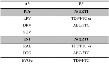
A maior diferença clínica entre ambas as infeções reside na progressão da doença, que é mais lenta no VIH-2 comparativamente ao VIH-1. A maioria dos indivíduos infetados pelo VIH-2 apresenta cargas virais indetetáveis e contagem normal de linfócitos T CD4+. Por outro lado, ao contrário do que sucede para o VIH-1, as opções terapêuticas disponíveis para tratar a infeção pelo VIH-2 são reduzidas. Todos os fármacos antirretrovirais foram desenvolvidos especificamente para o VIH-1, sendo a sua ação menos expressiva ou inexistente para o VIH-2. Por exemplo, o VIH-2 é resistente aos inibidores não nucleósidos da transcriptase reversa e apresenta diferentes níveis de suscetibilidade aos inibidores da protease (IPs). Neste contexto, os antagonistas do coreceptor CCR5, como o maraviroc (MVC), surgem como uma nova opção terapêutica para estes doentes. Contudo, a sua utilização requer a determinação do tropismo viral e, ao contrário do HIV-1, até aqui não existia nenhuma ferramenta informática que o permitisse fazer de forma adequada a partir das sequências da região V3.
O enfuvirtide (T-20) é o único inibidor de fusão aprovado, até ao momento, para o tratamento da infeção pelo VIH-1. Porém, este péptido tem atividade muito reduzida contra o VIH-2. Recentemente foi demonstrado que com um desenho adequado se podem produzir péptidos que inibem a fusão de VIH-2 e VIH-1, o que desencadeou a pesquisa de novos fármacos peptídicos para o tratamento das infeções por estes dois vírus.
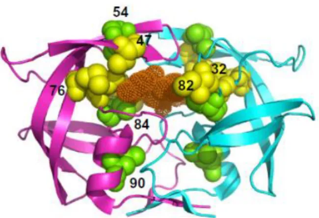
Os IPs são uma das principais classes de fármacos antirretrovirais utilizadas no tratamento da infeção pelo VIH-1. No entanto, o VIH-2 exibe suscetibilidade variável a estes fármacos e uma reduzida barreira genética de resistência.
viii
darunavir (DRV). Tendo em conta as opções terapêuticas limitadas e a seleção rápida de mutações de resistência no VIH-2, recomenda-se que o início do tratamento e a mudança dos esquemas terapêuticos nestes doentes devam ser realizados sob orientação de um teste de resistência. Por outro lado, desconhece-se ainda de que forma a diversidade da protease do VIH-2 afeta a resposta a longo prazo ao tratamento com IPs e de que forma a terapêutica com estes inibidores determina a evolução do VIH-2.
A entrada do VIH-2 no hospedeiro envolve a interação da glicoproteína de superfície do invólucro viral (SU) com o receptor CD4 e com os coreceptores de quimiocinas CXCR4 e CCR5 localizados nas células alvo, nomeadamente linfócitos T, macrófagos e outros tipos de células. Estirpes de VIH-2 que usam o CCR5 (variantes R5) são comuns em indivíduos assintomáticos enquanto vírus utilizadores do CXCR4 (variantes X4) são detetados apenas em indivíduos em fase avançada da doença e com contagens de linfócitos T CD4+ reduzidas.
Atualmente a informação existente relativa aos determinantes genéticos e estruturais da interação do VIH-2 com os coreceptores celulares ainda é muito limitada. Tal como no VIH-1, a utilização destes coreceptores pelo VIH-2 parece estar associada a alterações específicas na região V3 do invólucro. Por outro lado, estudos recentes indicaram uma associação entre o tipo de coreceptor utilizado pelo VIH-2 e a suscetibilidade à neutralização por anticorpos. No entanto, os determinantes do invólucro envolvidos nesta associação entre tipo de coreceptor e suscetibilidade à neutralização por anticorpos estão ainda por caracterizar.
Neste sentido, o objetivo geral deste trabalho foi caracterizar a suscetibilidade do VIH-2 aos inibidores da protease e inibidores de entrada e identificar determinantes virais do uso de coreceptores, tropismo celular e neutralização por anticorpos.
ix neutralizantes e no tropismo celular; 2) desenvolver um método genotípico para prever o tipo de coreceptor utilizado pelo VIH-2 com base na sequência de aminoácidos da região V3; 3) determinar a potência de ação de um novo péptido inibidor de fusão sobre o VIH-2 e 4) caracterizar a evolução da protease do VIH-2 em indivíduos infetados com ou sem experiência terapêutica prévia com IPs.
No primeiro capítulo desta tese é feita uma revisão dos conhecimentos atuais sobre a infeção por VIH-2 nos temas pertinentes para este trabalho, nomeadamente os fatores genéticos e biológicos que determinam o processo de entrada do vírus na célula do hospedeiro e as opções de tratamento para doentes infetados por VIH-2. O capítulo 2 faz referência aos objetivos e plano de trabalho da presente tese. Os restantes capítulos (3-6) descrevem o trabalho científico que deu origem a esta tese. Por último, no capítulo 7, são discutidos de forma integrada os resultados obtidos e realçadas as principais conclusões deste trabalho.
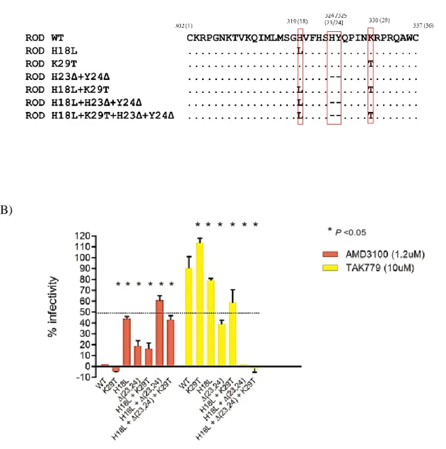
Para identificar os determinantes da região V3 do VIH-2 envolvidos na interação com os coreceptores celulares CCR5 e CXCR4 (Capítulo 3) foram efetuadas mutações por substituição nas posições 18 e/ou 19 e deleções nas posições 23 e/ou 24 da V3 do pROD10, um clone molecular infecioso do VIH-2ROD, o isolado X4 de referência. Os
clones mutados deram origem a seis vírus mutantes após transfecção de células 293T. Os ensaios celulares permitiram demonstrar que: 1) a conversão do fenótipo X4 em R5 no VIH-2ROD10 requer a substituição H18L e a deleção Δ(23,24); 2) os mutantes H18L e
H23Δ + Y24Δ são mais fáceis de neutralizar do que o VIH-2ROD e os outros mutantes por
plasma de indivíduos infetados pelo VIH-2; por outro lado a mutação K29T parece contribuir para aumentar a resistência à neutralização; 3) os mutantes K29T adquirem tropismo macrofágico sem comprometer a capacidade de replicação em linfócitos T CD4+; 4) os mutantes Δ(23,24) e H18L + Δ(23,24) adquirem tropismo macrofágico à custa de capacidade de replicação em linfócitos T CD4+.
x
com a fenilalanina em posição 20; 2) a deleção da H23 e Y24 leva à eliminação das folhas beta paralelas típicas da V3 e a uma perda de conteúdo aromático muito significativo o que pode comprometer a ligação a receptores celulares ou outras moléculas (ex. anticorpos); 3) a substituição K29T reduz a carga da V3 e elimina a ligação com a isoleucina em posição 27.
Coletivamente, estes resultados demonstraram que a V3 do VIH-2 é um determinante importante da ligação do vírus aos coreceptores celulares CCR5 e CXCR4, da suscetibilidade a anticorpos neutralizantes e da capacidade replicativa em linfócitos T CD4+ e macrófagos, e que estas características fenotípicas podem ser moduladas pela alteração de um único aminoácido. Estes resultados permitem atribuir à região V3 do invólucro do VIH-2 um papel crucial na patogénese da infeção por este vírus.
Até ao momento, o MVC é o único antagonista do coreceptor CCR5 aprovado para o tratamento da infeção pelo VIH-1. Estudos recentes têm demonstrado a sua eficácia também contra isolados de VIH-2. O início da terapêutica com MVC exige o conhecimento prévio do tropismo viral, dado que este fármaco pode potencialmente selecionar estirpes X4 minoritárias que estão associadas a maior capacidade replicativa, maior resistência aos anticorpos neutralizantes e a uma progressão mais rápida da doença. No entanto, ao contrário do VIH-1, ainda não existem testes genotípicos ou fenotípicos validados para a determinação do tropismo viral no VIH-2.
Nesse sentido, foi desenvolvido e validado um algoritmo para a determinação da utilização de coreceptores pelo VIH-2 com base na sequência da região V3. Este algoritmo deu origem a um serviço disponibilizado online semelhante ao existente para VIH-1 (geno2pheno[coreceptor-hiv2]) (Capítulo 4). O desenvolvimento e validação deste método genotípico para determinar o tropismo de VIH-2 requereu a análise de 126 sequências da região V3 obtidas a partir de indivíduos infetados pelo VIH-2, na sua maioria provenientes de Portugal, que apresentavam o perfil fenotípico definido para os coreceptores. A capacidade preditiva deste algoritmo foi ainda validada com base nas V3 mutadas produzidas e caracterizadas ao nível fenotípico no Capítulo 3. No conjunto, estes dados indicaram que o geno2pheno[coreceptor-hiv2] pode ser um instrumento útil na prática clínica, permitindo aos médicos uma melhor gestão dos doentes infetados pelo VIH-2 candidatos a terapêutica com MVC.
xi é idêntica à parte da região HR2 localizada no ectodomínio da glicoproteína transmembranar. O seu mecanismo de ação envolve a ligação à região HR1, o que impede a ligação natural desta região à HR2 e a formação da estrutura hexahelicoidal (3HR1:3HR2) que é fundamental para a fusão do vírus com a célula.
O 2P23 apresenta um desenho inovador na medida em que inclui dois resíduos, uma metionina e uma treonina, adjacentes ao domínio de ligação (pocket binding domain, PBD) da região HR2. Estes resíduos adotam uma estrutura específica designada por gancho M-T (M-T hook) que é importante para a estabilização da interação entre o PBD da região HR-2 e a cavidade hidrofóbica da região HR1. Esta interação é essencial para a estabilização da estrutura hexahelicoidal e para a fusão viral constituindo assim um alvo atrativo para o desenvolvimento de novos inibidores de fusão. Para além desta estratégia, a produção do 2P23 ainda envolveu a introdução de pontes salinas e resíduos cruciais para a ligação à região HR1 da glicoproteína transmembranar do VIH.
O 2P23 demonstrou ter uma potente atividade antiviral contra isolados primários de VIH-2 e VIH-1 (IC50 médio, 20.17 nM e 5.57 nM, respetivamente) e SIV (IC50 médio, 1.8 nM
para SIVpbj e 2.39 nM para SIV239). A sua atividade inibitória contra as seis variantes da
V3 (Capítulo 3) foi igualmente muito potente (IC50 médio, 15.38 nM) indicando
independência em relação à utilização de coreceptores. Em síntese, o 2P23 revelou ser um inibidor de fusão extraordinariamente potente contra diversos isolados primários de VIH-2, com diferentes perfis de utilização dos coreceptores, e poderá ser um fármaco promissor para desenvolvimento clínico futuro.
Os IPs são uma das principais classes de fármacos utilizadas no tratamento da infeção pelo VIH-2. Dada a prevalência significativa de VIH-2 em Portugal e a utilização frequente de IPs nestes doentes, torna-se essencial investigar a emergência de mutações de resistência nesta população e o seu impacto na resposta à terapêutica. No Capítulo 6 caracterizou-se a diversidade genética da PR e a resistência genotípica aos IPs em doentes infetados pelo VIH-2 residentes em Portugal e avaliou-se o seu impacto no resultado da terapêutica após oito anos.
Em 2007, foram colhidas amostras de sangue de 27 doentes infetados pelo VIH-2 com e sem terapêutica prévia, seguidos no Hospital de Santa Maria em Lisboa.
xii
apresentavam mutações associadas a resistência (exs. I54M, I82F, L90M) ao SQV, LPV, DRV.
Após oito anos, a análise genotípica da PR permitiu constatar: 1) a perda de mutações de resistência detetadas inicialmente em dois doentes, num dos casos associada a interrupção terapêutica; 2) a persistência de mutações de resistência num doente, como resultado de falência virológica e imunológica, em contexto de troca terapêutica e 3) o desenvolvimento de novas mutações de resistência em três doentes, associado a falências terapêuticas prévias.
Relativamente à diversidade genética da PR, verificou-se um aumento da diversidade em dois doentes tratados, virologicamente suprimidos e que apresentaram um aumento da contagem de T CD4+ comparativamente ao valor basal. Por outro lado, observou-se uma diminuição da diversidade genética da PR em três indivíduos (dois tratados e um não tratado) que apresentaram em algum momento do follow up cargas virais detetáveis. Estes resultados parecem evidenciar a persistência da replicação viral durante terapêutica antirretroviral a longo prazo, independentemente da supressão da carga viral plasmática. A manutenção da replicação viral poderá constituir a fonte de renovação das quasispecies provirais, levando a uma substituição gradual das variantes ancestrais ao longo do tempo. Neste estudo, também identificámos dois (15.4%) casos potenciais de resistência aos IPs em doentes sem terapêutica prévia. As mutações de resistência encontradas, L90M e I84V, foram também as mais prevalentes no subgrupo de doentes tratados. No conjunto, estes dados indicam que os testes de resistência baseados em ADN proviral podem ser úteis em doentes infetados pelo VIH-2 com cargas virais reduzidas ou indetetáveis e em indivíduos sem terapêutica prévia, e que a deteção precoce de resistência adquirida ou transmitida pode predizer a resposta à terapêutica nestes doentes.
Palavras-chave: Suscetibilidade do VIH-2 aos fármacos antirretrovirais; evolução no
VIH-2; determinantes do invólucro na utilização do coreceptor; determinantes do tropismo celular e dos anticorpos neutralizantes.
xiii The main aim of this work was to characterize the susceptibility of HIV-2 to protease and entry inhibitors and to identify viral determinants of coreceptor usage, cellular tropism and antibody neutralization. The specific objectives were: 1) to determine the contribution of amino acids residues in the V3 loop involved in CCR5 and CXCR4 use, susceptibility to antibody neutralization and cellular tropism; 2) to develop a genotypic method for the prediction of HIV-2 coreceptor usage based on V3 loop; 3) to evaluate the antiviral activity of a new short-peptide fusion inhibitor in HIV-2 and 4) to characterize the evolution and diversity of protease (PR) in HIV-2 infected patients treated and untreated with protease inhibitors (PIs).
In the first study (Chapter 3), site-directed mutagenesis was used to create amino acid substitutions in residues 18 and/or 29 and/or single deletions at positions 23 and 24 in V3 loop of pROD10, an infectious molecular clone of HIV-2ROD, the reference X4 isolate.
Cellular assays demonstrated that: 1) conversion from X4 to R5 phenotype in HIV-2ROD10
requires H18L substitution and the deletion Δ(23,24); 2) H18L and H23Δ + Y24Δ mutants are more easily neutralized than HIV-2ROD and other mutated viruses by plasma
from HIV-2 infected individuals; on the other hand, K29T substitution seems to contribute to increase resistance to neutralization; 3) K29T mutants acquire macrophage tropism without compromising replicative capacity in CD4+ T lymphocytes; 4) H18L + Δ(23,24) and Δ(23,24) mutants gained the ability to replicate in macrophages albeit at the cost of some capacity to replicate in CD4+ T cells.
Structural analysis by homology modelling showed that: 1) H18L substitution disrupts the interaction of histidine with methionine at position 15 and with phenylalanine at position 20; 2) deletion of H23 and Y24 leads to the elimination of the parallel β sheets presented in the V3 loop and the loss of the aromatic system which can compromise the binding of cellular coreceptors or other molecules (e.g. antibodies); 3) K29T substitution reduces the charge of V3 and leads to the loss of the interactions with isoleucine at position 27.
Collectively, these results demonstrated that V3 is an important determinant in HIV-2 coreceptor usage, susceptibility to antibody neutralization and replication capacity on CD4+ T cells and macrophages and that these phenotypic characteristics can be modulated by a single amino acid change in V3. These results support an important role for V3 in the pathogenesis of HIV-2 infection.
xiv
(geno2pheno [coreceptor-hiv2]). The development and validation of this tool was based on a data set of 126 samples from HIV-2 infected patients, most of them from Portugal, with phenotypic coreceptor usage annotations. Predictive accuracy was also validated based on the V3 mutants produced and phenotypically characterized in the previous chapter. Overall, these findings indicated that geno2pheno [coreceptor-hiv2] can be a useful tool in clinical practice, allowing better management of HIV-2 infected patients eligible for maraviroc (MVC).
In the third study (Chapter 5) a short-peptide named 2P23 was produced by combining a M-T hook structure, HIV-2 sequences and ‘salt-bridge’-based strategies. This peptide showed a potent antiviral activity against HIV-2 and HIV-1 isolates (mean 50% inhibitory concentration- IC50: 20.17 nM and 5.57 nM, respectively) and SIV (IC50: 1.8 nM for
SIVpbj and 3.29 for SIV239). This new fusion inhibitor also demonstrated a strong activity
against the V3 variants (Chapter 3) (IC50:15.38 nM), irrespectively of the coreceptor
phenotype. Thus, 2P23 is an ideal candidate for further clinical development due to its broad antiviral activity against several HIV-2 isolates, with different coreceptor tropism.
The last study (Chapter 6), involved the characterization of PR diversity and genotypic resistance to PIs of HIV-2 infected individuals living in Portugal and the evaluation of the impact of resistance mutations to PIs in treatment outcome eight years post-therapy. A high prevalence of PR mutations (e.g. I54M, I82F, L90M) associated to saquinavir (SQV), darunavir (DRV) and lopinavir (LPV) resistance, were detected in proviral DNA from these patients at baseline.
Eight years after study entry, the genotypic analysis identified: 1) loss of resistance mutations in two patients, that were initially detected at baseline, presumably as a consequence of treatment interruption; 2) long term persistence of resistance mutations in one individual as a result of virologic and immunologic failure, which might raise concern about transmission of drug resistance in the future and 3) development of new resistance mutations in three patients due to previous treatment failures.
xv the baseline. On the other hand, a reduction in PR genetic diversity was exhibited in three patients (two treated and one untreated), who presented detectable viral loads in at least one time point during the follow up. Due to small sample size it was not possible to investigate a potential relationship between PR genetic diversity and CD4+ T cell counts, presence of resistance mutations or/and treatment status. However, these results seem to indicate a persistent viral replication during long-term highly active antiretroviral therapy (HAART), regardless of plasma viral load. The maintenance of viral replication can act as a source of new proviral quasispecies, resulting in the gradual substitution of the ancestral variants over time.
Most importantly, we found two potential cases of transmitted drug resistance. However, due to the small sample size, additional studies with a higher number of patients are required to determine if primary drug resistance is a major problem in HIV-2 infected patients in Portugal.
Our findings suggest that proviral DNA may be useful in resistance testing in HIV-2 patients with low or suppressed viremia and in untreated patients, and that early resistance analysis of these archived viruses may predict treatment response.
Keywords: HIV-2 susceptibility to antiretroviral drugs; HIV-2 evolution; envelope
determinants of coreceptor usage; determinants of cell tropism and antibody neutralization.
xvii 6-HB Six-helix bundle 3D Three-dimensional 3TC Lamivudine μg Micrograms μl Microliters µM Micromolar ºC Celsius degree ABC Abacavir
AIDS Acquired Immunodeficiency Syndrome ART Antiretroviral therapy
ASA Accessible solvent area
ATV Atazanavir
AUC Area under the curve
AZT Zidovudine
bp Base pair
CA Viral capsid
cART Combined antiretroviral therapy
CD Circular dichroism
CDC Centers for Disease Control and Prevention
CHR C-terminal heptad repeat
CO2 Carbon dioxide
CRF Circulating recombinant form
CTL Cytotoxic T lymphocyte
d4T Staduvine
DC Dendritic cells
ddI Didanosine
D/M Dual/mixed population
DMEM Dulbecco’s minimal essential medium
DNA Deoxyribonucleic acid
dNTP Deoxyribonucleotide triphosphate
xviii
ECLs Extracellular loops
ECL2 Second extracellular loop
EFV Efavirenz
EI Entry inhibitor
ELISA Enzyme-Linked Immunosorbent Assay
ETV Etravirine
EVG Elvitegravir
FBS Fetal bovine serum
FDA Food and Drug Administration
FPR False positive rate
FPV Fosamprenavir
FTC Emtricitabine
GALT Gut associated lymphoid tissue
h Hour
HAART Highly active antiretroviral therapy
HIV Human Immunodeficiency Virus
HIV-1 Human Immunodeficiency Virus type 1 HIV-2 Human Immunodeficiency Virus type 2
HLA Human leukocyte antigen
HR-1 Heptad repeat 1
HR-2 Heptad repeat 2
IC50 50% inhibitory concentration IC90 90% inhibitory concentration
IDV Indinavir
IFN Interferon
Ig Immunoglobulin
IN Integrase
INI Integrase inhibitor
LPV Lopinavir
LTRs Long terminal repeats
xix
ml Milliliters
mm Millimeters
nm Nanometers
MOE Molecular operating environment program MPER Membrane proximal external region
MVC Maraviroc
Nabs Neutralizing antibodies
NC Nucleocapsid proteins
NFV Nelfinavir
NHR N-terminal heptad repeat
NIH National Institute of Health
NK Natural killer cells
nM Nanomolar
nm Nanometers
NNRTI Nonnucloside reverse transcriptase inhibitor NRTI Nucleos(t)ide reverse transcriptase inhibitor
NVP Nevirapine
PBD Pocket binding domain
PBMC Peripheral blood mononuclear cell
PBS Phosphate buffered saline
PCR Polymerase chain reaction
PDB Protein data bank
pDC Plasmacytoid dendritic cell
PI Protease inhibitor
PR Protease
PT Portuguese patients
RAL Raltegravir
RNA Ribonucleic acid
RPV Rilpivirine
RT Reverse transcriptase
xx
SIVcpz SIV from Pan troglodytes troglodytes chimpanzees SIVgor SIV from Western lowland gorillas
SIVsmm SIV from Cercocebus torgnatus atys sooty mangabeys
SQV Saquinavir
SU Surface glycoprotein
SVM Support vector machine
T-20 Enfuvirtide
TAM Thymidine analogue resistance mutation TCID50 50% tissue culture infectious dose
TDF Tenofovir
TDR Transmitted drug resistance
TLR Toll-like receptor
TM Transmembrane glycoprotein
TNF Tumor necrosis factor
TPV Tipranavir
U Units
xxi ACKNOWLEDGEMENTS i PREFACE iii RESUMO vii ABSTRACT xiii ABBREVIATIONS xvii CHAPTER I 1 General introduction 1
Discovery, origins and dissemination of HIV-2 3
HIV-2 epidemiology 4
Biology of HIV-2 5
Structure 5
Genomic organization 6
Envelope glycoproteins 7
Virus entry into the target cell 8
HIV-2 coreceptor usage and tropism 9
Pathogenesis and immune response in HIV-2 infection: differences for HIV-1 10
Innate and intrinsic immune responses against HIV-2 12
Adaptive immunity 14
Antiretroviral therapy and drug resistance in HIV-2 infection 17
Principles of antiretroviral therapy 18
When to start ART 18
What to start 21
Monitoring of treatment response 23
HIV-2 drug resistance 23
Mutation profiles conferring resistance to protease inhibitors 25
Activity of entry inhibitors on HIV-2 29
Transmitted drug resistance in HIV-2 31
Drug resistance testing 33
REFERENCES 35
CHAPTER II 61
Aims and work plan 61
References 66
CHAPTER III 69
Determinants of coreceptor usage, tropism and susceptibility to antibody neutralization
xxii
Material and methods 75
Results 79
Discussion 94
Conclusions 98
References 99
CHAPTER IV 103
A genotypic method for determining HIV-2 coreceptor usage enables epidemiological
studies and clinical decision support 103
Abstract 107 Background 108 Results 111 Discussion 120 Conclusions 123 Methods 124 References 134 CHAPTER V 145
A Helical Short-Peptide Fusion Inhibitor with Highly Potent Activity against HIV-1,
HIV-2, and SIV 145
Abstract 149
Introduction 150
Results 151
Discussion 166
Materials and Methods 168
References 171
CHAPTER VI 175
Resistance mutations to protease inhibitors in proviral DNA of HIV-2 infected
patients predict response to treatment 175
Abstract 179
Introduction 180
Material and methods 181
Results 183
Discussion 193
Conclusion 195
xxiii
Conclusions 221
CHAPTER I
3 Discovery, origins and dissemination of HIV-2
Acquired Immunodeficiency Syndrome (AIDS) is caused by two retroviruses, human immunodeficiency virus type 1 (HIV-1) and type 2 (HIV-2) [1, 2]. HIV-1 was isolated in 1983 and is responsible for the vast majority of HIV infections worldwide [2, 3]. HIV-2 was isolated in 1986 from two patients, one from Cape Verde interned at Claude Bernard Hospital in Paris, and another from Guinea-Bissau, interned in Hospital Egas Moniz in Lisbon with clinical symptoms similar to AIDS but negative serology for HIV-1 [1, 4]. HIV-2 is a lentivirus that belongs to the Orthoretrovirinae subfamily and the Retroviridae family [5]. This virus is closely related to simian immunodeficiency virus (SIVsmm) from sooty mangabeys monkeys (Cercocebus atys atys) that are found in the forests of the West Coast of Africa [6-8]. In contrast, HIV-1 descends from the SIVcpz found in Pan troglodytes troglodytes chimpanzees and from SIVgor that infects gorillas (Gorilla gorilla gorilla) [9-11].
HIV-1 can be divided into groups M, N, O and P, that resulted from tree cross-species transmissions from chimpanzees and one event transmission from gorillas, respectively [11-13]. HIV-2 resulted from at least nine independent transmissions from sooty mangabeys infected with SIVsm, originating nine groups termed A through I [7, 8, 14, 15]. Among these, only groups A and B cause epidemics, with group A being responsible for most HIV-2 infections worldwide. Isolates from group A are more predominant in Guinea-Bissau but are also found in other West African countries (e.g. Gambia, Ivory Coast and Cape Verde) whereas group B is more frequent in Ivory Coast and Ghana [16-21]. Groups C to I were only detected in isolates cases from Sierra Leone, Liberia and Ivory Coast [6, 14, 15, 22-26].
The date of introduction of HIV-2 groups A and B into the human population is estimated to be approximately 1940 and 1945, respectively [27-29]. Ivory Coast is the hypothetical geographic origin of HIV-2 group B, whereas the epicentre of group A remains to be defined. Some studies suggested Guinea-Bissau, based on serologic evidences, while others found SIVsmm lineages closely related to HIV-2 group A in Ivory Coast[7, 29]. More studies are needed to clarify the geographic origin of HIV-2 group A epidemic. HIV-2 group A has spread to Portugal from Guinea-Bissau and Cape Verde and to France from Ivory Coast and Senegal [29].
4
Within Europe, HIV-2 group A disseminated from Portugal to Luxembourg and the United Kingdom. Transmission of group A also occurred outside Europe in countries with historical links to Portugal such as Brazil, India and Mozambique [30, 31].
A circulating recombination form (CRF) of HIV-2 comprising sequences of groups A and B (designated as HIV-2 CRF01_AB) has been described in Cameroon, Ivory Coast and Japan [32-34].
HIV-2 epidemiology
Data on HIV-2 prevalence worldwide is quite old and more limited than for HIV-1. It is estimated that only about 1 to 2 million people are infected with HIV-2 in West Africa with most of them living in countries such as Guinea-Bissau, The Gambia, Senegal, Ivory Coast and Cape Verde [35]. Furthermore, HIV-2 prevalence is decreasing everywhere and some researchers estimate that HIV-2 will be extinct by the end of the century [36-39]. For instance, in the district of Caió in Guinea-Bissau the prevalence in adults declined from 8.3 % in 1990 to 4.7 % in 2007 [39]. The prevalence remained higher in older adults (age >45) while the decrease was more dramatic in young adults (15-35). In older adults the prevalence decreased from 22 to 12 % while young adults had a decrease from 3 to 0.9 %. The pace of decline was greater from 1997 to 2007 compared to 1990 to 1997. All of this occurred without antiretroviral therapy. In Gambia, HIV-2 prevalence declined from 7.0 to 4.0 % in 2001-2003 [40]. In contrast, both countries showed a rise in HIV-1 prevalence in the same period [39, 40]. HIV-1/2 dual infections are relatively common in West African countries, representing 0.3 to 1% of all HIV infected patients [41].
Outside West Africa, HIV-2 has been reported in The Americas, India and in several European countries, with Portugal and France being the countries with the highest prevalence of HIV-2 infected individuals [42-48]. In Portugal, at the end of 2015, an overall 1791 cases were associated with HIV-2 infection, corresponding to 3.3% of all notified HIV cases; HIV-1/HIV-2 dual infections were observed in 587 (1.1%) individuals [49]. The HIV-2 cumulative cases had an overall prevalence similar in both genders, with 878 cases in men and 913 in women. The majority was found in the age of 30 to 54 years at the time of their diagnosis.
The main mode of HIV-2 transmission was associated with heterosexual transmission (82%), with more than half of the individuals (61.5%) from Sub-Saharan Africa [49].
5 The number of new cases of HIV-2 infection in Portugal has been decreasing in the last twelve years, with 64 new infections in 2003 compared with 30 in 2015 [49].
Biology of HIV-2
Structure
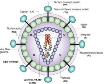
HIV-2 is an enveloped, spherical RNA virus with a diameter of around 110 nm (Figure 1) [50]. The virus is enveloped by a host cell derived phospholipid bilayer. The outer surface is covered with surface glycoproteins (SU) that are anchored to the transmembrane glycoproteins (TM) to form trimers in the mature virion [51]. The inner surface of the viral envelope is coated by the proteins (MA) and inside the matrix shell is a conical capsid core particle (CA) [52]. The capsid encapsulates two copies of single stranded RNA bound to the nucleocapsid proteins (NC) and also contains three essential viral encoded enzymes: protease (PR), reverse transcriptase (RT), and integrase (IN) and the accessory proteins Nef, Vif, Vpr and Vpx [53].
Figure 1. Schematic structure of the HIV particle. (Adapted from Robinson H. New hope for an aids vaccine. Nat Rev Immunol. 2002;2:239-50 [54]).
6
Genomic organization
The HIV-2 genome consists of two identical long single-stranded RNA molecules of 9800 nucleotides each, flanked by long terminal repeats (LTRs) at both ends (5’ and 3’) [50, 55]. It is organized into three main overlapping reading frames that comprise 9 genes; three major genes (gag, pol, env) encoding structural proteins or viral enzymes; two genes (rev, tat) for regulatory proteins and four (vif, vpx, nef, vpr) for accessory proteins (Figure 2) [50, 55].
The gag gene encodes the Gag (group specific antigen) precursor polyprotein (Pr55Gag) that is cleaved by viral protease during maturation of virion particles yielding four proteins: p6 (nucleocapsid), p6 (C-terminal protein), p16 (matrix) and p26 (capsid) [55, 56].
The Pol (polymerase) polyprotein is only expressed together with Gag as the Gag-Pol protein precursor (Pr160GagPol). GagPol precursor is cleaved by the viral protease into several enzymes that are required for virus replication: PR (p11), RT (p53) and IN (p34). The env gene encodes for the polyprotein precursor Pr140Env, which is processed by a cellular protease to form the surface envelope glycoprotein SU (gp125) and transmembrane envelope glycoprotein TM (gp36), which are necessary for HIV entry into the host cell [55, 56].
Figure 2. Genomic organization of HIV-2. (From Taveira N et al. Biologia Moelcular de VIH. In:
7
Envelope glycoproteins
The envelope glycoproteins (Env) mediate viral attachment and entry into target cells. The mature Env of HIV-1 and HIV-2 is arranged in trimeric spikes, comprising three glycoproteins SU (gp140/gp125) and three glycoproteins TM (gp40/gp36) that are non-covalently associated [58, 59]. The number and accessibility of Env spikes differ between HIV-1 and HIV-2. Compared to HIV-1, HIV-2 spikes are more stable and prominent after budding, while in HIV-1 the number of spikes decreases instantly after this process and during maturation [59-62].
The SU glycoprotein is composed of five constant (C1-C5) and five variable (V1-V5) regions. The constant domains correspond to the protein core while all variable regions, except V5, are exposed on the surface via disulphide bonds as large loops [63, 64]. The V3 loop of HIV-2, like HIV-1, is highly immunogenic, elicits antibody responses and seems to play an important role in coreceptor usage [65, 66]. On the other hand, the V4 and V5 loops are shorter and less glycosylated in HIV-2 than in HIV-1 [67].
The SU core is formed by an inner and outer domain. Both domains are linked by a bridging sheet. The inner domain is mainly formed by the C1 and C5 regions. This domain is hydrophobic and is responsible for the association between SU and TM. The outer domain is extensively glycosylated, and contains most of the antigenic determinants (neutralizing epitopes) and is implicated in the interaction between the SU and the cellular receptor (CD4) and coreceptors (mainly CCR5 and CXR4) [63, 64]. It was suggested that HIV-2 SU may sometimes adopt a CD4-induced conformation in its native state and thus may not require interaction with the receptor to induce conformational reorientation of the V1/V2 loops [68]. The TM glycoprotein consists of three major domains: an extracellular domain, a hydrophobic transmembrane domain and an intracytoplasmatic domain [69, 70]. The extracellular domain, or ectodomain, can be further divided into four segments: the hydrophobic fusion peptide at the N-terminus; two α-helices containing leucine zipper-like motifs (heptad repeat 1, HR1 and heptad repeat 2, HR2) and a membrane proximal external region (MPER). During virus entry into cells the HR1 and HR2 regions assemble into a six-helix structure consisting of a central parallel trimeric coiled-coil of the three HR1 helices, surrounded by the three HR2 helices in an anti-parallel way. The six-helix structure and the fusion peptide are essential for the fusion process between viral and host cell membranes.
The intracytoplasmatic domain of TM is required for Env incorporation into virions, during the maturation of new viral particles [64, 69, 70].
8
Virus entry into the target cell
Three different steps can be distinguished in the process that leads to HIV-2 entry into a target cell: binding of SU glycoprotein to the CD4 receptor, binding of SU to a coreceptor and finally the fusion of the viral envelope with host membrane [71].
The first step in viral infection is defined by the attachment of virus to the cell surface. This first interaction is mediated through the binding of the SU to a receptor CD4. After CD4 binding, SU undergoes conformational changes that rearrange its variable domains including the V1, V2, V3 and the constant region C4 and lead to the formation of the bridging sheet [63, 72]. These changes promote the reorientation and the exposure of a binding site in SU towards the target cell.
Although CD4 is the major receptor for HIV-2 as for HIV-1, some HIV-2 isolates can entry into cells independently of CD4 [73, 74]. The envelope SU glycoprotein of these isolates may have a more open structure comparing with HIV-1. It was proposed that the coreceptor binding site of CD4 independent isolates may be already formed and exposed and that this accessible conformation might facilitate the infection of target cells in the absence of CD4 [75, 76] .
Upon binding to CD4, the conformational changes mentioned before result in the interaction of V3 and eventually V1/V2 regions with a coreceptor, usually CCR5 or CXCR4. The exposure of coreceptor binding site in HIV-2 may be faster comparing to HIV-1, resulting in a more rapid fusion rate of the envelope with the membrane of the host cell [77]. It was suggested that differences in the cytoplasmic tail of the gp36/gp41 between both viruses might be the cause for the rapid rate of HIV-2 Env mediated-fusion. Both CCR5 and CXCR4 are seven-transmembrane G-protein coupled receptors. Each has an extracellular N-terminus, three extracellular loops (ECLs), three intracellular loops and a cytoplasmic C-terminus [78]. The two coreceptor regions that are required for the interaction with the viral SU, the N-terminal region and the second extracellular loop (ECL2), are the same for HIV-2 and HIV-1 infection [78].
The binding of SU to coreceptor triggers conformational changes in the TM glycoprotein that lead to the exposure of the fusion peptide.
The fusion peptide is then inserted into the host membrane, creating a structure denominated six-helix bundle, with HR1 and HR2 packed in antiparallel orientation. This structure brings the host cell membrane and viral envelope into close proximity and consequently a fusion pore is formed which allows the entry of viral core into the cytoplasm of the host cell [79].
9 Conformational alterations in gp41/gp36 required for viral entry in the host cell can be inhibited by fusion inhibitors. To date, enfuvirtide (T-20) is the only fusion inhibitor approved in treatment of HIV-1 infected patients. T-20 is based on the HR2 sequence of gp41 of HIV-1LAI isolate and prevents the formation of the six-helix bundle structure by
competitive binding to the HR1 region [80, 81]. However, despite its potent activity in HIV-1 isolates it presents a low genetic barrier to resistance and is not active on HIV-2 strains [82-85].
In the last years, second and third generation fusion inhibitors have been produced with increased antiviral activity against HIV-2 isolates, such as P3 and T-1249 [84, 86]. Still, there is an urgent need to produce novel fusion inhibitors with higher potency and stability than T-20, with strong antiviral activity against HIV-2 isolates and with a higher genetic barrier to resistance in order to expand treatment options for HIV-2 infection in the near future.
HIV-2 coreceptor usage and tropism
As for HIV-1, CCR5 and CXCR4 are the major coreceptors in vivo for HIV-2 [87, 88]. Variants that use the CCR5 coreceptor are termed R5, those that use CXCR4 are named X4, and those that use both are designated R5X4 (or dual tropic). Furthermore, a population of R5 and X4 variants is designated as dual/mixed (D/M) [89, 90].
Usually, CCR5 usage corresponds to a slow/low (viruses that replicate slowly and poorly), non-syncytium inducing phenotype while CXCR4 usage is associated with a rapid/high, syncytium inducing phenotype [89, 91]. Although HIV infects CD4+ T cells and peripheral blood mononuclear cells (PBMCs), CCR5 tropic strains tend to infect cells of the monocyte/macrophage lineage whereas X4 variants preferentially infect lymphocytic cell lines, according to the expression levels of CCR5 and CXCR4 in these target cells [89, 92].
Of note, in vitro some HIV-2 isolates obtained from patients in advanced disease stages have the ability to use a broad range of chemokine receptors: CCR1, CCR2b, CCR3, CCR6, CCR8, GPR15 (BOB), and CXCR6 (BONZO [87, 93-95].
The role of those alternative coreceptors in the pathogenicity of HIV-2 remains to be clarified [87, 88, 90].
10
R5 HIV-2 strains are common in asymptomatic patients or in acute stage of infection, while X4-tropic HIV-2 isolates have been found only in patients with advanced disease, low levels of CD4+ T cells and higher viral loads [66, 96-100].
In HIV-1 infection, switch from CCR5 to CXCR4 occurs in 50% of the infected individuals and has been associated with accelerated depletion of CD4+T cell counts and progression to AIDS [91, 101]. However, in contrast to HIV-2, X4 HIV-1 variants are more sensitive to plasma antibody neutralization comparing to R5 strains [102].
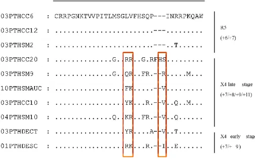
The transition from R5 to X4 phenotype in HIV-2 seems to implicate specific alterations in the V3 region and perhaps also in V1/V2 region of SU glycoprotein. Particularly in V3, a global net charge superior to 6 and the presence of mutations in positions 18, 19 and amino acid insertions at position 24 were associated to CXCR4 usage [65, 66, 75, 97, 100, 103-106]. These alterations have an impact on the structural conformation of V3. In fact, it was shown that R5 viruses are characterized by the absence of a secondary structure in the V3 region while transition to X4 tropism is characterized by the acquisition of a secondary structure (β-hairpin structure) in the V3 loop [65, 66, 97]. These alterations on the structure and conformation of the V3 may prevent the efficient binding of neutralizing antibodies (Nabs) to this region thus leading to escape and resistance to Nabs [65, 66, 97].
In HIV-1, V3 glycosylation has also been reported to influence coreceptor usage [107, 108]. Although HIV-2 has been reported to have lower number of glycosylation sites in V3 than HIV-1, the impact of these on coreceptor usage is still unknown [66, 75, 109].
Pathogenesis and immune response in HIV-2 infection: differences for HIV-1 HIV-2 infection is characterized by a longer asymptomatic phase and slower progression towards AIDS, when compared with HIV-1 [110, 111].
Clinically, a significant proportion of HIV-2 infected individuals (~80%) have higher CD4+ T cell counts and lower or undetectable plasma viral loads, in the absence of therapy, than that seen in HIV-1 infection [111-115].
HIV transmission occurs mainly across mucosal tissues. In this context, the dendritic cells (DC) may play a role, capturing the virus and spreading the infection to lymph nodes and secondary lymphoid tissue to present the virus to CD4+ T cells [116-118].
11 Therefore, the acute phase of HIV-1 infection is associated to a massive depletion of CD4+ T cells, especially in gut associated lymphoid tissue (GALT), as a consequence of several mechanisms, including direct viral infection, apoptosis, activation-induced cell death and host cytotoxic responses [119, 120]. In contrast, HIV-2 infection does not affect gut mucosal integrity and CD4+ T cells number despite local viral replication [121]. The viral set point establishes the beginning of the chronic phase and is associated with the rate of disease progression in untreated patients [122]. Viral set point in HIV-2 is 30 fold lower than in HIV-1 (median, 2500 versus 70000 RNA copies/ml, respectively) [123].The chronic phase (clinical latency) is the asymptomatic stage of HIV infection, with a median of duration 8-10 years in HIV-1, and 20 years or more in HIV-2 in the absence of treatment [124]. This stage is characterized by a persistent immune activation, which is manifested by high levels of CD4+ and CD8+ T cell apoptosis, polyclonal B cell activation with hypergammaglobulinemia, increased cell turnover of T cells, monocytes and natural killers (NK) cells [110, 122, 125]. This results in immunological abnormalities including, poor cell renewal, senescence and cellular exhaustion [122]. Several mechanisms have been proposed to be involved in the establishment and maintenance of HIV-associated chronic immune activation [126-128]. The innate and adaptive responses of the immune system against HIV replication and viral antigens may play an important role in this context. Moreover, the effect induced by specific viral proteins, including Tat, Env and Nef and the presence of opportunistic infections that are reactivated more frequently in these individuals (e.g. cytomegalovirus and Epstein-Barr virus) have also been suggested to be involved in HIV induced immune activation [126, 128].
Additionally, HIV response to the increased levels of type I interferons and pro-inflammatory mediators, the loss of the integrity of the gastrointestinal tract, with the consequent microbial products translocation from the GALT and the massive depletion of memory CD4+T cells are other potential players associated to chronic immune activation in HIV infection [126-128].
Immune activation is strongly linked to disease progression in HIV infection. The levels of immune activation are similar between HIV-1 and HIV-2 infected patients, when matched for the same degree of CD4+ T cell depletion [129, 130].
12
As mentioned before, in HIV-2, progression to AIDS is less frequent than in HIV-1, however, the clinical manifestations are very similar [131, 132]. One prospective study demonstrated that the probability of AIDS-free survival at five years after seroconversion, was near 100% in HIV-2 versus 67% in HIV-1 infected patients [133]. In the same way, mortality rates in HIV-2 infected individuals are lower compared with HIV-1, being only about two thirds of that for HIV-1 infected patients [134, 135].
A detectable viral load at baseline significantly predicts the rates of disease progression as determined by a decline in CD4+ T cell count or death [113, 136]. However, a substantial proportion of untreated HIV-2 infected patients (13%-46.5%) displays undetectable viral loads [137-141]. Therefore, as with HIV-1 infected patients, HIV-2 individuals with high viral loads undergo rapid CD4+ T cell count declines and death, while those who present low or undetectable HIV-2 RNA viral loads have decreased or no disease progression [136].
In HIV-2 infected patients, CD4+ T cell count rises as a response to an effective antiretroviral therapy [142-144]; however this response appears to be lower than in HIV-1 infected patients [114, 145, 146].
Innate and intrinsic immune responses against HIV-2
The innate immune response comprises several cellular and humoral components, such as cytokines, complement proteins, DCs, macrophages and NK that interact and cooperate to induce a robust immune response against pathogens until the adaptive response is established [147, 148].
HIV infection triggers innate immune receptors, including toll-like receptors (TLR), like TLR-7, TLR-8, and TLR-9, inducing the activation of DCs and the secretion of type 1 interferons (IFN) and tumor necrosis factor α (TNF- α) [147-149]. IFN and TNF-α play a role in the inhibition of viral replication, enhancing antiviral activity of immune cells (NK, T and B cells and macrophages) and recruiting other cells of the immune system to the sites of infection [147, 150].
Plasmacytoid DC (pDC) are one subset population of human DCs that specializes in the production of IFN- α upon TLR9 stimulation [151, 152]. The responsiveness of HIV-1 and HIV-2 infections to TLR-9 is defective in the advanced disease stage, along with CD4+ T cell loss [153].
13 On the other hand, the levels of circulating pDCs are diminished in HIV-2 infection, despite the absence of viremia, and are associated with CD4+ T cell depletion and immune activation [154].
Consequently, the production of IFN-α is decreased in HIV-2 infection [154]. The depletion of pDC in HIV-2 infected individuals with undetectable viremia might result from other mechanisms besides direct viral infection [154, 155]. Moreover, monocyte-derived dendritic cells and myeloid DCs were also shown to be less sensitive to HIV-2 infection, suggesting a preservation of these cells’ function through infection [155-157]. However, despite this less sensitivity, a progressive loss in circulating levels of myeloid DCs was observed in advanced disease stages in association with increase in viral load, CD4+ T depletion and immune activation [125].
NK cells are another important component of innate response. These cells secrete inflammatory chemokines such as IFN-γ and TNF-α and can recognize HIV infected targets leading to cytolysis of the infected cells [158]. Unlike HIV-1, HIV-2 asymptomatic patients show higher levels of NK cytotoxicity, similar to that found in uninfected controls [159]. However, the cytolytic and chemokine response of NK cells deteriorates with CD4+ counts decline, reaching similar levels to those seen in HIV-1 infection [159].
Important differences exist between the responses elicited by HIV-1 and HIV-2 against the four major host restriction factors, TRIM5-α, APOBEC3G, SAMHD1 and tetherin [160]. These proteins integrate the so called intrinsic branch of immunity that constitute the first line response to HIV infection and other viruses and are often blocked by specific viral proteins, such as Gag, Vif, Env and Vpx [160].
TRIM5-α is a member of the tripartite motif family of proteins that destabilizes the viral capsid core leading to premature uncoating, perturbing subsequently reverse transcription [161]. HIV-2 is more susceptible to restriction by TRIM5-α than is HIV-1, although there are strain specific variations, depending on motifs in the viral capsid [162, 163].
APOBEC3G belongs to the cytidine deaminases family [164]. In the absence of Vif, this protein, is packaged into virions and induces G to A hypermutation in the viral DNA, leading to degradation of the nascent proviral DNA [165, 166]. Compared with HIV-1, HIV-2 seems to be more resistant to APOBEC3G [167].
SAMHD1 contains nuclease and deoxyribonucleoside triphosphate phosphohydrolase (dNTPase) activity [168].
14
This enzyme lowers the concentration of deoxyribonucleotide triphosphate (dNTPs) in nondividing cells such as DCs, monocytes, macrophages and resting CD4+ lymphocytes thereby blocking HIV reverse transcription [169].
This effect could be more pronounced on HIV-2 replication in these cells because the RT of HIV-2 is less processive than HIV-1 and requires higher concentration of dNTPs to work properly [170-173].
However, the accessory protein Vpx, which is present in HIV-2 and related SIV lineages but not in HIV-1, degrades SAMHD1 [174]. It was suggested that HIV-2 would trigger a more efficient immune response relative to HIV-1 by productively infecting DC [110, 175]. However, a recent study showed that, HIV-2, like HIV-1, does not efficiently infect monocytes derived dendritic cells in vitro, suggesting that other factors not linked to SAMHD1 blockade may disturb HIV-2 infection in this cell population [156].
Tetherin, also known as bone marrow stromal antigen 2 (BST-2) or CD317 is a type II transmembrane protein that prevents virus release by inserting its N-terminal transmembrane domain in the plasma membrane and its GPI-linked C terminus in the virus envelope lipid bilayer. The tethered virus is then endocytosed [160]. HIV-1 and HIV-2 counteract tetherin by two very different mechanisms. In HIV-2, the envelope gp36 glycoprotein blocks its activity by interacting with the tetherin cytoplasmatic ectodomain, whereas in HIV-1, the anti-tetherin activity is mediated by the protein Vpu. Sequestration of tetherin by HIV-1 Vpu and HIV-2 Env in the endoplasmic reticulum prevents its transit to the plasma membrane. Tetherin proteasomal degradation is then induced by its interaction with the SCF-β-TRCP complex [176-178].
Adaptive immunity
Cellular immune response
HIV infects several types of immune cells, such as CD4+ T lymphocytes, DCs, monocytes/macrophages [122, 147]. CD4+ and CD8+ T cells are the most important players in HIV infection, with the former being responsible for the activation of diverse innate and adaptive immune cells and the latter mediating cell killing and secreting antiviral factors in order to control viral infection [179].
15 CD8+ T cells (cytotoxic T lymphocytes-CTL) play a critical role in the cellular immune response to HIV infection, since the initial decline in viral load, in acute infection, is assumed to be attributable to the activity of these cells [180, 181].
CTL are able to recognise viral determinants, at the surface of the infected cells, in the context of antigen presentation by human leukocyte antigen (HLA) class I and, subsequently lyse these cells [179, 182]. Additionally, CD8+ T cells secrete chemokines to control infection, particularly MIP-1a (CCL3), MIP-1b (CCL4) and RANTES (CCL5), that bind to HIV coreceptors on the surface of CD4+ T cells, inhibiting viral entry [179, 182, 183].
The degree of polyfunctionality of CD8+ T cells seems to be higher in HIV-2 infected patients, with production of higher levels of IFN-γ, TNF-α and other cytokines relative to HIV-1 infected patients [184, 185]. The heterogeneity and promiscuity of TCR usage is higher in HIV-2 infected patients, leading to a more efficient response in these patients comparing with HIV-1 infected individuals [186].
CD4+ T cells are preferentially infected by HIV, and depleted from the host as disease progresses [120, 187, 188]. Naïve CD4+T cells are activated after recognition of viral determinants through HLA class II, differentiating into specific subtypes and releasing cytokines (like IL-2). These cells have multiple functions, playing an important role in induction and maintenance of CTL and macrophages and in maturation of B cells [122, 188, 189].
In the majority of HIV-2 infected patients, qualitative and quantitative features of CD4+ T cells function seem to be preserved [190]. In HIV-2 infection, these cells appear to be more polyfunctional, with improved proliferative capacity, secreting a wider range of cytokines (namely IL-2 and IFN-γ), compared with HIV-1 [110, 184, 190, 191].
Antibody responses to HIV-2 infection
The humoral immune response is mediated by antibodies, which belongs to a family of globular proteins named immunoglobulins (Ig) that are secreted by B cells [192, 193]. Antibodies consist of two heavy and two lights chains linked by disulphide bonds and noncovalent interactions. Each chain is composed by two variable and two constant regions at the amino- and carboxyl-terminal end, respectively. The antigen-binding sites are located in the variable regions of both light and heavy chains [192, 194].