UNIVERSIDADE DE LISBOA
Faculdade de
Ciências
ASYN and Tau Interaction: New Drug
Target for Neurodegenerative Diseases
Gianmario Ciaccioli
Doutoramento em Bioquímica
Especialidade de Biotecnologia
2014
Tese co-orientada pela Doutora Patricia Calado e pela
Prof. Doutora Margarida D. Amaral, especialmente
elaborada para a obtenção do grau de doutor em Bioquímica,
especialidade de Biotecnologia
2014
UNIVERSIDADE DE LISBOA
Faculdade de
Ciências
ASYN and Tau Interaction: New Drug
Target for Neurodegenerative Diseases
Gianmario Ciaccioli
The experimental work presented was done at Bioalvo, TEC LABS Centro de Inovação, FCUL and at the Cellular and at the Molecular Neuroscience Unit, Instituto de Medicina Molecular, FMUL. The financial support was given by the European Community‘s Seventh Framework Programme FP7/2009 under grant agreement no. 238316 and from the FCT (Fundação para a Ciência e a Tecnologia) under grant agreement no. SFRH/ BI/51091/2010.
O trabalho experimental constante de a presente tese foi realizado na Bioalvo, TEC LABS Centro de Inovação, FCUL e na Unidade de Neurociência Celular e Molecular, Instituto de Medicina Molecular, FMUL. O apoio financeiro foi dado pela Comunidade Europea Seventh Framework Programme FP7/2009 contrato no. 238316 e pela FCT (Fundação para a Ciência e a Tecnologia) contrato no. SFRH/BI/51091/2010.
A impressão desta dissertação foi aprovada pelo Concelho Científico
da
Faculdade de Ciências de Lisboa em reunião de ____________________
Todas as afirmações efectuadas no presente documento são da exclusiva
responsabilidade do seu autor, não cabendo qualquer responsabilidade à
Faculdade de Ciências de Lisboa pelos conteúdos nele apresentados.
Table of Contents
Acknowledgments ... iii Abstract ... iv Resumo ... v Abbreviations ... x Chapter I. Introduction ... 1 1. Brain Diseases ... 11.1. Neurodegenerative Diseases Overview ... 1
1.2. Aging Brain: The Biggest Risk Factor in Neurodegeneration ... 2
2. Neurodegeneration Mechanisms ... 4
2.1. Failure of Neural Network ... 4
2.2. Protein Aggregation and Degradation ... 6
2.3. Mitochondrial Dysfunction and Oxidative Stress ... 11
2.4. Neuroinflammation ... 13
2.5. Neuronal Loss ... 14
3. The Role of ASYN in Neurodegeneration ... 17
3.1. ASYN Structure and Function ... 17
3.2. ASYN Implications in Neurodegenerative Diseases... 19
3.2.1. ASYN Expression ... 20
3.2.2. ASYN, Aggregation Mechanisms and Clearance ... 21
3.2 3. ASYN and ER/Oxidative Stress ... 25
3.2.4. ASYN, Cytoskeleton and Synaptic Activity ... 26
3.2.5. ASYN and Cell-to-Cell Propagation ... 27
3.3. ASYN Physiopathology in Yeast Models ... 28
4. The Role of Tau in Neurodegeneration ... 31
4.1. Tau Structure and Function ... 31
4.2. Tau Implications in Neurodegenerative Diseases ... 34
4.2.1. Tau, Oxidative Stress and Clearance ... 36
4.2.2. Tau and Cell-to-Cell Propagation ... 37
4.3. Tau Physiopathology in Yeast Models ... 38
5.1. ASYN and Tau Interactions ... 40
5.2. A Dual Target for Therapeutic Intervention ... 43
6. Goal of the Project: Identification of Modulators of ASYN and Tau Interaction ... 44
Chapter II. Results ... 46
1. A Powerful Yeast Model to Investigate the Synergistic Interaction of ASYN and Tau in Neurodegeneration ... 46
2. Identification of Modulators of ASYN and Tau Synergistic Toxicity by GWS and HTS Assays ... 62
Chapter III. General Discussion, Future Perspectives and Conclusions ... 70
1. Discussion ... 70
2. Conclusions ... 73
Acknowledgments
Alla mia famiglia, che mi ha sempre supportato in ogni mai scelta. (To my family, for always supporting me and being present) A Silvia “S’agapò tora ke tha s’agapò pantote” Oriana e Alekos A PhD is a long and challenging journey, during this time met a lot of amazing people who enriched my persona and encouraged my personal growth. I thank my supervisor Patricia Calado for all the scientific support, for her constant presence and for her wise advices. For always being honest and supportive, for her positive attitude, for sharing her personal experience during difficult moments and for always believe in my work.
I thank Helena Vieira for trust in me and for the opportunity to work whit her great team.
I thank my co-supervisor in the FCUL Magrarida D. Amaral for accepting me as student and for all her support.
I thank my co-mentor Tiago Outeiro for the opportunity to work in his laboratory and for the lively scientific discussions.
I thank my co-supervisor Ana Martins for her continuous scientific and personal support, for sharing the frustrations of my work and for guiding me during the most difficult steps of my PhD.
I thank Gonçalo Andrade, my first Portuguese friend and colleague, he introduced me to the world of yeast. He is a fantastic person, with a big heart and great sense of humour.
I thank all my colleagues from Bioalvo who in one way or another contributed to my work. Especially Ricardo Pineiro, Catia Rodrigues, Marta Cerejo, Marta Abrantes and Maria Antonia Pereira. From Tiago’s lab (UNCM) I am thankful to Sandra Tenreiro, Elisa Basso, Susana Gonçalves and Leonor Fleming for all the scientific and personal support given.
Abstract
Neurodegenerative diseases are among the most complex and puzzling human disorders and in the last century the number of people affected by neurodegenerative disorders is increasing year after year. These devastating disorders currently do not have any effective therapies or treatments, thus are a social and economic burden for modern society and novel therapeutic strategies need to be developed for these disease states.
Synucleinopathies and tauopathies regroup a wide number of neurodegenerative disorders characterized by the presence of abnormal protein aggregates respectively composed of alpha-synuclein (ASYN) or tau protein. Several reports revealed a consisting overlapping between these two groups of disorders and that the interactions between ASYN and tau may be a relevant disease component that enhances the pathological cascade and spreads the neuronal damage. Budding yeast have been largely used to perform studies on the physiopathology of synucleopathies and tauopathies and are a powerful tool for rapid screening assays that have resulted in the identification of several promising therapeutic drugs and targets.
In this work we used yeast as a model system to reproduce the synergistic cytotoxic effect mediated by the co-expression of the human ASYN and tau proteins that recapitulates some of the pathological features observed in patient’s brain. The model was used to perform genome-wide screening and high-throughput assays to identify both genes and natural extracts able to modulate the synergistic cytotoxic interactions of ASYN and tau.
In the end, we were able to identify 5 different S. cerevisiae genomic fragments containing a total of 25 different complete genes and 11 natural extracts able to interfere with the reported synergistic cytotoxicity. These modulators have the potential to be further explored in other appropriate disease models and might be relevant for academic groups and companies working on neurodegeneration engaged in drug discovery.
Resumo
As doenças neurodegenerativas representam uma das patologias humanas mais complexas e desafiadoras, sendo que no último século o número de pessoas afetadas tem crescido anualmente. Estas doenças devastadoras não têm atualmente nenhuma terapia ou tratamento eficaz, pelo que são um fardo social e económico para a sociedade moderna tornando urgente o desenvolvimento de novas estratégias terapêuticas para estes estados patológicos.
O termo sinucleinopatias agrupa várias doenças neurodegenerativas diferentes em que a característica patológica marcante é a presença de agregados fibrilares insolúveis da proteína alfa-sinucleína (ASYN), designados por corpos de Lewy, em populações específicas de células do cérebro. Este grupo de patologias inclui a doença de Parkinson, a demência com corpos de Lewy, a atrofia multissistémica, muitos casos de doença de Alzheimer (variante da doença de Alzheimer com corpos de Lewy), neurodegeneração com acumulação de ferro tipo I, falência autonómica pura, e um subtipo de tremor essencial. Na maioria dos casos as sinucleinopatias são doenças idiopáticas, envolvendo mutações poligénicas, interações génicas e de estilo de vida. Em 1997 o gene da alfa-sinucleína foi o primeiro gene que se descobriu estar associado à doença de Parkinson.
De igual forma, as tauopatias representam outro grupo de doenças neurodegenerativas caracterizadas pela presença de agregados neurofibrilares compostos maioritariamente pela proteína tau anormalmente hiperfosforilada. Patologias como a doença de Alzheimer, degeneração lobar frontotemporal, demência frontotemporal com parkinsonismo ligado ao cromossoma 17, paralisia supranuclear progressiva e a degeneração córtico-basal são exemplos de tauopatias. O estado de fosforilação da proteína tau tem um papel central na etiologia das tauopatias, contudo um elevado número de mutações no gene tau foi também identificado e associado com algumas destas doenças.
As sinucleinopatias e as tauopatias partilham mecanismos moleculares comuns conducentes à neurodegeneração e os processos patogénicos através do qual as proteínas ASYN e tau induzem morte celular estão relacionados com auto
agregação levando à formação de espécies oligoméricas potencialmente citotóxicas que por sua vez conduzem a alterações em vários processos celulares, nomeadamente na sinalização celular dependente de cálcio, atividade mitocondrial, estrutura do citoesqueleto, sistema de controlo de qualidade de proteínas, propagação célula a célula e ativação da resposta imune. Mais ainda, recentemente foram descobertas muitas interações diretas e indiretas entre estas duas proteínas, sendo de salientar a identificação de toxicidade sinergística aquando da co-expressão de ASYN e tau em modelos de mamíferos e de levedura. Tendo em conta todas estas analogias na fisiopatologia das sinucleinopatias e das tauopatias e a citotoxicidade sinergística entre ASYN e tau, estas duas proteínas podem representar um duplo alvo para o desenvolvimento de novas abordagens terapêuticas para o tratamento de um vasto número de doenças neurodegenerativas.
Apesar da ausência de um sistema nervoso os modelos de levedura são amplamente utilizados para abordagens às doenças neurodegenerativas, tendo em conta que muitos dos processos envolvidos na progressão patológica, tais como atividade mitocondrial, regulação da transcrição, tráfico intracelular e controlo de qualidade de proteínas, são muito conservados entre as leveduras e os humanos. Adicionalmente, cerca de 30% dos genes humanos envolvidos no desenvolvimento de muitas doenças humanas têm ortólogos funcionais em levedura. As células de levedura são também usadas para o rastreio em larga escala de bibliotecas de compostos devido a vantagens como a avaliação de compostos num ambiente fisiologicamente relevante e a seleção negativa imediata de compostos tóxicos ou com baixa permeabilidade membranar. Mais ainda, estão disponíveis coleções de estirpes de levedura mutadas por deleção, juntamente com coleções de sobre-expressão que constituem ferramentas poderosas para ensaios de análise genómica - genome-wide screening.
No trabalho descrito nesta tese utilizámos a levedura como sistema modelo para reproduzir e caracterizar o efeito citotóxico sinergístico mediado pela co-expressão das proteínas ASYN e tau humanas. O modelo foi depois usado como uma ferramenta para a descoberta de genes e compostos que medeiam a interação
tóxica entre ASYN e tau, possibilitando a identificação de novos alvos para intervenção terapêutica.
Começámos por promover a expressão de ASYN e tau em levedura a partir de um vetor epissomal bi-direcional. Os resultados obtidos mostraram que a presença de ASYN afeta a solubilidade da proteína tau, aumentando a fração de proteína insolúvel/agregada em associação com um aumento significativo na fosforilação de tau nos epítopos patogénicos S396/404, o que se sabe levar à formação de agregados de tau insolúveis. Mais ainda, mostrámos que no nosso modelo a presença de ASYN conduz ao aumento de agregação de tau no epítopo S396/404 via Rim11, o ortólogo de levedura de GSK3B. Estas descobertas estão alinhadas com outros relatos em que se provou que o epítopo patológico S396/404 é um substrato típico de GSK3B e que a ASYN tem a capacidade de estimular diretamente a fosforilação de tau via GSK3B, fazendo parte de um complexo heterotrimérico contendo ASYN, tau e GSK3B. Apesar do aumento na fosforilação e agregação de tau promovido pela co-expressão de ASYN, não foram observadas diferenças sinergísticas no fenótipo de crescimento do nosso modelo epissomal de levedura. Contudo, esta foi a primeira evidência que a ASYN tem capacidade de induzir a fosforilação e agregação de tau em levedura. Avaliámos de seguida o efeito da co-expressão de ASYN e tau em levedura recorrendo a uma abordagem experimental diferente, baseada na integração estável de uma cópia de cada transgene no genoma da levedura. Neste cenário, foi observado um forte efeito citotóxico sinergístico no crescimento da levedura, associado com um aumento da fosforilação de tau no epítopo S396/404 e a um aumento da fração insolúvel de proteína tau. De salientar que esta citotoxicidade sinergística é eliminada aquando da remoção de RIM11, demonstrando que o fenótipo obtido é resultante da cooperação entre ASYN e tau. Estes resultados sugerem que a hiperfosforilação de tau no epítopo S396/404 mediada por ASYN pode dar origem a espécies oligoméricas citotóxicas de tau, uma vez que esta citotoxicidade sinergística só é observada na estirpe de levedura que apresenta níveis mais elevados de tau fosforilada, em correlação com a presença de tau na fração proteica insolúvel.
Em resumo, as nossas estirpes de levedura, epissomal e integrativa, recapitulam muitos dos aspetos mais relevantes das interações de ASYN e tau na patologia humana e são ferramentas poderosas para ensaios de rastreio rápidos. Em particular, a estirpe integrativa, que revela um fenótipo sinergístico tóxico devido à co-expressão de ASYN e tau, foi usada para a realização de um genome-wide screening (GWS) e de um high-throughput screening (HTS) com o objetivo de identificarmos genes e compostos que possam futuramente ser alvos para o desenvolvimento de novas terapias para doenças neurodegenerativas.
O GWS foi realizado usando a colecção “Yeast Genomic Tiling Collection Assay Ready DNA”, que consiste em cerca de 1500 clones, cada um contendo um segmento único do genoma da levedura S. cerevisiae num vetor shuttle (E. coli – levedura) e fornecida como uma pool de DNA num tubo único. O GWS resultou na identificação de 5 fragmentos genómicos diferentes com um total de 25 genes completos que, quando sobre-expressos, têm a capacidade de diminuir fortemente a citotoxicidade sinergística mediada por ASYN e tau. Apesar dos nossos esforços iniciais para identificar os genes responsáveis por esta recuperação de fenótipo não terem sido bem sucedidos, a análise destes fragmentos genómicos deve ser alvo de atenção futura com vista a compreender quais os genes individuais ou interação de genes que medeia este efeito benéfico. O HTS foi realizado utilizando a coleção proprietária LUSOEXTRACT, que consiste em 3932 extratos naturais provenientes de 1206 organismos isolados de ecossistemas aquáticos e terrestres Portugueses únicos. A análise final do HTS resultou em 11 extratos naturais com capacidade para melhorar o efeito citotóxico mediado pela expressão concomitante de ASYN e tau, sendo que estes extratos devem ser alvo de estudos posteriores neste e noutros modelos celulares de doença.
A identificação de moduladores da toxicidade sinergística mediada por ASYN e tau é relevante tanto para grupos académicos que trabalham em neurodegeneração como para empresas que desenvolvem programas de identificação de novos fármacos. Temos a esperança que os resultados do presente trabalho possam no futuro ter um impacto positivo nos doentes, famílias
e prestadores de cuidados de saúde que lidam diariamente com a falta de alternativas terapêuticas para as doenças neurodegenerativas.
Abbreviations
AD AJPR ALP ALS AP-1 ASYN ATP AVs Aβ Alzheimer’s diseaseautosomal recessive juvenile parkinsonism autophagosome-lysosome pathways
amyotrophic lateral sclerosis jun proto-oncogene
alpha-synuclein protein adenosine triphosphate autophagic vacuoles amyloid beta
CADASIL cerebral autosomal dominant arteriopathy with subcortical infarcts and leukoencephalopathy
CDK5 cyclin-dependent kinase 5
CMA chaperone-mediated autophagy
CNA central nervous system
DAMPs danger-associated molecular patterns
DLB dementia with Lewy bodies
ER endoplasmic reticulum
ERAD endoplasmic-reticulum-associated degradation
FTD frontotemporal dementia
FTDP-17 frontotemporal dementia with parkinsonism linked to the chromosome 17q21-22
FTLD frontotemporal lobar degeneration
GSK3B glycogen synthase kinase 3 beta
GWAS genome wide association studies
GWS genome-wide screening HD Huntington’s disease HNE 4-hydroxy-2-nonenal HTS high-throughput screening JNK c-jun kinases LB Lewy bodies
MAPKs mitogen-activated protein kinase superfamily
MAPT microtubule associated protein tau
MARK microtubule affinity regulating kinase
MCI mild cognitive impairment
MPTP 1-methyl-4-phenyl-1,2,3,6-teteahydropyridine
mRNA messenger RNA
MSA multiple system atrophy
MT microtubule
MTBD microtubule binding domain
NAC non-amyloid beta component
NBIA neurodegeneration with brain iron accumulation
NFTs neurofibrillary tangles
NMDA N-methyl-D-aspartate
PAF pure autonomic failure
PCD programmed cell death
PD Parkinson’s disease
PDPKs proline-directed protein kinases AMP-dependent protein kinase PKA PQC RABS ROS rRNA rRNA
protein quality control
ER-to-Golgi vesicular trafficking proteins reactive oxygen species
ribosomal RNA
SAPKs stress-activated protein kinases
SNARE soluble N-ethylsaleimide-sensitive fusion protein attachment protein receptor
SNCA synuclein, alpha gene
STEP striatal-enriched protein tyrosine phosphatase
TKs tyrosine kinases
TLRs Tool-like receptors
tRNA transport RNA
UPR unfolded protein response
Chapter I. Introduction
1. Brain Diseases
1.1. Neurodegenerative Diseases Overview
According to the Alzheimer’s Foundation of America, dementia is a general term that describes a group of symptoms—such as loss of memory, judgment, language, complex motor skills, and other intellectual functions caused by the permanent damage or death of the brain's nerve cells, or neurons. The group of disorders causing dementia includes Alzheimer’s disease (AD), Parkinson’s disease (PD), Huntington’s disease (HD), dementia with Lewy bodies (DLB), frontotemporal dementia (FTD), frontotemporal lobar degeneration (FTLD), amyotrophic lateral sclerosis (ALS), and cerebral autosomal dominant arteriopathy with subcortical infarcts and leukoencephalopathy (CADASIL). AD is the most common cause of dementia in people over 65 and represents about 60% of all dementias, whereas vascular dementia caused by stroke or blockage of blood supply accounts for about 30% [1-3]. Epidemiologic studies predict that the number of people with dementia will double each 20 years in developed and developing countries, with a rate three to four times higher in developing areas than in developed regions, as consequence of increased life expectancy and exposure to many other risk factors (Figure 1).
Figure 1: Worldwide dementia prevalence and projection in developed and developing countries. A) Red countries have a prevalence estimated to be higher than 5%, which is similar
to the one estimated in developed countries (in grey). Blue countries have a prevalence of dementia lower than 3% while in green countries cases of dementia have been reported but
prevalence or incidence are unknown. No information is available from white countries. Red spots show locations of families with neurodegenerative disorders causing dementia [3]. B) The plot is representative of a study were it was estimated that 24 million people had dementia in 2001 and this amount will double every 20 years to reach 42 million by 2020 and 81 million by 2040 [2].
Aging is well known to be the greatest risk factor for this group of disorders, however the neuronal loss caused by intracellular oxidative stress accumulation in elderly peoples is much lower than the one observed in chronic neurodegenerative process [4]. All the neurodegenerative diseases are mainly characterized by an initial abnormal neuronal activity that progressively leads to neuronal cell death [5,6] due to the impairment of many intracellular processes including protein degradation and mitochondrial biology [7,8]. Another common hallmark of several neurodegenerative diseases is the presence of plaques and other atypical protein aggregates composed by normal or mutated disease-related proteins with different intra or extra cellular localization. Depending on the disease subtype, the distribution of these aggregates can be mainly cytosolic, predominantly intranuclear, in the endoplasmic reticulum (ER) or extracellular [9], but their direct contribute on the pathology progression is still under debate [9,10].
1.2. Aging Brain: The Biggest Risk Factor in Neurodegeneration
Neurodegenerative diseases share a common risk factor, the aging of the brain. In most cases aging and cognitive decline walk side by side, although the molecular mechanisms involved in aging are still unclear. Another important issue to address is how the physiological brain aging gives rise to neurodegenerative disorders. In normal elderly human population the cognitive decline—also called mild cognitive impairment (MCI)—is represented by delayed recall of verbal information, regression of working memory, short-term memory, reduced spatial memory and processing speed which is the most affected, then normal aging people take longer to learn new information [11-15]. Other cognitive functions such as long-term memory, implicit memory, attention span, vocabulary and verbal knowledge are well preserved during the aging process and emotional components of memory seem to be even improved
especially in people over 65 [16]. The memory functions compromised during aging may be the result of activation changes in the hippocampus and prefrontal cortex brain regions as revealed by functional imaging analysis [17,18]. Activation of the contralateral hemisphere was also shown in normal aging brains, representative of the normal compensatory response known as “loss of hemispheric asymmetry” that is lost in MCI and in AD [19]. AD human brains revealed an over-time accumulation of protein aggregates together with a progressive massive neuronal loss in the medial temporal lobes, in particular in the entorhinal cortex in contrast with the normal aging brains where a mild cell loss was observed only in the prefrontal cortex (Figure 2).
Figure 2: Brain structure and pathology progression. In MCI, hippocampus and prefrontal
cortex are the affected regions marked by neuronal loss accompanied by a diffuse moderate presence of amyloid plaques composed by Aβ peptide and neurofibrillary tangles composed by tau protein. Remarkable general volume loss and selective neuronal cell death in temporal lobe in association with widespread massive presence of protein aggregates was observed only in AD human brains [4,20].
Loss of synaptic function was also observed in the frontal cortex of normal aging brain [21]. Synaptic plasticity is a calcium-dependent function and reduced mRNA levels of calcium channel subunits and key proteins in the calcium signal pathways have been demonstrated in the aging prefrontal cortex [22]. Several microarray studies on human aging brain have been performed to evaluate changes in gene expression and in the frontal cortex about 4% of the active genes are regulated during the aging process [22,23]. Genes involved in memory, learning, synaptic plasticity, vesicle protein transport and mitochondrial function were significantly down-regulated while genes involved in stress response, in
particular antioxidants, DNA repair and inflammation, were up-regulated (Figure 3). Thus, age-related molecular changes at several levels expose neuronal cells to oxidative stress and biological dysfunction, compromising the entire biological systems of the brain. In accordance, the risk of AD onset increases 14-fold from the age of 65 to 85, afflicting about 47% of people over 85 [24]. Moreover, the presence of abnormal protein aggregates composed of amyloid-β peptides (Aβ) in AD, of tau protein in FTLD and of alpha-synuclein (ASYN) protein in PD is considered a potential contributory factor [10], but the current animal models do not explain the interconnections between aging and diseases onset.
Figure 3: Aging and neurodegeneration. During the aging process changes in gene
expression (left panel) might contribute to impaired intracellular pathways rendering the neurons more sensitive to several stress insults. This age-dependent neuronal vulnerability may represent the link between aging and neurodegeneration [4].
2. Neurodegeneration Mechanisms
2.1. Failure of Neural Network
The early impairment of neural activity plays a crucial role in the manifestation of neurodegenerative disorders. The evidence of neural network failure is the manifestation of impressive fluctuation in functional abilities– disabilities within the same day in people affected by neurodegenerative
disorders which significantly correlate with neuropsychological and
electrophysiological measures [25-27]. Functional reversible fluctuations could be the result of impaired intra–extra cellular processes not related to significant
neuronal death, such as molecular signalling, synapses activity, neuronal activity and neural network (Figure 4) [28-30]. This thesis is strongly supported by many studies in transgenic models expressing normal or mutant proteins related to AD, PD, HD or other neurodegenerative diseases. Results obtained showed how impaired behaviour and neuronal deficit observed without neuronal loss could be restored or prevented upon removal of the diseases related proteins [31-34].
Figure 4: Neurodegenerative disorders affect neural activities at many levels.
Neurodegenerative disorders can disrupt molecular pathways, synapses and local circuits in specific brain regions, as well as higher-order neural networks. Abnormal network activities may result in a vicious cycle, impairing the integrity and functions of neurons and synapses, for example, through aberrant excitation or inhibition [5].
For instance, in AD the cellular mechanisms involved in network failure are the ones linked to:
• Activity-regulated genes such as those encoding for cytoskeletal proteins, long-term potentiation, memory formation and immediate-early proteins forming the transcription factor AP-1 [35-39].
• Cell membrane receptors including cell adhesion proteins, glutamate receptors, nicotinic acetylcholine receptors and NMDA receptors [35,40-44].
• Signalling cascades regulated by several kinases and phosphatase like the mitogen-activated protein kinase superfamily (MAPKs), cyclin-dependent kinase 5 (CDK5), tyrosine kinases (TKs), calcineurin phosphatase and striatal-enriched protein tyrosine phosphatase (STEP) [35,40,43,45-48]. • Synaptic integrity controlled by presynaptic terminals and postsynaptic
dendritic spines [49-51].
Dysregulation of the described pathways triggers the activation of complex compensatory mechanisms that recruit different neuron subtypes to restore the normal activity [53,54]. This compensation could explain the slow progression of most neurodegenerative disorders and why so many neurons can die before the manifestation of relevant clinical symptoms. Progressive failure of these compensatory mechanisms may contribute to the remarkable daily fluctuations.
2.2. Protein Aggregation and Degradation
The cellular environment is composed by millions of proteins packed in their respective subcellular compartments. Maintaining a proper protein homeostasis under intrinsic and environmental conditions is essential for long-term cells health and to preserve the correct biological functions. Protein homeostasis involves transcription regulation, translation, folding, trafficking, processing, assembly–disassembly, localization and degradation. Many neurodegenerative diseases are characterized by the accumulation of misfolded proteins in insoluble inclusions with different anatomical distribution depending on the disease (Figure 5) [55].
Figure 5: Anatomical location of protein aggregates. Macroscopic and microscopic changes
observed on the most common neurodegenerative disorders [55].
AD and PD are respectively the first and second most common age-related neurodegenerative diseases characterized by the presence of protein aggregates. In AD the extracellular amyloid plaques are generated from the sequential
cleavage of the amyloid precursor protein (APP) by β-secretase and γ-secretase, giving rise to Aβ peptides of 40 and 42 amino acids residues called Aβ40 and
Aβ42 [56-58]. Numerous heritable mutations in the APP are linked to AD onset
during the first fifth decade of life, while the majority of the sporadic cases of AD develop clinical onset in people of 70 years old or more [24]. The other major hallmark in AD is the presence of neurofibrillary tangles (NFTs) which are intracellular insoluble fibrillar deposits formed by hyperphosphorylated tau protein [59,60]. Furthermore, more than 50 different mutations in the microtubule associated protein tau (MAPT) gene have been associated to familial forms of frontotemporal dementia with Parkinsonism linked to chromosome 17 (FTDP-17) [61,62]. PD is characterized by the presence of Lewy bodies (LB) which are intracellular cytoplasmic aggregates composed by the presynaptic ASYN protein and the cells affected are mainly the dopaminergic neurons in the substantia nigra [61]. The majority of PD cases are sporadic, however duplication and triplication of the synuclein-alpha (SNCA) locus gene as well point mutation in the ASYN proteins (A30P, E46K and A53T) have been identified in early-onset PD patients [63,64].
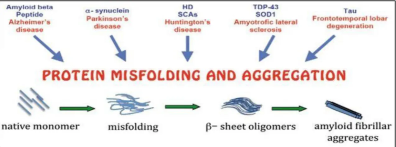
All these aggregates are composed by misfolded or unfolded disease-related proteins organized in a well-defined structure or amorphous, in both cases insoluble and refractory to proteolysis [65]. It is currently known that genetic mutations, environmental factors and different stress conditions are all able to induce protein misfolding and aggregation (Figure 6).
Figure 6: Misfolding and aggregation. Protein aggregation is a natural phenomenon occurring
in living organisms, although in several neurodegenerative disorders the disease related proteins start to self-aggregate in oligomeric intermediates that evolve into fibrillar structures.
Misfolding and aggregation can be promoted by genetic mutations, intra–extra cellular concentration, abnormal post-translational modification, proteolyitic cleavage, aging and environmental factors [66,67].
Recent studies revealed that those protein aggregates may diffuse from one affected cell to one normal cell, acting as seeds to initiate protein misfolding and aggregation in a process knew as “prion-like diffusion” [68]. In general it seems that the aggregation ability correlates with the cytotoxicity, but the direct implication of soluble monomer, oligomers or larger protein aggregates with the pathology progression is still under debate [69-71]. Several contrasting results have been observed and produced in the last century since Alois Alzheimer in 1907 discovered for the first time the presence of amyloid plaques in post-mortem brains from demented patients [72]. Two early hypotheses have been formulated to correlate neurodegenerative diseases with proteins aggregates: A) human neurodegenerative disease causes fibrillar deposits, but protein aggregation has no causal role; B) fibrillar protein deposits cause neurodegenerative disease. Several evidence argues against both hypotheses, as it was observed that some subtypes of neurodegenerative disorders are characterized by the absence of protein aggregates, as happens in the genetic prion disorder Gerstmann–Straussler syndrome, in sporadic ALS and in autosomal recessive juvenile parkinsonism (AJPR) [73-75]. Furthermore, dissociation between the severity of the symptoms and the number and the size of amyloid deposits in post-mortem AD brains was proved [76,77], and amyloid plaques were also found in non-demented elderly peoples [78]. Contrasting results have also been produced in neurodegenerative models where, in some cases, symptomatic reversal occurs upon removal of the aggregation-prone proteins and correlate with the disappearance of intracellular tangles [79-81] while in other cases fibrillary deposits are not directly related to diseases symptoms [82-84]. Moreover, amyloid fibrils can continue to grow and multimerize to form larger aggregates that evolve into an aggresome. It has been suggested that the aggresome is a protective structure formed to regroup potential proteasome-resistant toxic aggregates that will be then eliminated by autophagy [65,85]. However, even if it is not currently possible to directly correlate the
amount of a discrete protein aggregate to a human disease, the intracellular pathways involved in the maintenance of a proper protein homeostasis are largely studied as potential therapeutic targets for the treatment of neurodegenerative disorders. The first line of defence against misfolded proteins prone to aggregation are the molecular chaperones that can stabilize or refold such protein species and are also associated with the protein quality control (PQC) system in order to promote the elimination of those proteins that can no longer be refolded [86-90]. The PQC is regulated by two main distinct pathways, the ubiquitin-proteasome system (UPS) and the autophagosome-lysosome pathways (ALP). The successive failure of these protein degradation pathways, as cause or consequence of early pathological alterations in affected neurons, might represent a key step in the pathological cascade that gives rise to spreading neurodegeneration. Within the UPS pathway the key structure is the proteasome, a barrel-shaped multiprotein complex able to hydrolyse not only cytosolic and nuclear soluble proteins but also misfolded proteins inside the ER through a process known as endoplasmic-reticulum-associated degradation (ERAD) (Figure 7) [91,92].
Figure 7: UPS system. Proteasomal degradation is ubiquitin (Ub) dependent. Proteins tagged
with chains of four or more ubiquitins are shuttled to the proteasome by various proteins. The ubiquitin conjugation requires three different subtypes of enzymes: the ubiquitin-activating enzyme (E1), ubiquitin-conjugating enzyme (E2) and ubiquitin-ligase enzyme (E3). In the proteasome, proteins are hydrolysed to peptides, which are then released into the cytosol and further broken down by peptidases [93].
The target signal for protein degradation is the presence of a chain composed by four or more ubiquitin molecules covalently bound at the C-terminal region [93]. The polyubiquitinated targeted proteins need to be unfolded to pass through the
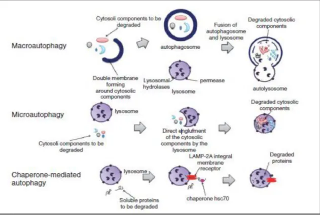
proteasome barrel pores to be hydrolysed, thereby oligomeric and aggregated proteins are resistant to UPS clearance [87]. Most of the proteins involved in neurodegeneration, such as ASYN or tau, are UPS dependent for their clearance before they start to self-aggregate [9,91]. When a cytosolic aggregation-prone protein becomes a poor proteasome target the ALP pathways succeed as major clearance route. Three different types of autophagy (literally self-eating) have been described: macro-autophagy, micro-autophagy and chaperone-mediated autophagy (CMA) (Figure 8). The components degraded by these roots may range from entire organelles, such as damaged mitochondria, to proteins aggregates, soluble peptides or microbes that can be specifically or non-specifically targeted [94].
Figure 8: Autophagic pathways: In macro-autophagy the cytosolic components to be
degraded are included in double layer vacuoles called autophagosome that then fuse with a lysosome, giving rise to an autolysosome, where the intracellular components are degraded by acid hydrolases. Micro-autophagy is characterized by the direct engulfment of the intracellular components to be degraded by the lysosome itself. In CMA only soluble proteins are degraded by the lysosome, the internalization occurs via the lysosomal-associated membrane protein LAMP-2A receptor and an accessory chaperone, the heat shock cognate protein Hsc70 [95-97].
Each autophagy pathway has individual characteristics, but they all appear to be interconnected and display compensatory up-regulation upon failure of one of
them. Cells respond to a blockage of CMA by activating macro-autophagy in a constitutive manner [98] while CMA is enhanced when macro-autophagy is dysfunctional [99]. The macro-autophagy pathway plays a critical role in the selective elimination of aggregated proteins in mammalian cells, a process known as aggrephagy in which exclusive proteins are required for substrate selection and targeting [88,89].
In summary, a common hallmark in many neurodegenerative diseases is the presence of intra or extra-cellular protein aggregates. All these proteins are degraded by UPS and/or ALS pathways, but persistent high intracellular proteins levels, post-translational modification, mutations or self-aggregation might render them resistant to the UPS-ALS mediated proteolysis, creating an inhibition–accumulation loop that can promote aggregation. Although, the PQC efficiency undergoes an age-dependent reduction and only a small fraction of elderly people develop PD or AD with the presence of intracellular protein aggregates, suggesting that other mechanisms are involved in maintaining a proper protein homeostasis.
2.3. Mitochondrial Dysfunction and Oxidative Stress
Mitochondria are key regulators of cell survival and death [100], play a central role in aging and interact with many specific proteins implicated in genetic forms of neurodegenerative diseases. Mitochondria possess many copies of their own circular DNA (mtDNA) that encode for 13 components of the respiratory chain, 2 rRNAs and 22 tRNAs to support the intra-mitochondrial protein synthesis [101]. Inherited mutations in mtDNA cause a variety of diseases affecting tissues with high energy requirements such as brain and muscles and it is well established that mtDNA accumulates mutations and large-scale deletions with aging that correlate with decline of mitochondrial function [100,102]. Mitochondrial insults, including oxidative damage itself, can cause imbalance between net production and removal of reactive oxygen species (ROS), resulting in ROS accumulation [103]. ROS encompass variety of partially reduced metabolites of oxygen such as superoxide anions, hydrogen peroxide and
hydroxyl radicals which are generated as by-products of cellular metabolism, primarily in the mitochondria. When cellular production of ROS overwhelms its antioxidant capacity, damage to cellular macromolecules such as lipids, protein, and DNA may ensue [104]. Several enzymatic and non-enzymatic defence pathways are involved to prevent the accumulation of ROS. These defence mechanisms are not always adequate to counteract the production of ROS, resulting in what is termed a state of oxidative stress [105]. Oxidative stress has been implicated in a wide variety of disease processes including neurodegenerative disorders and is believed to be a major factor in aging [106]. At the cellular level, oxidant injury elicits a wide spectrum of responses ranging from proliferation to growth arrest, senescence or cell death [105]. Thus, mitochondrial accumulation of mutations and net production of ROS might contribute to the physiological decline and aging-related neurodegeneration. Many neurodegenerative diseases are characterized by mitochondrial dysfunctions, displaying increased oxidative stress, decreased synaptic activity, reduced energy supply, impaired turnover, defective transport, fragmentation and cell death mediated by activation of the apoptotic cascades upon cytosolic release of cytochrome c and other pro-apoptotic mediators (Figure 9) [107-114].
Figure 9: Mitochondrial dysfunctions are associated with several genes related to
neurodegenerative disease. Impairment of mitochondrial functions and activity leads to increased production and accumulation of ROS, mtDNA mutations accumulation, cytochrome c release with subsequent apoptosis activation, cell death and neurodegeneration [115].
Therefore, mitochondrial biology holds a central role in the neurodegenerative disorders progression, hence net ROS turnover and energy supply are key events
to maintain a proper neuronal activity and cell survivor is strictly dependent on their integrity.
2.4. Neuroinflammation
Patient brains affected by many different chronic neurodegenerative disorders show increased levels of pro-inflammatory molecules as a consequence of immune response activation [116-122]. The immune response can be divided in innate and adaptive. In the central nervous system (CNS) the innate immune response is mediated by the resident macrophages called microglia. The main function of these cells is to contrast infections caused by bacteria or virus and remove necrotic and apoptotic cells [123]. The effector cells of the adaptive immune response are the T cells. Together with microglia, T cells can help to recover brain damage during neurodegeneration trough a mechanism that still remains unclear [124]. However, impeded microglia activity or persistent microglia activation leading to a systemic inflammation may contribute to neuronal damage (Figure 10).
Figure 10: Microglia-mediated neuron damage. In response to disease-specific stimuli, such
as protein aggregates, microglia can become deleteriously activated to produce a catalogue of factors, like ROS and cytokines that are toxic to neurons. Neuronal damage/death can also activate microglia to produce these toxic factors. This continual and self-perpetuating cycle of neuronal damage/death followed by microglial activation is commonly called reactive microgliosis and may be an underlying mechanism of the progressive nature of diverse neurodegenerative diseases [125].
In normal conditions T cells activity is repressed by both microglia and neuronal cells, while activated microglia and damaged neurons lose the ability to suppress the inflammation response [126]. The innate immune response cells recruit the adaptive immune cells by secreting various cytokines and by expressing
co-stimulatory molecules. Not only micro-organisms but also endogenous signals coming from damaged or stressed tissue (danger-associated molecular patterns, DAMPs) can activate the immune response within the CNS interacting with the Tool-like receptors (TLRs) expressed by the microglia cells [127]. Some DAMPs, including heat shock proteins, chromatin, ATP and modified or misfolded proteins have adjuvant and pro-inflammatory activity [128]. Notably, both ASYN monomers and tau oligomers are able to induce microglia activation and T cells proliferation [129,130]. Moreover, microglia does not appear to be involved in the removal of protein aggregates or degenerating synapses despite its phagocytic potential [131,132] and preventing the microglia proliferation leads to a delay in the onset behavioural symptoms and prolonged survivor [133]. Animal models of chronic neurodegeneration to deeply study the effects of a persistent inflammation are still missing. However, in the murine prion disease characterized by the accumulation of misfolded protein, microglia is persistently activated as a result of many factors which may include loss of inhibitory contact with neuronal ligands, accumulation of misfolded proteins, presence of neuronal debris and other unidentified routes [134]. It is clear that systemic inflammation is a contributor factor in neurodegeneration [135], thus the use of anti-inflammatory drugs might be a reasonable therapeutic approach to contrast the disease progression.
2.5. Neuronal Loss
Impaired intra–extra cellular processes are the prelude of neuronal cell death in chronic neurodegenerative diseases. Many evidences of activated programmed cell death (PCD) pathways have been collected from post mortem brain analysis of patients affected by neurodegeneration [136-139]. PCD plays an important role in the development of nervous system and three different forms of PCD have been described: apoptotic, autophagic and cytoplasmic [140-143]. These different pathways are activated in response to critical events and neurodegenerative diseases are associated with insults able to activate suicide mechanisms such as accumulation of misfolded proteins, DNA damage, ER
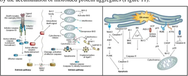
stress, inflammation or mitochondria damage [144]. Apoptotic pathways are largely investigated in neurodegeneration, in particular in diseases characterized by the accumulation of misfolded protein aggregates (Figure 11).
Figure 11: Apoptotic pathways. Three distinct mechanisms are known to trigger apoptotic
activation: (1-extrinsic) a death receptor–ligand mediated pathway, (2-intrinsic) a mitochondrial pathway involving the production of active caspases and (3-ER mediated) an endoplasmic reticulum-mediated pathway [144].
The ER is the central organelle for the biogenesis and trafficking of membrane and secretory proteins. It primarily serves as a cellular adaptive mechanism, activating cellular pathways that increase the chaperones mediated protein-folding capacity and reducing the protein influx into the ER by inhibiting gene expression at multiple levels [145]. Proteins that fail to achieve proper conformation are removed from the ER through the ERAD process [146]. Accumulation of misfolded proteins, inhibition of protein degradation or other insults to the ER lead to ER stress, which activates the unfolded protein response (UPR) [147]. Prolonged ER stress or defective UPR mechanisms, result in the activation of apoptotic suicide mechanisms contributing to the progression of neurodegenerative pathologies associated with misfolded proteins [9,91,93,148-150]. Misfolded proteins are also able to aggregate in oligomers and fibrils that interact with many critical cellular targets related to PCD activation. For instance, the disease associated ASYN and Aβ proteins are both able to promote cell death through apoptosis and flogistic processes activation [151-156]. Autophagic PCD represents another important aspect linked to cell death in neurodegenerative disorders. It was initially described as a cell death mechanism but in the latest years many studies revealed how this pathway is essential for
survival when cells are exposed to a wide variety of stress insults including nutrient deprivation, growth factor withdrawal, oxidative stress, infection, and hypoxia [157]. Besides the autophagy pro-survivor function, a persistent activation of this pathways leads to cell death by initiation of a self-digestion process. In progressive neurodegenerative disorders the aggregation and accumulation of misfolded proteins in intra and/or extra cellular aggregates correlates with an abnormal accumulation of intracellular autophagic vacuoles (AVs)—index of impaired autophagy—in degenerating cells. The direct contribution of the disease related Aβ or tau proteins to impaired autophagy is still under investigation, however impaired autophagy initiation in AD brains has been described [158] and in neurodegenerative models autophagy inhibition leads to intracellular Aβ accumulation [159]. Moreover, a defective lysosomal clearance of autophagic substrates due to impaired transport of AVs to lysosomes in association with NFTs deposits has been also established [160,161]. Duplication or triplication of the SNCA locus gene is sufficient per se to cause PD [64] and ASYN overproduction directly impairs autophagy blocking the formation of omegasomes which are the autophagosome precursors [162]. Specific ASYN post-translational modifications and mutations are also able to inhibit the CMA pathway by blocking the substrate internalization into the lysosome and thus the subsequent degradation [163,164]. Moreover, an indirect autophagy inhibition has also been revealed in the autosomal-recessive form of early-onset PD related to impaired mitochondria turnover [165,166]. Hence, impaired autophagic response might render cells more sensitive to stress conditions inducing cell death [167,168]. In general, complex relationships between autophagy and cell death have been described and in neurodegenerative disorders the specific factors that make autophagy neuroprotective rather than neurotoxic or how impaired autophagy leads to cell death are still enigmatic.
3. The Role of ASYN in Neurodegeneration
3.1. ASYN Structure and Function
The SNCA gene is located on chromosome 4q21 and encodes for a 140
amino acid protein called ASYN. ASYN has been defined as “natively unfolded” because it does not have a defined secondary structure in aqueous solution, although it can assume a α-helical secondary structure upon binding to negatively charged lipids such as the phospholipids present on cellular membranes [169]. The ASYN protein can be divided in three distinct regions: A) amino terminus region, from 1 to 60 amino acid residues, containing the apolipoprotein lipid-binding motifs, which forms the α-helical structure, B) central hydrophobic region, from residues 61 to 95, also called non-Aβ component (NAC), which confers the β-sheet potential and C) carboxyl terminus region, negatively charged and unfolded prone (Figure 12) [170].
Figure 12: ASYN protein structure and disease-related mutations. The peptide structure
can be divided in three main regions, A) amino terminus region, B) NAC region, necessary for aggregation and C) carboxyl terminus region.
Two different ASYN alternative transcript variants exist but their physiological role remains unclear [171-173]. The synuclein family group includes ASYN together with β-synuclein and γ-synuclein and all of them are mainly expressed in the brain, generally localized in the presynaptic terminals. The protein structure is similar for all, except for ASYN which is the only one possessing the NAC region [174]. Some evidence exists about the role of β and γ-synuclein in neurodegeneration [175-177], but a detailed characterization to further assess their potential contribution to neurodegenerative disease progression is still needed. ASYN is highly expressed in the CNS accounting for about 1% of total cytosolic proteins and it is actively transported from the cell body, along axons,
to synaptic termini [178,179]. Small amounts of ASYN protein have been detected also in glial cells [180], while abundant amounts of ASYN protein are detectable in erythrocytes and platelets but the physiological function exerted in these cells remains unknown [181]. During the neuronal cell development ASYN expression is progressively induced depending on the neuronal phenotype, however ASYN localizes to the presynaptic terminals only in a later stages of synaptic development, while it is absent when they first form [182-184]. Moreover, ASYN expression is modulated in critical conditions altering the synaptic plasticity or leading to injury [183,185,186], it is implicated in the size regulation and amount of the presynaptic vesicular pool and might have a chaperone activity to other presynaptic membrane proteins [187-189]. The main function of ASYN appears to be the control of neurotransmitter release by a chaperone-like function, in synergy with CSPα, in the assembly of SNARE complex (Figure 13) [188,190].
Figure 13: Role of ASYN and CSPα in the synaptic vesicle cycle. a) Binding of the carboxyl
terminus of ASYN to the amino terminus of synaptobrevin (VAMP) primes subsequent SNARE complex assembly. b) Binding of Hsc70 to SNAP-25 recruits SGT and CSPα to form a chaperone machine that promotes a SNAP-25 conformation compatible for SNARE complex formation. c) SNARE complex assembly drives membrane fusion and neurotransmitter release [191].
The SNARE proteins (Soluble NSF (N-ethylmaleimide-sensitive fusion protein) Attachment protein REceptor) consist of more than 60 members and are essential for the fusion of synaptic vesicles with the presynaptic membrane [192]. In neuronal cells the formation of the assembled complex is a critical step to mediate the neurotransmitter release through interactions between SNARE proteins localized on vesicles and those on target membranes [193]. Besides the regulation of neurotransmitter release, because of its membrane binding ability and association with synaptic vesicles, it is possible to assume that ASYN might be also involved in endo or exocytosis mechanisms, essential for neuronal function and survival.
3.2. ASYN Implications in Neurodegenerative Diseases
The term synucleinopathies regroups different neurodegenerative disorders characterized by the presence of insoluble fibrillary aggregates of ASYN protein, called LB. The distribution of the pathology at the cellular and regional level is different in each disease. This group of disorders includes PD, many cases of AD (called LB variant of AD), multiple system atrophy (MSA), DLB, neurodegeneration with brain iron accumulation (NBIA) type I, pure autonomic failure (PAF) and a subtype of essential tremor [194,195]. In most cases, synucleinopathies are sporadic diseases caused by multifactorial processes in which genetic, environmental and lifestyle factors culminate in overall risk [196,197]. The first link between PD and genetic defects was proved in 1997 when the mutation G209A in the SNCA gene, resulting in an A53T amino acid change in the ASYN protein, was associated with autosomal-dominant familial cases of early-onset PD in Italian and in some Greek kindred [198]. Later, two more mutations in the SNCA gene corresponding to A30P and E46K amino acid changes [199,200], together with duplication or triplication of the SNCA locus and Rep1 microsatellite expansion, were associated with autosomal-dominant forms of familial PD [64,201-203].
3.2.1. ASYN Expression
The correlation between ASYN protein levels in synucleinopathies pathogenesis arises from the association of SNCA gene duplication or triplication
and REP-1 polymorphisms, which leads to increased SNCA transcriptional
activity, with familial forms of PD. Little is known about the mechanisms involved in SNCA transcription regulation. Recently, a signalling pathway involving the ERK/PI3 kinases that mediates the SNCA transcriptional activity has been identified [204,205]. Is has also been reported an ASYN-mediated sequestration from the nucleus of the methylation factor Dnmt1 leading to decreased SNCA methylation and enhanced transcription creating a feed-forward loop [206]. Epigenetic factors are also involved in SNCA transcription regulation and abnormal SNCA gene methylation, causing enhanced SNCA expression, has been observed in PD brains [207,208]. Familial point mutations in the SNCA gene are also involved in SNCA expression. It has been observed that the expression of mutant alleles is suppressed over time through mechanisms involving histone methylation and in particular the presence of the A53T mutation enhances the expression of the wild-type allele in a compensatory manner. Nevertheless, the total levels of ASYN mRNA are above the normal controls suggesting, in this case, that enhanced wild-type ASYN expression is responsible for the disease [209-211]. Moreover, in synucleinopathy models, when ASYN is phosphorylated in S129 it preferentially localizes to the nucleus, reducing the histone acetylation affecting the general mechanisms of gene transcription and promoting neurodegeneration [212-214]. There is a general agreement that ASYN causes neurodegeneration through a gain-of-function when it exceeds a certain level. Concordantly, ASYN protein levels are increased in the substantia nigra of aging brains and a significantly increased SNCA expression was observed in surviving nigral neurons coming from PD brains [215,216]. However, in synucleinopathies models, neurotoxic effects can occur upon a severe reduction of ASYN protein levels [217,218]. Hence, the loss-of-function as consequence of the endogenous ASYN sequestration by oligomerization and inclusions formation causing neuronal damage and in particular, impaired
synaptic neurotransmitter release, might be involved in synucleinopathies progression as well.
3.2.2. ASYN, Aggregation Mechanisms and Clearance
The presence of LB in synucleinopathies is the final step of ASYN self-aggregation process, which starts with intracellular ASYN accumulation and gives rise to dystrophic neurites, defined as aberrant neuritic sproutings, swollen dendrites, and/or swollen axons [219]. The aggregation process starts with the formation of soluble oligomeric forms of ASYN protein aggregates in β-sheet structure (protofibrils) that gradually become insoluble and evolve into fibrils forming mature LB (Figure 14), although the exact neurotoxic soluble and/or insoluble oligomeric species still remains to be identified. Persistently increased intracellular ASYN protein level can promote the formation of ASYN containing aggregates [220]. ASYN oligomerization is also enhanced by interaction with other proteins, fatty acids and post-translational modifications such as phosphorylation, oxidation, nitrosylation, glycation or glycosylation.
Figure 14: Electron microscope pictures of ASYN aggregates. a-b) Protofibrils round or
elliptical shaped. c-d) Fibrils in β-sheets structure amyloid like. e-f) Round shaped LB inclusions ASYN containing (ASYN immunohistochemistry is indicated by the arrow in e) [221].
Phosphorylation is the most studied modification as in LB there is a conspicuous amount of ASYN hyperphosphorylation at S129 [222]. The effects of S129 hyperphosphorylation on ASYN aggregation and cytotoxicity are still controversial [223-227] but currently it seems that hyperphosphorylation in S129, together with S87, occurs mainly in mature LB [228]. Besides, ASYN phosphorylation in these specific epitopes leads to reduced axonal transport speed, as observed for the A30P and A53T mutants, which is a plausible explanation for the formation of perikaryal and neuritic aggregates [179]. ASYN oxidation is another important modification causing oligomerization. Metal ions, dopamine and mitochondrial dysfunctions leading to increased ROS levels all drive to ASYN oxidation and oligomerization [229-235]. ROS are also associated with the peroxidation of cellular membrane lipids and lipoprotein. One of the most important products of lipid peroxidation implicated in the pathogenesis of many neurodegenerative disorders is the highly reactive aldehyde 4-hydroxy-2-nonenal (HNE) [236-239]. ASYN oxidation mediated by HNE generates toxic stable soluble oligomers and inhibits their conversion into insoluble fibrils supporting the soluble oligomers toxicity theory [240]. ASYN nitration, similarly to oxidation, induces comparable effects promoting fibril formation and within the LB there is an extensive accumulation of nitrosylated ASYN [241,242]. Another significant ASYN modification is the carboxyl terminus truncation that generates aggregation-prone fragments [243,244]. Calpain is one of the most important calcium-dependent enzymes involved in the ASYN cleavage, both proteins localizes at the presynaptic terminals [245,246] and increased intracellular calcium influx mediated by ASYN oligomers could generate a feed-forward loop between calpain-mediated ASYN cleavage and oligomeric species formation [247,248]. ASYN oligomerization can also occur upon interaction with other proteins or fatty acids. The most notable protein-protein interactions are with tau and synphilin-1 that directly promote ASYN aggregation enhancing the formation of insoluble inclusions [249-251]. Polyunsaturated fatty acids interact with ASYN as well, enhancing oligomerization and neurotoxicity [252,253] and sequestration of the arachidonic
fatty acid away from the SNARE complex by ASYN has an inhibitory effect on neuronal transmission [254]. In synucleinopathy models based on ASYN variants lacking the NAC region, essential for aggregation, no toxic effects have been reported [255]. Moreover, protective effects have been observed in synucleinopathy models upon treatment with reagents preventing or neutralizing ASYN oligomers [256,257] and upon overexpression of heat shock chaperones which assist the refolding of aggregation prone proteins [258,259]. Thus, contrasting the formation of cytotoxic ASYN oligomeric species might be a plausible therapeutic approach to treat patients affected by synucleinopathies. Within the cellular environment the appropriate intracellular protein turnover is maintained by the UPS, CMA and macro-autophagy pathways, all involved in ASYN degradation. Soluble unfolded ASYN monomers are targeted for the UPS system and the CMA, whereas ASYN oligomers and ASYN containing aggresomes are targeted for macro-autophagy and lysosomal degradation [164,260-262]. LB are mainly composed by highly ubiquitinated ASYN protein, but also proteasomal subunits, ubiquitinating and de-ubiquitinating enzymes and proteasome activators are abundantly present in these aggregates, asserting an involvement of the UPS pathway in the clearance of ASYN [263-267]. The first evidence of UPS impairment was observed in the substantia nigra of PD brains where the proteasomal activity was significantly decreased in comparison with age-matched controls [268,269]. This evidence correlates with decreased gene expression of several proteasomal subunits only in the substantia nigra and in the cortex of patients, while in other brain regions the expression was unchanged or even increased [268,270-273]. In synucleinopathy models a direct inhibitory effect of ASYN on proteasome activity has been reported in a wide number of studies. Persistently increased intracellular ASYN protein levels leads to impaired proteasome function creating a vicious circle between UPS impairments and ASYN accumulation [154,274-280]. Besides, induced proteasome inhibition elicits a dose-dependent neurodegeneration with formation of ubiquitin and ASYN positive inclusions [281-283] and acute UPS inhibition was shown to induce compensatory clearance pathways by up-regulating autophagic flux
[284,285]. Moreover, by genome wide association studies (GWAS), mutations in PARK2 (an E3 ligase also involved in the proteasome activity) and PARK5 (an ubiquitin hydrolase) represent a risk factor for monogenic forms of PD [74,286]. However, some patients exhibit a normal or enhanced proteasome activity in the brain regions affected by the presence of LB, suggesting that region-specific perturbation of proteasome function is unlikely to explain the cause of neurodegeneration [287,288].
Together with the UPS system, CMA and macro-autophagy are the two compensatory pathways involved in the ASYN clearance that ends with the degradation of the targeted proteins inside the lysosomes. In CMA the lysosome internalization of targeted proteins occurs via the lysosomal-associated membrane protein LAMP-2A receptor [95-97]. Mutant A53T and A30P ASYN were shown to strongly bind the CMA receptor LAMP-2A but unable to be internalized for degradation, thus acting as CMA inhibitors [163]. A similar inhibitory effect on LAMP-2A occurs upon dopamine-induced conformational changes in wild-type ASYN [164]. However, contrasting results exist in relation to the levels of the CMA receptor LAMP-2A. These were found to be reduced in the substantia nigra and amygdala of PD patients and increased in the temporal cortex of DLB patients [289,290]. Defective CMA was also observed in familial PD patient’s brains carrying mutant LRRK2 protein which interfere with the organization of the CMA translocation complex that could results in ASYN accumulation and other PD related proteins degraded by this pathway [291]. On the other hand, the involvement of macro-autophagy in synucleinopathies was first proved by ultrastructural examination of neurons in the substantia nigra of PD patients revealing accumulation of AVs, which is consistent with either overproduction or impaired vacuoles turnover [292]. This finding was supported by increased autophagosome markers and decreased lysosomal markers in degenerating neurons, validating the presence of abundant and dysfunctional autophagosomes [289,293-297]. Persistent increased intracellular ASYN levels are able to inhibit macro-autophagy in a very early stage, blocking the formation of omegasomes, which are the autophagosome precursors, via Rab1 inhibition
and Atg9 mislocalization [162]. Moreover, lysosomal dysfunctions also affect the secretion of ASYN via exosomes [298], which could have implications for the cell-to-cell spreading and disease progression [299]. Collectively, all these findings show the ability of ASYN to inhibit all the clearance pathways involved in the maintenance of a proper intracellular protein turnover, an impairment that might be crucial in the pathology progression. Thus, restoring the proper PQC system activity could be a plausible approach to arrest or reduce the disease spreading.
3.2.3. ASYN and ER/Oxidative Stress
In synucleinopathy models, ER stress together with Golgi fragmentation leading to cell death is an early event that occurs in concomitance with the appearance of ASYN soluble oligomeric species [300,301], arguing that the primary target of ASYN-mediated neurotoxicity might be the ER-Golgi compartment. The ER-to-Golgi trafficking seems to be impeded through a direct ASYN-mediated mechanism, blocking the vesicle docking and fusion at a pre-Golgi step and inhibiting the assembly of the ER/pre-Golgi SNARE complex [302]. The proteins involved in the ER-to-Golgi vesicular trafficking (RABS) resulted to be the major class of modulators of ASYN toxicity [303]. Hence, in synucleinopathy models overexpression of RABS involved in the synaptic vesicular functions, in particular RAB1, prevent the dopaminergic toxicity to which neurons are very sensitive [304,305].
The degeneration of dopaminergic neurons in the substantia nigra correlates with impaired mitochondrial activity leading to increased oxidative stress and consequent cell death. Accumulation of point mutations and deletions in mtDNA, causing mitochondrial dysfunctions, have been observed in the brain of PD patients [306], while several mtDNA polymorphisms and haplotypes are associated with the risk of PD onset [307]. In synucleinopathy models ASYN directly interacts with the inner mitochondrial membrane causing mitochondrial fragmentation, activity impairment, increased ROS production and cell death due to the activation of the apoptotic cascade mediated by the mitochondria release of cytochrome c [156,308,309]. Moreover, by GWAS
required for the mitochondrial activity causing mitochondrial dysfunctions are associated with familiar forms of PD [310]. In detail, parkin and PINK1 proteins are associated with the mitochondrial outer membrane and prevent cell death by inhibiting cytochrome c release and caspases activation [311,312], mutations or deficiency cause oxidative stress, selective complex I impairment and altered synaptic functions [313]. DJ-1 is mostly localized in the cytosol, but also in the nucleus and associated with mitochondria [314]. Its biological implications are extremely diverse, it can act as a negative apoptosis regulator [315] or a potential ROS scavenger sensor activating neuroprotective pathways [316]. Mutations in DJ-1gene are associated with increased oxidative stress and apoptosis [317]. HTRA2 is a mitochondrial quality control protein and when released in the cytosol acts as a pro-apoptotic factor [8], mutations induce mitochondria swelling and decreased membrane potential [318]. Finally, LRKK2 is a protein largely present in the cytoplasm but also associated with the mitochondrial outer membrane. PD patients carrying the G2019S mutation exhibit reduced mitochondrial membrane potential and total intracellular ATP levels accompanied by increased mitochondrial elongation and interconnectivity [319], suggesting an important role in the regulation of mitochondrial homeostasis. Furthermore, specific inhibitors of the mitochondria complex I transport chain, such as pesticides and MPTP (1-methyl-4-phenyl-1,2,3,6-teteahydropyridine), are also able to induce neurological changes similar to PD, reconfirming the key role of mitochondria activity and oxidative stress in the disease progression [310]. Hence, restoring the normal mitochondrial activity might be helpful to contrast the progressive neuronal loss observed in patient brains.
3.2.4. ASYN, Cytoskeleton and Synaptic Activity
The cytoskeleton is a cellular scaffold composed by a network of microtubules and plays important roles in both intracellular transport, such as the movement of vesicles and organelles, and cell division. ASYN has been shown to bind and modulate actin polymerization, thus indirectly affecting intracellular trafficking [320]. A direct interaction between ASYN and microtubules has been reported in many studies [321] but weather this interaction leads to enhanced or


![Figure 5: Anatomical location of protein aggregates. Macroscopic and microscopic changes observed on the most common neurodegenerative disorders [55]](https://thumb-eu.123doks.com/thumbv2/123dok_br/18238115.878591/23.893.131.767.662.974/anatomical-location-aggregates-macroscopic-microscopic-observed-neurodegenerative-disorders.webp)