328 Radiol Bras. 2018 Set/Out;51(5):328–333
Radiological findings of breast involvement in benign
and malignant systemic diseases
Aspectos radiológicos do envolvimento mamário em doenças sistêmicas benignas e malignas
Renato Augusto Eidy Kiota Matsumoto1, Juliana Hiraoka Catani1, Mirela Liberato Campoy2, Arthur Magalhães Oliveira1, Nestor de Barros1
Matsumoto RAEK, Catani JH, Campoy ML, Oliveira AM, Barros N. Radiological findings of breast involvement in benign and malignant systemic diseases. Radiol Bras. 2018 Set/Out;51(5):328–333.
Abstract
Resumo
Although the primary purpose of periodic mammograms in screening programs is to identify lesions suspected of being carcinomas, the findings are often related to systemic (benign or malignant) diseases, rather than breast cancer. Although the involvement of breast structures in systemic diseases is unusual, it can be included in the differential diagnosis of masses, skin changes, calcifica
-tions, asymmetry, and axillary lymphadenopathy. The main diagnostic entities that can be associated with such involvement are diabetes, chronic kidney disease, heart diseases, connective tissue diseases, HIV infection, lymphoma, leukemia, and metastases from primary tumors at other sites. In many cases, information related to knowledge and treatment of chronic diseases is not avail
-able to the radiologist at the time of evaluation of the mammography findings. The purpose of this essay is to offer relevant pictorial information to the general radiologist about systemic diseases involving the breast, expanding the range of differential diagnoses in
order to avoid unnecessary invasive procedures.
Keywords: Breast; Systemic diseases; Collagen disease; Lymphoma; Metastases.
Embora o objetivo primário da realização periódica da mamografia nos programas de rastreamento seja a identificação de lesões suspeitas para carcinoma mamário, muitas vezes as alterações encontradas não estão relacionadas ao câncer de mama, e sim, a doenças sistêmicas benignas e malignas secundárias de outros sítios. O envolvimento das estruturas mamárias nas doenças sis
-têmicas é incomum, mas pode ser incluído no diagnóstico diferencial de nódulos, alterações cutâneas, calcificações, assimetrias e linfonodomegalias axilares. As principais entidades diagnósticas que podem estar associadas ao acometimento mamário são o diabetes, a nefropatia crônica, as cardiopatias, as colagenoses, as infecções pelo vírus HIV ou parasitas, o linfoma, a leucemia e as metástases de tumores primários de outros órgãos. Muitas vezes as informações relacionadas ao conhecimento e/ou tratamento de doenças crônicas não estão disponíveis para o radiologista no momento da avaliação da mamografia. O objetivo deste ensaio é oferecer informações iconográficas relevantes a respeito de doenças sistêmicas com envolvimento mamário, permitindo ampliar o leque de diagnósticos diferenciais e evitar eventuais procedimentos invasivos desnecessários.
Unitermos: Mama; Doenças sistêmicas; Colagenoses; Linfoma; Metástases.
Study conducted in the Department of Radiology of the Hospital das Clínicas da Faculdade de Medicina da Universidade de São Paulo (HC-FMUSP), São Paulo, SP, Brazil.
1. Department of Radiology, Hospital das Clínicas da Faculdade de Medicina da Universidade de São Paulo (HC-FMUSP), São Paulo, SP, Brazil.
2. Faculdade de Medicina da Universidade de São Paulo (FMUSP), São Paulo, SP, Brazil.
Correspondence: Dr. Renato Augusto Eidy Kiota Matsumoto. HC-FMUSP – De -partamento de Radiologia. Avenida Doutor Enéas de Carvalho Aguiar, 269, Cer-queira César. São Paulo, SP, Brazil, 05403-010. E-mail: [email protected].
Received July 20, 2016. Accepted after revision December 6, 2016.
parasitic diseases, and connective tissue diseases (e.g., der-matomyositis, scleroderma, and systemic lupus erythema-tosus). Within that context, patients may present, clinically, with skin changes, palpable masses and skin thickening. Malignant systemic diseases with secondary manifesta-tions in the breasts can include lymphoma, leukemia, and metastases from primary cancer at other sites.
The initial diagnostic flow chart involves the analy-sis of the clinical history and previous treatments. When these tools are used in conjunction with the mammogra-phy and ultrasound findings and yet do not result in a de-finitive diagnosis, percutaneous biopsy can be performed. The objective of this article is to present the most common systemic diseases affecting the breasts, as well as their ra-diological manifestations.
DIABETES
Diabetic mastopathy is an uncommon entity, occur-ring mainly in young women with a long history of type I
INTRODUCTION
diabetes, and affects less than 15% of insulin-dependent patients(1). Although the cause is not well known, it is
re-lated to an increase in the amount of collagen, increasing the extracellular matrix in the setting of hyperglycemia(2).
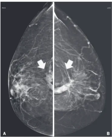
On mammography, it manifests as focal asymmetry or a solid mass, usually in the retroareolar region, without ac-companying calcifications (Figure 1). The sonographic ap-pearance is a hypoechoic mass with indistinct or spiculated margins, with pronounced posterior acoustic shadow, and no vascularity on the Doppler evaluation(3), as illustrated
in Figure 2. Those presentations raise the possibility of malignancy, and, consequently, percutaneous biopsy is recommended. During the biopsy procedure, the lesion is often hard, which hampers its sampling.
HEART DISEASES
There are two main aspects of heart diseases with man-ifestation in the breasts(3): arteriopathy and edema. Arterial
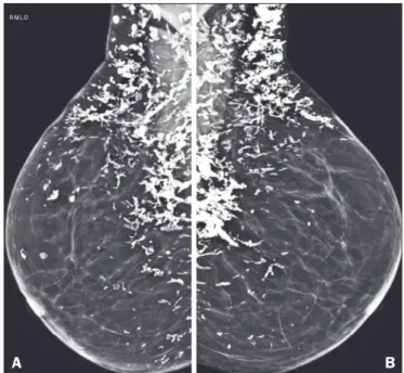
calcifications are common and do not cause diagnostic dif-ficulties in mammography (Figure 3), unless they are incipi-ent, in which case they can mimic linear suspicious calcifi-cations. It is not well established in the literature whether the detection of arterial calcifications is related to increased cardiovascular risk. It is intuitively assumed that calcifica-tions and peripheral arteries are a consequence of ongoing cardiovascular disease and are associated with risk factors for coronary artery disease, and this assumption is supported by some studies showing a positive association between the presence of vascular calcifications and cardiovascular
dis-ease(4). As can be seen in Figure 4, the edema manifests as
skin thickening, vein engorgement, and increased fibroglan-dular tissue density on mammography, whereas it manifests as increased echogenicity of superficial fatty planes and hy-poechoic fluid collections on ultrasound(3).
CHRONIC KIDNEY DISEASE
The imaging findings most commonly seen in chronic kidney diseas are related to its pathophysiology. Due to Figure 2. Ultrasound showing an irregular, spiculated, hypoechoic mass, with posterior acoustic shadowing, with no flow on Doppler evaluation. Percutane-ous biopsy of the mass resulted in a diagnosis of perilobular lymphocytic infiltrate, consistent with diabetic mastopathy.
Figure 3. A 58-year-old female patient presenting with multiple vascular cal-cifications on mammography.
RMLO LMLO
A B
volvement is rare and manifests in two main forms: axillary lymphadenopathy and tuberculous mastitis. When there is lymph node involvement, the lymph nodes are enlarged, the cortex is hypoechoic, and there can be calcifications. In mastitis, ultrasound shows abscess formation represented by complex (solid-cystic) masses or fluid collections (Fig-ure 7). Granulomas may also appear as irregular masses fluid retention, there are radiographic findings similar to
those of congestive heart failure, with increased fibroglan-dular density, thickening of trabeculae, and skin thicken-ing(3). Calcifications in the medial layer of the arteries can result in prominent vascular calcifications. Secondary hyperparathyroidism can give rise to coarse, mainly cuta-neous, calcifications. An arteriovenous fistula for dialysis results in prominent venous collaterals in the ipsilateral breast (Figure 5). As a consequence of the medications used in patients undergoing renal transplantation, fibro-adenomas can be commonly seen in women taking cy-closporine(5) and infectious processes can result from the immunosuppressive state. In men with chronic kidney disease, the drop in serum testosterone levels may cause gynecomastia.
HIV
Axillary lymph node enlargement and infectious pro-cesses can be seen in HIV-infected individuals. The lymph nodes tend to present hyperdense and with larger dimen-sions, although nonspecific. On ultrasound, the lymph nodes show diffuse, symmetrical cortical thickening. Breast composition is also affected by HIV-associated lipo-dystrophy, because there is a lower proportion of adipose tissue, resulting in a breast with a greater density on mam-mography. In HIV-infected patients, there may be filling of the breast with autologous adipose material, promoting areas of fat necrosis (Figure 6).
GRANULOMATOUS DISEASES
Granulomatous diseases include tuberculosis and mastitis. In systemic tuberculosis, breast or axillary
in-LMLO RMLO
Figure 5. History of chronic kidney disease in the creation of a left arteriove-nous fistula. Mammogram showing a vascular prominence in the left breast.
A B
Figure 6. Mammogram of a 42-year-old female patient with a history of HIV infection, currently receiving antiretroviral therapy, who presented with bilat-eral areas of fat necrosis. The patient had a history of adipose tissue graft in the breasts due to lipodystrophy caused by HIV infection.
R C C
A
L C C
B Figure 4. Mammogram, in mediolateral oblique views, of a 57-year-old
fe-male patient presenting with an increase and accentuation of the trabecular breast tissue, accompanied by diffuse bilateral thickening of the skin. These findings are associated with decompensation of congestive heart failure.
LMLO RMLO
accompanied by edema of the adjacent fat tissue(3,6). In
these situations, it is difficult to make an accurate diagno-sis, given that it is often impossible to exclude a malignant lesion on the basis of imaging findings alone and a biopsy is therefore necessary.
PARASITIC INFECTIONS
Filariasis is a parasitic infection that can involve the breasts, caused by the helminth Wuchereria bancrofti. The main clinical manifestations occur as a consequence of obstruction of the lymphatic vessels by the presence of ac-tive or calcified worms. In the breast, the larva penetrates the lymphatic vessels and causes lymphangitis, fibrosis, and changes in the lymphatic drainage, resulting in global or focal asymmetry accompanied by trabecular and skin thickening. The larvae can later present as linear or ser-pentine calcifications(7), as depicted in Figure 8.
CONNECTIVE TISSUE DISEASES
Connective tissue diseases are a heterogeneous group of diseases characterized by inflammatory processes in the connective tissues. They include systemic lupus ery-thematosus, scleroderma, dermatopolymyositis, and mixed connective tissue disease. The most common findings are bilateral axillary lymph node enlargement, skin thickening, and calcifications. In systemic lupus erythematosus, it is common to find skin thickening with multiple subcutane-ous nodules, incipient linear calcifications that later be-come more numerous and coarse, representing areas of fat necrosis(6,8), as can be seen in Figure 9. Scleroderma
mani-fests as thickening of the skin, trabecular thickening of the fibroglandular tissue, and coarse superficial calcifications (Figure 10). Dermatopolymyositis typically presents as cu-taneous and dystrophic calcifications (Figure 11).
LYMPHOMA/LEUKEMIA
Secondary involvement of the breasts by lymphoma is uncommon, mainly due to the rarity of lymphoid tissue.
Secondary lymphomas are associated with prior or con-comitant systemic lymphoma and are more common than primary lymphomas. The most common subtype is diffuse large B-cell non-Hodgkin lymphoma. Secondary lympho-mas manifest as lympho-masses, as well as focal or global asym-metry. The masses are oval or round, with circumscribed or microlobulated margins (Figure 12), mimicking benign lesions(7).
Leukemic infiltration of the breasts is extremely rare, being most common after bone marrow transplantation. Figure 8. Mammogram, in a left mediolateral oblique view, of a 53-year-old female patient, under treatment for filariasis, who presenting with serpentine calcifications in the axillary tail of the breast.
Clinically, there are palpable masses; on mammography, the masses are rounded, microlobulated, and hyperdense, whereas they are hypoechoic or solid-cystic (complex) on ultrasound(9).
METASTASES
Secondary lesions in the breast are uncommon, due to the limited arterial supply. The main types of primary cancer are melanoma, thyroid cancer, and ovarian cancer. Mammography shows masses with benign characteris-tics—oval, circumscribed, and not calcified—as depicted in Figure 13. Ultrasound shows masses that are oval or round, hypoechoic with posterior acoustic shadowing, due to the high cellularity, and presenting as calcifications in ovarian cancer (Figure 14) or thyroid cancer. The nodules are usually located in the superficial planes and are often palpable(10).
Figure 10. Mammogram, in mediolateral oblique views, of a patient clinically diagnosed with scleroderma, showing several dystrophic calcifications, pre-dominantly in the upper quadrants of the breasts.
R M L O
A B
Figure 9. A 41-year-old female patient, diagnosed with systemic lupus ery-thematosus and under rheumatology follow-up, who presented with coarse, dystrophic calcifications in the retroareolar region of the right breast.
Figure 11. Mammogram of 69-year-old female patient, clinically diagnosed with dermatomyositis, showing bilateral dystrophic calcifications.
A B
This is an open-access article distributed under the terms of the Creative Commons Attribution License.
CONCLUSION
Although the breast is not a common site of lesions caused by systemic diseases, its involvement can occur af-ter benign or malignant changes. Knowledge of the main changes found on breast imaging can increase the range of differential diagnoses of an imaging change and occasion-ally avoid an unnecessary invasive procedure.
REFERENCES
1. Gouveri E, Papanas N, Maltezos E. The female breast and diabetes.
Breast. 2011;20:205–11.
2. Dorokhova O, Fineberg S, Koenigsberg T, et al. Diabetic mas
-topathy, a clinicopathological correlation of 34 cases. Pathol Int. 2012;62:660–4.
3. Cao MM, Hoyt AC, Bassett LW. Mammographic signs of systemic disease. Radiographics. 2011;31:1085–100.
4. Chadashvili T, Litmanovich D, Hall F, et al. Do breast arterial calci
-fications on mammography predict elevated risk of coronary artery disease? Eur J Radiol. 2016;85:1121–4.
5. Son EJ, Oh KK, Kim EK, et al. Characteristic imaging features of breast fibroadenomas in women given cyclosporine A after renal transplantation. J Clin Ultrasound. 2004;32:69–77.
6. Dilaveri CA, Mac Bride MB, Sandhu NP, et al. Breast manifesta
-tions of systemic diseases. Int J Womens Health. 2012;4:35–43. 7. Bastarrika G, Pina L, Vivas I, et al. Calcified filariasis of the breast:
report of four cases. Eur Radiol. 2001;11:1195–7.
8. Masood S, Davis CL, Kubik MJ. The clinical significance of recog -nizing distinct morphologic features of systemic diseases on breast
biopsies. Adv Anat Pathol. 2012;19:217–9.
9. Surov A, Holzhausen HJ, Wienke A, et al. Primary and secondary
breast lymphoma: prevalence, clinical signs and radiological
fea-tures. Br J Radiol. 2012;85:e195–205.
10. Lee SH, Park JM, Kook SH, et al. Metastatic tumors to the breast: mammographic and ultrasonographic findings. J Ultrasound Med. 2000;19:257–62.
Figure 14. Ultrasound showing a rounded nodule with circumscribed, hy-poechoic margins, containing some echogenic foci (calcifications), and pos-terior acoustic shadowing in the lower inner quadrant of the left breast. Analy-sis of a percutaneous biopsy of the mass revealed that it was secondary to an ovarian carcinoma.