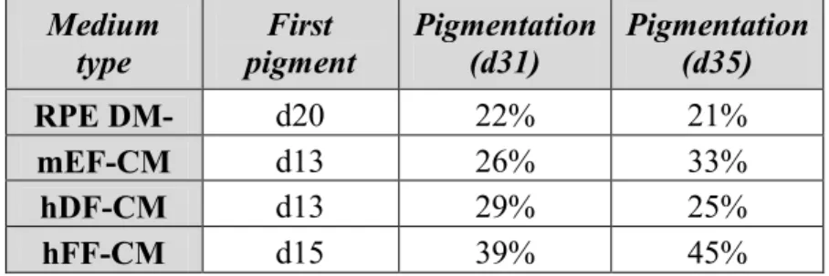
Title: The effect of soluble factors secreted by different types of fibroblasts on the differentiation of human embryonic stem cells into retinal pigment epithelial cells. Analysis of growth factor secretion showed that hDF and mEF secreted much more activin A than hFF, whereas TGF- secretion was greatest in hFF.
INTRODUCTION
REVIEW OF THE LITERATURE
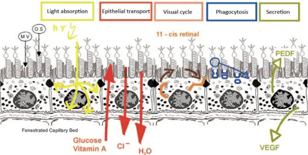
- RETINAL PIGMENT EPITHELIUM
- Retinal degeneration
- Cell replacement therapies
- STEM CELLS
- Human embryonic stem cells
- DIFFERENTIATION OF hESC S TOWARDS RPE CELLS
- Cell culture conditions
- Characterization of hESC-RPE cells
- FEEDER CELL LAYERS AND hESC S
- Mouse embryonic fibroblasts
- Fibroblasts of human origin
- SOLUBLE FACTORS AND RPE
- Activin A, bFGF and TGF-
- Other soluble factors
This leakage damages photoreceptors and rapidly leads to loss of vision if left untreated (Chakravarthy et al., 2010). It is unclear how dysfunction of BEST1 relates to accumulation of lipofuscin (Xiao et al., 2010). This effect can be attributed to the apparent increased deposition of ECM in RPE cells cultured in the presence of TGF-2 (Kubota et al., 2006).
AIMS OF THE STUDY
MATERIALS AND METHODS
CELL CULTURE
- Stem cells
- Feeder cells
- Collection of conditioned media
- Differentiation culture
- Maturation of hESC-RPE
Coating was performed by adding 10 ml of 0.1% gelatin (in ddH2O, sterilized by autoclaving) to each flask and after a 2 hour incubation at room temperature, excess gelatin was removed and flasks were rinsed twice with Dulbecco's phosphate buffered saline (DPBS). (Lonza Group Ltd). When all fibroblast cultures reached confluence, they were detached using TrypLE Select (Life Technologies): culture media was aspirated and cells were rinsed twice with DPBS, 5 ml of prewarmed TrypLE Select was added to each flask and allowed to run at 37 step. °C for about 15 min. Once the cells detached from the plastic, 5 ml of the appropriate pre-warmed culture medium was added to each flask and the resulting single-cell suspensions were collected in 15 ml Falcon tubes.
Single-cell suspensions were centrifuged at 1000 rpm for 3 min, after which the supernatants were aspirated and the cell pellets were resuspended in 1 ml of the appropriate culture medium. Each single-cell suspension was treated with radiation (40 Gy) in order to inactivate the mitotic activity of the cells. For a period of 10 days, the media were collected from the culture dishes and replaced with 16 ml of fresh DM-RPE.
Differentiation in a 6-well plate was performed using each type of CM (mEF-CM, hDF-CM and hFF-CM) and RPE DM- served as a control. To replace the medium, the plates were tilted until clusters of cells settled to the bottom of each well and approximately 80% of the medium was gently aspirated.
EVALUATION OF DIFFERENTIATION
- qPCR
- RT-PCR
- Immunocytochemistry
- In vitro phagocytosis assay
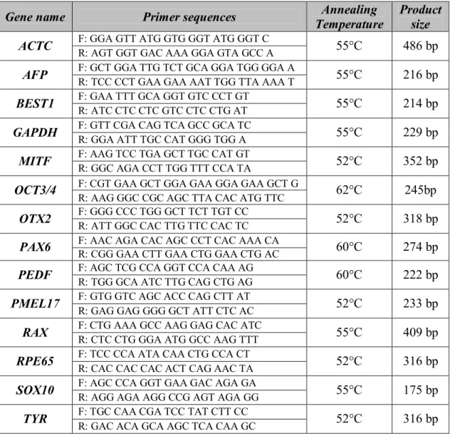
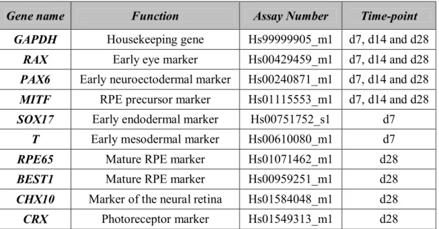
Sequence-specific 20x TaqMan gene expression assays (Applied Biosystems Inc.) used for this purpose are presented in Table 4.1, along with the functions of the genes, assay numbers, and times at which their expression was analyzed. All samples and controls were run as triplicate reactions in optical 96-well plates with the 7300 Real-time PCR system (Applied Biosystems Inc.) as follows: 2 min at 50 °C, 10 min at 95 °C, and 40 cycles of 15 s at 95°C and 1 min at 60°C, with the measurements taken during the last step of each cycle. In this way, the normalized gene expression of each target gene is compared to that of the calibrator.
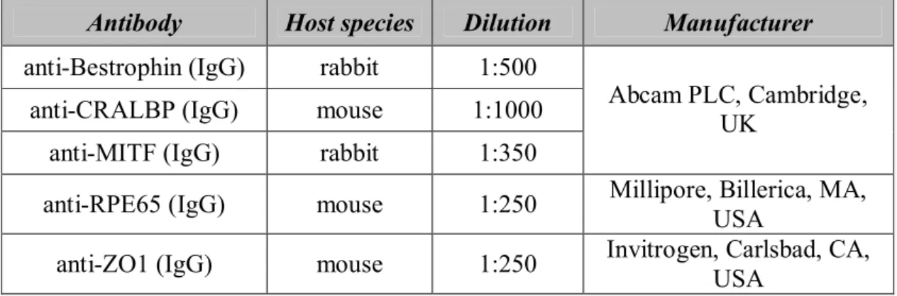
Gels were run for 50 min at 90 V and bands were visualized with a quantity of one 4.5.2. Each piece was treated for 1 hour with one or two of the primary antibodies listed in Table 4.3 – anti-CRALBP and anti-MITF were used as dual staining and the remaining antibodies as single staining. FITC stock solution (50 µg/µl) was diluted to 1 µg/µl with 0.1 M NaHCO3 and 4 µl of the resulting dilution was added to each POS sample and incubated for 1 hour.
After two washes with DPBS, they were treated with 0.2% Trypan Blue for 10 minutes at room temperature - to quench the signal from unphagocytosed POS - and then washed with DPBS until clear. Permeabilization was performed with 0.1% Triton X-100 for 10 minutes at room temperature, after which cell aggregates were washed twice with DPBS.
ENZYME-LINKED IMMUNOSORBENT ASSAYS
RESULTS
- CELL MORPHOLOGY AND PIGMENTATION
- GENE EXPRESSION IN SUSPENSION CULTURE
- CHARACTERIZATION OF MATURE hESC-RPE
- Gene expression
- Protein localization
- Assessment of functionality
- ANALYSIS OF KEY GROWTH FACTOR SECRETION
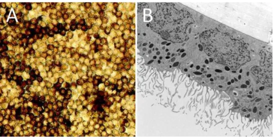
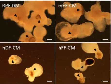
In mEF-CM, cobblestone morphology appeared earlier and spread faster than in other cultures, although pigmentation remained quite weak until the end of the experiment (Figure 5.2F). Nevertheless, areas not obscured by stratification had clear RPE morphology and were pigmented (Figure 5.2H). The most prominent and widespread pigmentation was observed in hDF-CM starting from d17 onwards (Figure 5.2G).
Good cell morphology was observed in the center of the monolayer despite its weak adhesion properties (Figure 5.2E). As seen in Figure 5.3, expression of PAX6 continues to increase after 28 days in differentiation culture, while expression of RAX at the latter time point decreased in cells grown in mEF-CM and hFF-CM. This pattern is observed in Figure 5.4 - gene expression is slightly down-regulated after 7 days in differentiation culture, but reaches a 10-20 fold increase after 28 days, regardless of culture conditions.
As seen in Figure 5.5, expression levels of both genes are low in cells grown in RPE DM- or mEF-CM, whereas those grown in hDF-CM express high levels of SOX17 but not T. Finally, after 28 days in suspension culture ( Figure 5.10), several differences in levels of gene expression were observed. In images taken at a higher magnification, different amounts of pigment granules are visible in each cell (Figure 5.12).
A representative image where an internalized POS is visualized using orthogonal section is presented in Figure 5.13.
DISCUSSION
EVALUATION OF DIFFERENTIATION
At the end of the suspension culture phase, the strongest pigmentation was in hFF-CM, especially in relation to the size of the aggregates: they were generally small and separate from each other, and either almost completely pigmented or not pigmented at all. The amount and size of cell aggregates in each medium were approximately equal at the start of the study, meaning that cell density was not the issue in this case. This will then lead to insufficient distribution of soluble factors and nutrients to the cells located further from the surface of the aggregates.
Perhaps this phenomenon could be minimized by dividing the cell aggregates into multiple wells, thus reducing the cell density in each of the culture conditions. During this phase of the study, cell aggregates were randomly selected from each suspension culture at three time points and differences in expression levels of a range of genes were quantified. Unfortunately, this was not possible within the time frame of this study due to the fact that the amount of available hESCs of the same cell line and passage was limited.
Based on the results of the qPCR assays, mEF-CM appears to be somewhat better at promoting differentiation of hESCs to RPE cells during the first few weeks in differentiation culture. Second, expression of the early eye marker RAX began to decrease after 28 days in differentiation culture, which is expected to occur when early neural progenitors progress to RPE-specific progenitors.
CHARACTERIZATION OF MATURE hESC-RPE
This suggests that cell aggregates differentiated in mEF-CM contained the highest amount of hESC-RPE cells after 28 days in suspension culture. The hESC-RPE monolayers under all culture conditions developed cell morphology intrinsic to mature RPE cells. The hESC-RPE monolayer in RPE DM- appeared to have the weakest adhesion properties as it began to detach from the substrate, while the others remained attached until the end of the experiment.
Perhaps if a larger amount of pigmented cells were obtained from aggregates of differentiated cells in DM- RPE, a better quality hESC-RPE monolayer would have been formed. However, it is clear that hESC-RPE cells constituted the majority of the monolayers, as evidenced by successful immunocytochemical stainings and in vitro phagocytosis assays, as discussed below. In all likelihood, differences in quality between hESC-RPE monolayers of different culture conditions were due to technical problems rather than culture conditions.
Finally, although stainings were successful in all samples, it is possible that prolonged maturation of hESC-RPE monolayers in adherent culture would further improve their quality. It is also possible that binding and uptake of POS by hESC-RPE can be improved by varying the incubation time during which cells are allowed to interact with POS.
SOLUBLE FACTORS SECRETED BY FIBROBLASTS
There is evidence that increasing numbers of POS are internalized by hESC-RPE over time, and the incubation time can be as long as 20 h (Carr et al., 2008). It is possible that performing the assay on cells in adherent culture would give better results that would be easier to visualize. The fact that PEDF was undetected in all four media is inconsistent with the studies that found this growth factor in mEF-CM using proteomics (Lim & Bodnar, 2002; Chin et al., 2007).
The commercial ELISA kit used in this study was human specific, which could easily explain why PEDF was undetected in mEF-CM, given that the mouse and human homologues of this protein are not identical. Measurement of PEDF concentration could be used in the characterization of mature hESC-RPE cells - they should secrete PEDF and therefore its concentration would be high in hESC-RPE-CM. Therefore, it would be possible to compare different culture conditions by comparing the levels of PEDF secretion.
The aim of this small-scale growth factor analysis was to get an idea of how different the secretion of soluble factors really is in different types of fibroblasts. Of course, no individual growth factor is exclusively responsible for how media conditioned by fibroblasts affect differentiation.
CONCLUSION
Molecular characterization and functional analysis of phagocytosis by human embryonic stem cell-derived RPE cells using a novel human retinal assay. Effects of extracellular matrix and neighboring cells on the induction of human embryonic stem cells into retinal pigment epithelial or retinal progenitors. Derivation and comparative evaluation of retinal pigment epithelium from human embryonic stem cells using transcriptomics.
Proteome analysis of nutrient layer-conditioned medium of mouse embryonic fibroblasts supporting human embryonic stem cell growth. Autologous retinal pigment epithelium and choroid transplantation in the treatment of age-related neovascular macular degeneration. Comparative evaluation of different human nutrients for prolonged undifferentiated growth of human embryonic stem cells.
Effects of eight growth factors on the differentiation of cells derived from human embryonic stem cells. Embryonic stem cells adopt a primitive neural stem cell fate in the absence of extrinsic influences.