Bone morphogenetic proteins (BMP) are a group of growth factors that have been linked to various cancers, including breast cancer. BMPs have been noted to have different effects on breast cancer cells, reducing proliferation in some, and conversely increasing migration and invasion in others. Methods: The breast cancer cell lines (estrogen receptor-positive BT-474, MCF-7, MDA-MB-361, T-47D and ZR-75-30, and the estrogen receptor-negative MDA-MB-231) were cultured in estrogen-free medium for three days before treatment with 17β-estradiol (E2, 100 nM) or vehicle control for 24 and 48 h.
Introduction
Literature Review
- Breast cancer
- Estrogen and breast cancer
- Normal functions of estrogen in the body
- Effects of estrogen on breast cancer development and growth
- Endocrine therapies for breast cancer
- Bone morphogenetic proteins
- BMP signaling
- BMPs in cancer
- BMPs in breast cancer
- Estrogen and BMP interactions
- Cross-talk between TGF-β and estrogen
- Cross-talk between BMPs and estrogen
Estrogen receptor (ER) and progesterone receptor (PR) status in breast cancer patients has been determined to predict response to endocrine therapy (Shah et al. 2014). Gene expression profiles are now used to classify breast cancer into luminal A (ERα+, PR+, low proliferation rate), luminal B (ERα-, PR+, high proliferation rate), HER2 overexpressed, and triple negative carcinoma (abbreviated as TNC). ; ERα-, PR-, HER2-) (Huang et al. 2015). The association between ERα and ERβ status and breast cancer survival outcome has been demonstrated in several studies (Huang et al. 2015).
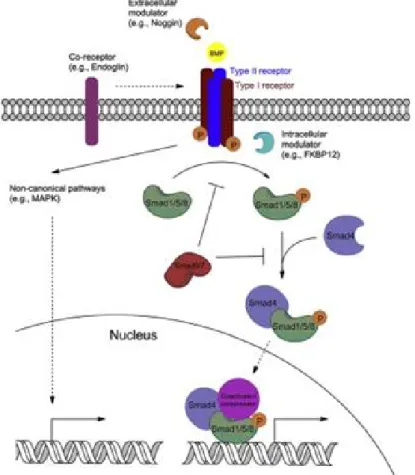
Bone morphogenetic proteins (BMPs) are a group of extracellular signaling molecules belonging to the TGF-β superfamily (Alarmo and Kallioniemi 2010; Carreira et al. 2014). In many cancer types, different BMP ligands appear to be aberrantly expressed (Ehata et al. 2013). Osteoprogeneric cells, osteoblasts, chondrocytes, platelets and endothelial cells produce BMPs in bones (Carreira et al. 2014).
BMPs work in conjunction with other growth factors in a complex cell signaling system (Carreira et al. 2014). The SMAD-dependent BMP signaling pathway has been shown to induce breast cancer cell invasion and bone metastasis (Katsuno et al. 2008). Downregulation of BMP6 has been shown to increase cell proliferation and chemoresistance, implying that BMP6 inhibits growth and migration (Lian et al. 2013).
Decreased expression of BMP7 has been suggested to confer a specific bone metastatic potential in human breast cancer cells (Buijs et al. 2007). BMP2 has been shown to inhibit E2-induced proliferation of MCF-7 breast cancer cells by inducing expression of the cyclin kinase inhibitor p21 (Ghosh-Choudhury et al. 2000). The effect appears to be mediated by positive inhibition of cell cycle regulatory proteins (Ghosh-Choudhury et al. 2000).

Objectives
Materials and Methods
Gene expression analysis
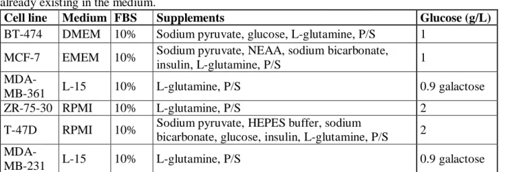
- Cell lines
- Stripping
- Estrogen treatment
- RNA extraction and cDNA synthesis
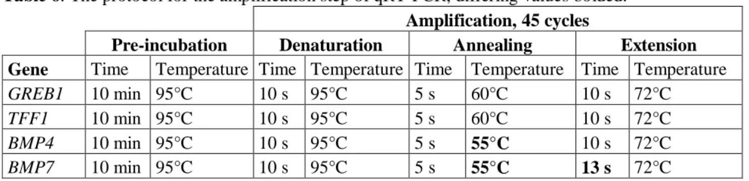
- Quantitative real-time polymerase chain reaction (qRT-PCR)
- Analysis of gene expression data
The cells were cultured in the estrogen-free medium for three days, after which estrogen treatment or vehicle treatment medium was applied. After 24 hours and 48 hours in the E2 or vehicle medium, the cells were lysed with the lysis buffer from the RNA Plus RNA extraction kit (Macherey-Nagel, Düren, Germany) and three parallel wells were pooled. For qRT-PCR analysis, the RNA samples were synthesized into cDNA with the Invitrogen SuperScript® III First-Strand Synthesis System for RT-PCR (Thermo Fisher Scientific, Waltham, MA, USA), using random hexamers as primers.
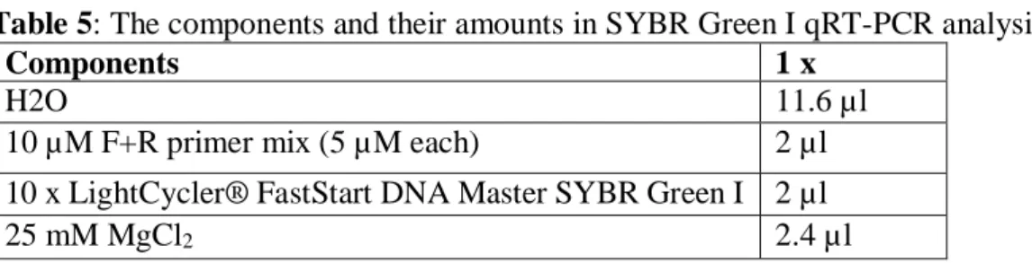
qRT-PCR analysis was performed using the Roche LightCycler® 2.0 device, with software version 4.05 (Roche, Basel, Switzerland). In the SYBR Green I-based assay, the LightCycler® FastStart DNA Master SYBR Green I kit from Roche was used. At the beginning of the SYBR Green I assay, the components (shown in Table 5) were mixed to prepare a reaction mixture.
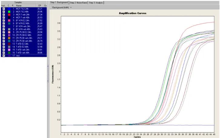
18 µl of the reaction mixture was pipetted into each LightCycler® capillary and then 2 µl of the sample cDNA (1:10 dilution) was added. Ct values (threshold cycle) for each gene in all samples and corresponding controls were acquired in the qRT-PCR analysis. This exponential curve is obtained by measuring the accumulating fluorescence during each cycle, which in turn is comparable to the amount of double-stranded DNA generated in the reaction.
The start of the straight line is extended to intersect with a horizontal crossing line, creating an intersection (i.e., the threshold cycle, Ct).
Functional experiments with estrogen and BMP4
- Cell culture
- Estrogen and BMP4 treatment
- Cell proliferation assay
- Statistical analyses for functional experiments
For experiments with six parallel samples, the Mann-Whitney test was used to assess the statistical difference between differently treated samples.
Results
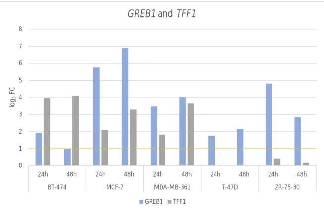
Effects of E2 treatment on BMP expression
Biological replicates for the E2 treatment were assessed with similar results, and technical replicates for qRT-PCR were also performed.
Effects of E2 and BMP4 treatment on cell proliferation
The vehicle-treated cells made raft-like formations, but with apparently fewer cells, as did the cells treated with both estrogen and BMP4. Both estrogen and estrogen- and BMP4-treated cells spread and formed a confluent layer of cells at the bottom of the well, whereas in estrogen-free environment the cells grew in patches. The cells were counted on day 0 (after 72 hours of estrogen depletion) to determine the actual amount of cells in the wells, because the estrogen depletion negatively affected cell growth.
The count of BT-474 cells at day 7, seen in Figure 9A, showed that the vehicle control and BMP4-treated cells were the least proliferative, at approximately 70,000 cells/ml, when the cells originally were at 80,000 cells/ml. ml was sown. . With the combination of E2 and BMP4 treatment, the cell count was significantly higher than with the vehicle or BMP4-treated cells, but significantly less than with only E2-treated cells. The BT-474 cell proliferation was reduced 57% when treated with both E2 and BMP4 compared to only E2-treated cells.
The differences in cell numbers are statistically significant between all but vehicle and BMP4 treatments. The estrogen depletion did not seem to affect T-47D as much, so the difference in cell counts between vehicle and BMP4-treated cells was significant. The estrogen treatment significantly increased proliferation, while combined E2 and BMP4 treatment reduced it by 65% compared to the E2 treatment.
There was no significant change between E2 and BMP4 combination treatment, and BMP4 only treated cell counts, but significant differences could be detected between other treatments.

Discussion
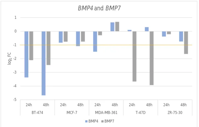
Estrogen inhibits the expression of BMP4 and BMP7 in a cell line dependent manner
Also, as expected, the ER-negative MDA-MB-231 cell line showed very low expression of both GREB1 and TFF1 and did not show any changes after E2 treatment. BMP4 expression after E2 treatment has not been studied before, at least not in the studies found. In particular, BMP4 expression was reduced after E2 treatment in three of the five ER-positive cell lines: BT-474 MCF-7 and MDA-MB-361.
BT-474 was the only cell line in which gene expression was downregulated at both time points and only further downregulated over time. BMP7 expression was found to be downregulated after E2 treatment in the MCF-7 cell line (Takahashi et al. 2008), but no other cell lines were evaluated. Taken together, E2 appears to have different effects on BMP expression in different cell lines.
The only cell line in which the expression of both BMP4 and BMP7 was markedly reduced was BT-474. Different cell lines are derived from different individuals, so their genetic background can be highly variable (Heiser et al. 2009). Several different factors are required to regulate transcription, and the expression of these factors varies by cell line.
Moreover, factors other than just E2 are required to regulate BMP expression, and perhaps some of these factors were not present in those cell lines in which E2 did not have a significant effect.
BMP4 treatment attenuates the effects of estrogen on breast cancer cell growth
BMP4 has not been studied in this respect, but it has been shown to inhibit breast cancer cell growth in several breast cancer cell lines (Ampuja et al. When treated with both estrogen and BMP4, both cell lines showed significant reductions in cell proliferation, as in a previous study using of BMP6 and BMP7 (Takahashi et al. 2008) There was no significant difference in proliferation between vehicle and BMP4 in BT-474 cells, but in T-47D cells, BMP4 treatment significantly reduced cell proliferation.
In T-47D, treatment with E2 and BMP4 reduced growth to the same level as treatment with BMP4 alone. In BR-474, however, cells treated with E2 and BMP4 proliferated more than cells treated with BMP4 alone, probably due to the poor tolerance of the cell line to the "stripping" medium. Interestingly, microscopic images of T-47D cells taken on day 5 show that the cells treated with E2 and BMP4 have expanded, and there appear to be more cells than in the well-treated BMP4, although in fact there are almost the same number of cells.
The effect is not seen with BT-474 cells. 2010) show that migration actually decreases in the T-47D cell line after BMP4 treatment. Addition of estrogen affected the morphology of both BT-474 and T-47D cells, and the cells looked more like normal (when cultured under normal conditions). Otherwise, the cells grew quite poorly in the vehicle-treated wells, and treatment with BMP4 only seemed to exacerbate the suffering.
Overall, combined E2 and BMP4 treatment of BT-474 and T-47D cells shows reduced growth compared to cells treated with E2 alone.
Future aspects
This is an important finding that may have clinical relevance for the treatment of breast cancer patients with ER-positive tumors.
Conclusion
Bone morphogenetic protein-2 induces cyclin kinase inhibitor p21 and retinoblastoma protein hypophosphorylation in estradiol-treated MCF-7 human breast cancer cells. First evidence supporting a potential role of the BMP/SMAD pathway in estrogen receptor-positive breast cancer progression. Bone morphogenetic protein signaling enhances invasion and bone metastasis of breast cancer cells through the Smad pathway.
Parallel inhibition of cell growth and induction of cell migration and invasion in breast cancer cells by bone morphogenetic protein 4. Downregulation of BMP6 increases cell proliferation and chemoresistance via activation of the ERK signaling pathway in breast cancer. Functional interaction of fibroblast growth factor-8, bone morphogenetic protein, and estrogen receptor in breast cancer cell proliferation.
Estrogen mediation in breast tumor formation involves both estrogen receptor-dependent and independent genotoxic effects. Bone morphogenetic protein 6 (BMP6) and BMP7 inhibit estrogen-induced proliferation of breast cancer cells by suppressing p38 mitogen-activated protein kinase activation. Induction of estrogen receptor alpha-36 expression by bone morphogenetic protein 2 in breast cancer cell lines.