To be presented with the permission of the Faculty of Health Sciences, University of Eastern Finland for public examination in Kuopio University. In addition, significant ergonomic challenges were observed using some of the reported HSI solutions.
ACKNOWLEDGEMENTS
Jarkko Pesonen, I am grateful for your constant support and our friendship since elementary school; it means a lot to me. Kristian Tarkkio, thank you, our friendship since the beginning of medical school has made these years much more fun and memorable.
LIST OF ORIGINAL PUBLICATIONS
CONTENTS
HELICoid HypErspectral Imaging Cancer Detection HGG High Grade Glioma HSI Hyperspectral Imaging HgCdTe Mercury Cadmium.
1 INTRODUCTION
DEVELOPMENT OF MICROSURGICAL TECHNIQUES
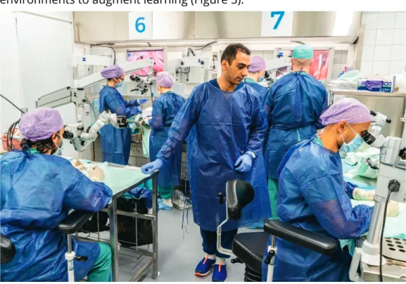
In Finland, the Helsinki Live course was an internationally recognized demonstration course where neurosurgeons worldwide observed and discussed complex neurosurgical operations performed by the masters, such as Yaşargil in the years 2001-2003. In the 2020s, several educational resources are available for learning anatomy, including digitized anatomical books by the pioneer Rhoton and others [12], but also more and more videos, virtual content and increasingly sophisticated simulation models [13,14].
MODERN MICRONEUROSURGICAL TISSUE ANALYSIS
TOWARDS INTRAOPERATIVE HYPERSPECTRAL ANALYSIS Hyperspectral imaging (HSI) is an optical imaging modality that can
2 REVIEW OF THE LITERATURE
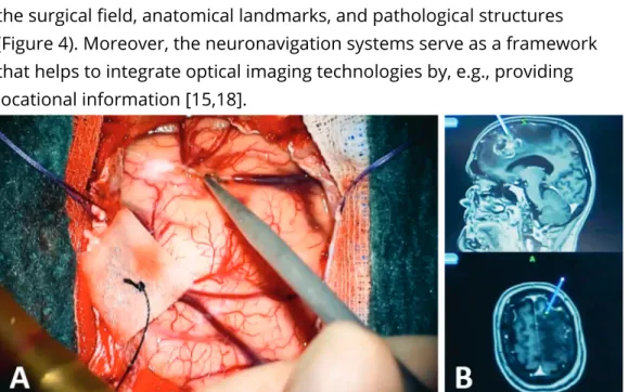
NEUROSURGICAL TISSUE NAVIGATION
- Surgical anatomy and technical skills
- Neuroradiology and neuropathology
- Intraoperative neuronavigation and radiography
- Intraoperative ultrasound
- Intraoperative neurophysiological monitoring
- Fluorescence-guided surgery
- Multimodal tissue navigation
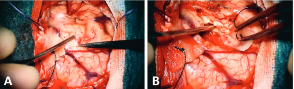
Intraoperative ultrasound uses ultrasound waves to image subcortical or deep targets, such as tumors (Figure 5). In addition to the conventional fluorescent probes, various experimental probes, such as nanoparticles, peptide, affibody, antibody, and.
OPTICAL TISSUE DETECTION IN NEUROSURGERY
- Biophotonic principles
- Neurosurgical optical imaging tools
- Optical properties of neuroanatomical tissues
- Experimental optical tissue detection techniques
Schematic of the illumination path and the optics (A) and illustration of OPMI Pentero 900 (Carl Zeiss, Germany) before surgery (B). The main determinants of the optical properties of CNS tissue are the absorbers and scatterers, mainly oxygenated and deoxygenated.

INTRAOPERATIVE HYPERSPECTRAL IMAGING
- Principle of hyperspectral imaging solutions
- Industrial and medical hyperspectral imaging
- Surgical applications of hyperspectral imaging
- Hardware in hyperspectral imaging systems
- Analysis of hyperspectral images
- Medical hyperspectral imaging databases
- Challenges of surgical hyperspectral imaging
Finally, HSI can be used to observe fluorescence beyond the capabilities of the human eye. PCA reduces the original data set (blue) to a reduced data set (green), which explains most of the variance without losing a significant amount of data. The information from the kernels is propagated through the layers to the fully connected layer, which maps the extracted features into the final output, i.e. the classification (Figure 19) [204].
A detailed description of the structure and principles of CNNs is presented in [212], and current deep learning concepts for HSI medical applications are described in [213]. Illumination requirements are specific to the hyperspectral camera, i.e., the light source should ideally cover the entire imaging wavelength range equally to provide adequate data.

HYPERSPECTRAL IMAGING APPLICATIONS IN NEUROSURGERY
- Neurosurgical hyperspectral imaging systems
- Neuro-oncology
- Neurovascular
- Fluorescence-guided surgery
- Additional indications
Unlike the HELICoiD demonstrator, the system was covered with a sterile drape, as is routinely done. In addition to two case series, case reports of HGG HSI analysis using an RF classifier [149] and PCA [220] have been reported. Hyperspectral images were acquired at 5–16 seconds during superficial temporal artery to middle cerebral artery bypass surgery.
In a proof-of-concept research by Laurence et al., the authors showed that spectral imaging can. Since tumor cell colonies are typical for HGGs, the authors suggested that HSI could be used to identify these subgroups, but histological confirmation is warranted [232].

3 AIMS OF THE STUDY
4 MATERIALS AND METHODS
STUDY I – SPECTRALLY TUNABLE NEURAL NETWORK- ASSISTED SEGMENTATION OF MICRONEUROSURGICAL
Both tissues bear complex segmental anatomy and are key elements in temporal bone microsurgery [258,259]. The single-output fiber optic bundle (Thorlabs OSL2FB, Thorlabs, Inc.) was used to direct the light to the input port of the LCTF device. Mann-Whitney U test was used to assess the differences in spectral reflectance between the grouped images of the tissues, as the assumption of normality was not met for each comparison.
Each of the four types of tissue sample annotations and the corresponding spectral signatures are given in Figure 2. In our study, the optical spectra of well-defined neuroanatomical tissue samples showed modest normal variance at most.

STUDY III – TOWARDS CLINICAL HYPERSPECTRAL IMAGING (HSI) STANDARDS: INITIAL DESIGN FOR A
Introduction
The data were collected using a large line-scan HSI system operating in the visible and near-infrared range from 400 nm to 1000 nm. Accurate anatomical annotations, localization information, or associated magnetic resonance imaging (MRI) scans are not included in the database. Similarly, variant shooting modes lead to different image correction requirements, e.g. different tolerance for movement in the scene in line-scan and snapshot HSI systems.
The database was obtained using wavelength scan and line scan hyperspectral cameras operating in the US. The spectral videos were captured in the range of 380-1000 nm and the still images in the range of 450-950 nm by using snapshot-based hyperspectral cameras and liquid crystals with adjustable filters.
Materials and methods
Metadata description that enables multidisciplinary and clinically relevant reuse of captured HSI data.
A. Intraoperative microneurosurgical HSI system
The camera uses a snapshot technique and a CMOS sensor to capture images in the visible light (VIS) and near infrared (NIR) regions with a resolution of 1024 x 1024 pixels. Thorlabs, Inc., USA) were used to calibrate the system against ambient illumination. Software: Camera manufacturer's software (Senop HSI-2) was used to control data acquisition and determine wavelength ranges and exposure times.
Initially, we recorded a wide range of wavelengths in small increments of 3-5 nm to obtain a full spectrum of non-target reflectance of various tissues in the operating area. Based on preliminary analysis of optimal spectral bands, the wavelength range was narrowed for the target analyte, eg, hemoglobin.

B. Phases of HSI acquisition
Postoperative HSI data processing: Postoperative contrast MRI scans were routinely obtained to evaluate the degree of tumor removal and archived in the HSI database. These data and intraoperative RGB videos were used to annotate and label the hyperspectral images for further development of a practical tissue classification model.
C. Microneurosurgical HSI database
An experienced neuropathologist examined the tissue samples, graded the tumors, and submitted digital histological images to the HSI database. Anatomical annotation and labeling: RGB images of the operating microscope were matched in alignment and scale with the corresponding HSI images. Expert annotations of tissue areas were transferred as labels to hyperspectral images using custom-made software.
Multimodal data incorporation: We collected clinically relevant data and metadata to be included in the HSI database in collaboration with HSI researchers and clinicians. Anatomical and brain mapping data are included as MRI images, including the craniotomy area (Elements. Fibertracking 2.0.0.188 RELEASE, Brainlab Elements).
Results
The shortest wavelength bands showed high noise and bands above 750 nm showed a low signal-to-noise ratio due to insufficient exposure time relative to light intensity.
A. HSI database architecture
Crow's feet notation is used to annotate one-to-one, one-to-many and many-to-many relationships between models. Typical relationships, one-to-one, one-to-many, and many-to-many, while abstract, are still used to relate model instances to each other. An instance of another model can define a foreign key (FK) that references the primary key of another model instance forming a relationship.
This means that an instance of the "Patient" model can be connected to multiple instances. The patient is also linked to multiple hyperspectral images linked to prospective imaging settings, RGB reference images, and annotations.

B. Illustrative case
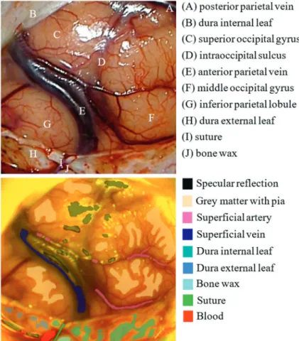
Anatomically annotated operating microscope RGB reference image (top) and the corresponding HSI mask with marked cortical healthy tissue areas (bottom) of the patient ID 0001 after opening the dura. The mean spectral signatures and their standard deviations of the four HSI images of the patient ID 0001 are illustrated in Fig. The selected tissue spectra were collected from different phases of the operation, e.g. after the opening of the dura (Fig. 3.) .
However, the number of pixels of the meningioma class was low compared to the other classes due to flickering of the brain during imaging. Data from preoperative and postoperative MRI scans are very important for mapping intraoperatively visualized anatomy and possible changes such as swelling or infarction of brain tissue.
Discussion and conclusion
To address the focus issues, e.g., no-reference image quality assessment algorithms have proven to be viable options to implement in the future [310]. We aim to expand the HSI indications to e.g. various brain tumors, aneurysms, cranial nerves and spinal nerves, as well as to monitor cortical blood flow and perfusion, in our ongoing HSI data collection program. When sufficient data is collected, we will be able to establish robust HSI models and enhance the operating surgeon's visual field with highlighted features of various tissues.
In the future, DICOM-comparable standards for HSI data are essential for the development of clinically valid intraoperative HSI solutions. Standardization can benefit clinical HSI by leveraging existing enterprise network and workflow services and enabling image and metadata interoperability, as in the case of.
5 DISCUSSION
- DETECTION OF MICROANATOMICAL TISSUE ALTERATIONS In the first study, the spectral behavior of the internal carotid artery (ICA)
- HYPERSPECTRAL IMAGING IN NEUROSURGERY
- CLINICAL HYPERSPECTRAL IMAGING DATABASES AND STANDARDS
- STRENGTHS AND LIMITATIONS
- Societal impact
- CLINICAL IMPLICATIONS
- FUTURE PROSPECTS
In particular, the identification of the facial nerve is a central task in posterior fossa operations, such as vestibular schwannomas [316], but also in peripheral nerve surgery [317]. Similarly, hyperspectral detection of microanatomical injuries, such as ruptures in the adventitia of the artery, could help predict iatrogenic complications such as vasospasm. They had no sterile covering and required separate lighting units, or in the case of the HELICoiD system, a stepper motor [203,214].
As this dissertation illustrates, evaluating a patient's HSI data requires detailed knowledge of the anatomy, gross and microscopic pathology, and limitations of the HSI system. The main limitations of machine learning methods are the large computational requirements, the required amount and quality of training data, and the black box problem [323,324].
6 CONCLUSIONS
Feedforward artificial neural network-based colorectal cancer detection using hyperspectral imaging: a step towards automated optical biopsy. In vivo use of hyperspectral imaging to develop a non-contact endoscopic diagnosis support system for malignant colorectal tumors. Hyperspectral imaging solutions for metabolic and hemodynamic monitoring of brain tissue: past, present and future developments.
The HELICoiD project: a novel use of hyperspectral imaging for real-time brain cancer detection during neurosurgery. Real-time computing of intraoperative hyperspectral imaging for brain cancer detection using multi-GPU platforms. P04.20 Hyperspectral imaging for real-time brain tumor identification and delineation during neurosurgery.
Comparison of hyperspectral imaging and fluorescence angiography for the determination of the transection margin in colorectal resections - a comparative study.
SAMI PUUSTINEN