β-TCP/PLCL composites reliably supported the viability, proliferation and osteogenic differentiation of hASCs in vitro. Characterization and in vitro and in vivo evaluation of β-TCP/PLCL composites foamed with supercritical CO2 for bone applications.
Bone tissue
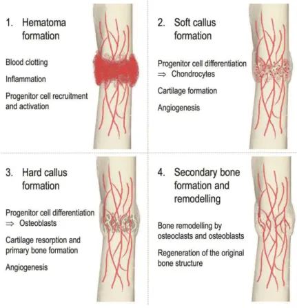
Bone healing
The hematoma serves as the initial framework for the regeneration process, and stem and inflammatory cells begin to migrate to the site of the fracture, where they are activated. The progenitor cells begin to differentiate into chondrocytes, which are the main players in the formation of the cartilage soft callus, which mainly contains collagen and proteoglycans.
Vasculogenesis and angiogenesis
Then, when tissue oxygen levels rise, EC migration ceases (Rouwkema & Khademhosseini, 2016). Arteriogenesis is a maturation phase where the diameter and thickness of the vessel wall increase (Rouwkema & Khademhosseini, 2016).
Cells and differentiation in bone tissue engineering
- Adult stem cells
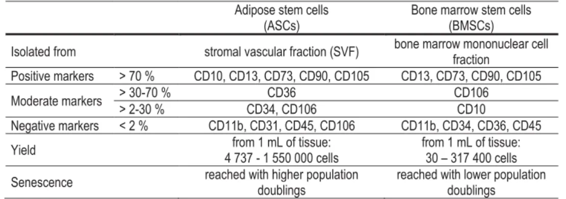
- Adipose stem cells
- Bone marrow stem cells
- Osteogenic differentiation
- Identification of osteogenic differentiation
- Endothelial differentiation
- Identification of endothelial differentiation
- Human umbilical vein endothelial cells modelling
In addition, matrix stiffness has been shown to influence the differentiation of human adipose stem cells (hASC) and human BMSC (hBMSC) (Wen et al., 2014). In addition, topographic cues combined with VEGF stimulus have been shown to induce endothelial differentiation of hBMSCs (Kukumberg et al., 2017).
Biomaterials in bone tissue engineering
- Scaffold requirements
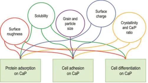
- Calcium phosphates
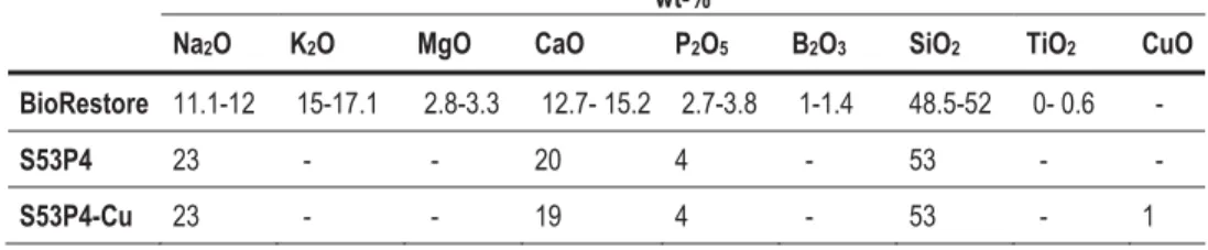
- Bioactive glasses
- Aliphatic polyesters
- Composites
CaP ceramics usually consist of hydroxyapatite (HA) or tricalcium phosphate (TCP) or both known as biphasic CaP (BCP) (Campana et al., 2014; Turnbull et al., 2017). In addition, S53P4 has been shown to induce VEGF release in human fibroblasts in vitro (Detsch et al., 2014).

Bone in vitro models
They then seeded monocytes into the constructs and supplemented the culture system with medium that directed the monocytes to an osteoclast line for an additional 3 weeks (Rossi et al., 2018). The bone model created was able to demonstrate the deposition and remodeling of the bone matrix, which successfully formed the basis for their research related to bone homeostasis (Rossi et al., 2018).
Animal models in bone tissue engineering
Bone formation in vivo
The authors concluded that β-TCP resulted in more pronounced bone formation when compared to Infuse® and an equal level of bone formation as autologous bone graft (H. Yuan et al., 2010). The authors concluded that hBMSCs combined with CaP microparticles and encapsulated in a platelet-rich plasma promoted bone formation and showed higher bone maturation when compared with hASCs, SVF cells or rhBMP-2 (Fennema et al., 2018).
Clinical landscape in bone engineering
Regulation related to commercialization of medical
The regulation of medical devices confirms that there are only safe and functional devices on the market. On the other hand, when targeting the United States (USA) market, medical devices must comply with FDA regulations.
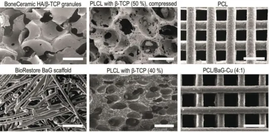
Biomaterial scaffolds used in the study
- Commercial scaffolds
- ScCO 2 foaming of β-TCP/PLCL composites
- Scaffold characterization
- Mechanical testing of scaffolds
- Hydrolytic degradation of composites in vitro and in vivo
- Ion release from PCL/BaG-Cu composites
The Cu2+ doped BaG/PCL composites in study IV were 3D printed with direct ink writing as described in the original publication. In study III, mechanical testing of scCO2-cured β-TCP/PLCL composites was performed for both pristine and pre-compacted scaffolds (Ø = 8 mm, h mm) both dry at RT and in a soln. aqueous at 37 °C with Instron Electropuls E1000 (High Wycombe, UK) as described in the original publication.
Cell isolation, characterization and culture
- Human adipose stem cells
- Human bone marrow stem cells
- Human umbilical vein endothelial cells
- Cell characterization
To verify the mesenchymal origin of hASCs (studies I-III) and of hBMSCs (study IV) and to characterize the isolated HUVECs, the surface marker expression of the cells was determined by a fluorescence-activated cell sorter (FACSAria; BD Biosciences, Erembodegem) , Belgium) to reads 1-3 as described in the original publications. Their surface marker profile verified the mesenchymal origin of the isolated cells (Bourin et al., 2013; Dominici et al., 2006).
Optimization of endothelial medium for hASCs
Based on the results of the comparison, which is described in more detail in the original publication, EM was selected for the 3D experiments in Study II. Surface marker expression of hASCs and hBMSCs cultured in HS or FBS used in studies I-IV.
Cell culture in different scaffolds
The hASCs seeded on day 7 were cultured in EM for 5 days before cell seeding in scaffolds. In EM cocktail group, hASCs were cultured in EM for 5 days before cell seeding 60,000 cells in the scaffolds on day 0. The hASCs seeded on day 7 were cultured in OM for 5 days before cell seeding on day 7.
Cell viability and proliferation
In Study III, the hASCs were seeded into β-TCP/PLCL composites at a density of 510 cells/mm3 in 50 μL medium. Then, 20,000 HUVECs were seeded into the scaffolds and the medium was changed to EGM-2 (Lonza, Basel, Switzerland) for the remainder of the experiment. OR, USA) as described in the original publications. In study IV, hBMSC viability was also quantitatively assessed using a lactate dehydrogenase (LDH) activity assay (Abcam, Cambridge, UK) as described in the original publication.
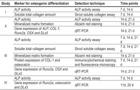
Analysis of osteogenic and endothelial differentiation in vitro
Proliferation was analyzed quantitatively in studies I–IV at different time points by determining the total amount of DNA by the CyQUANT Cell Proliferation Assay Kit (Molecular Probes, Eugene, OR, USA; Invitrogen) as described in the original publications. Endothelial (PECAM-1, vWF and CD144) and osteogenic marker proteins (COL-1 and OCN) were detected by immunocytochemistry using an indirect staining method in studies II-IV as described in the original publications. Quantitative real-time reverse transcription polymerase chain reaction (qRT-PCR) analysis was used to evaluate the relative expression of osteogenic and endothelial genes in studies II-IV as described in the original publications.
Analysis of bone formation in vivo
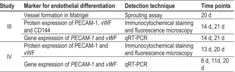
Markers, methods, and time points used to analyze endothelial differentiation and vascularization in studies III and IV.
Statistical testing
In Studies II and III, a Mann-Whitney U test with Bonferroni correction was used as described in the original publications. In study IV, a Kruskal-Wallis test with Mann-Whitney U post hoc test and Bonferroni correction was used as described in the original publication.
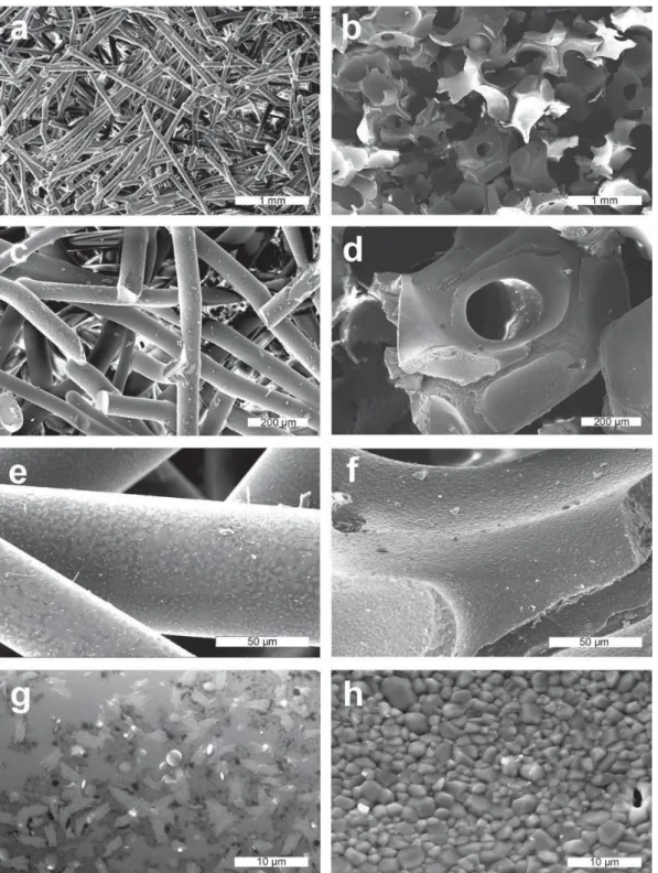
Scaffold characterization
- Scaffold architecture and surface topography
- Mechanical properties of the scaffolds
- Hydrolytic degradation of β-TCP/PLCL composites
- Bioactivity and ion dissolution profile of PCL/BaG-Cu
In study IV, printed PCL/BaG-Cu composites (4:1, 2:1 and 1:1) showed enhanced mechanical properties compared to the PCL framework. A higher concentration of Cu2+ in SBF was measured with PCL/BaG-Cu (1:1) compared to other composites. In contrast, the P concentration remained high in the PCL/BaG-Cu (4:1) composite during the first two weeks.
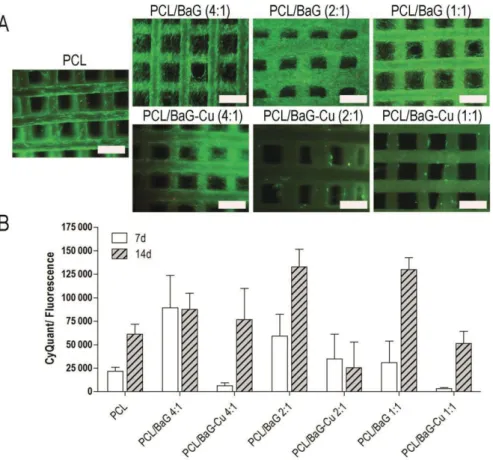
Cell viability and proliferation
Most likely due to high Cu2+ concentrations, hBMSC proliferation was inhibited in PCL/BaG-Cu (2:1) and PCL/BaG-Cu (1:1) composites during the 14-day experiment. According to live/dead staining, the higher copper content had an inhibitory effect on hBMSC amount on scaffolds (Fig. 8A). In contrast to live/dead staining results, the Cu2+ released from PCL/BaG-Cu (2:1) and PCL/BaG-Cu (1:1) composites did not induce increased LDH levels in the culture medium.
Osteogenic differentiation
- Alkaline phosphatase activity
- Total soluble collagen quantification
- Production of mineralized matrix
- Immunocytochemical staining of osteogenic markers
- Osteogenic gene expression
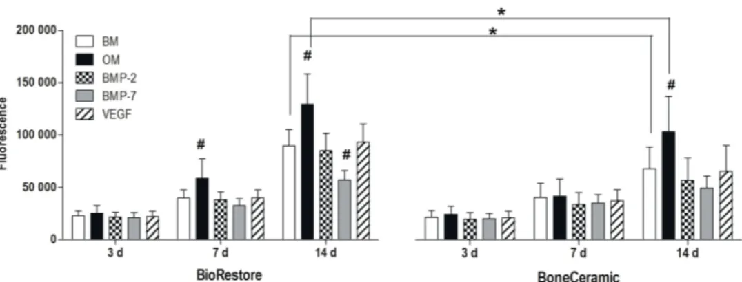
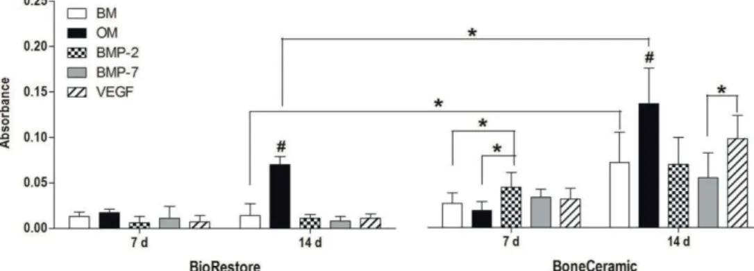
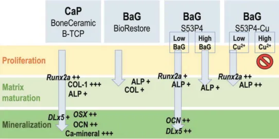
In study II, ALP activity analysis showed significant induction in the EM group compared to BM or OM at 14 and 21 days. In contrast, BMP-2 in BoneCeramic after 7 days induced significantly higher collagen amount compared to BM and OM. However, the expression of Runx2a and OSX was elevated in OM compared to BM at all time points.
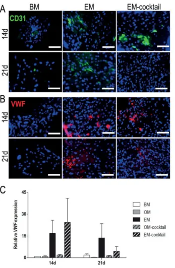
Endothelial differentiation of hASCs in β-TCP/PLCL composites
The gene expression of vWF varied between donors, but the trend between them was similar: vWF expression was increased in EM and EM cocktail compared to the other groups at 14 and 21 day time points (Fig. 16C).
Vascularization in PCL/BaG-Cu composites
The effect of β-TCP/PLCL composites on bone formation in vivo
Scaffold architecture and properties affect cell and tissue response in
In addition, the composites in Study III were pre-compressed, resulting in an increased display of β -TCP on the composite surface compared to the pristine composite. The higher ceramic content in the composite and the demonstration of β -TCP in Study III resulted in the formation of an intensely colored and continuous mineralized matrix on the surface of the composite also in BM. In study IV, PCL/BaG and PCL/BaG-Cu composites supported the formation of tubular structures in hBMSC+HUVEC co-.
The relevance of mechanical properties of BTE scaffolds and the
This increase in modulus may be due to the reorganization of the polymer chains in the aqueous environment at 37. This finding allows the study and observation of the degradation process as well as the subsequent structural changes of the composite in vitro in the future. In comparison, the molecular weight of non-foamed scaffolds after 12 weeks of incubation at 37 °C was at the same level as that of foamed scaffolds (Ahola et al., 2013).
OM induction is more effective than exogenously added BMP-2,
The authors also demonstrated that hASCs were more sensitive to BMP-2 when cultured in HS-based medium in contrast to FBS-based medium (Vanhatupa et al., 2015). Similar to study I, BMP-7 has been shown to have a negative effect on the osteogenic differentiation of mouse BMSCs (Wongwitwichot & Kaewsrichan, 2017). Another study observed a negative effect of BMP-7 on osteogenic differentiation of human periodontal ligament stem cells, however, they also observed an inductive effect of BMP-7 on hMSCs of osteogenic differentiation (Ern et al., 2017) .
Previously, Mathieu and colleagues (2006) concluded that in scCO2 treatment, a higher ceramic content reduces the porosity and increases the average pore size of the composite (Mathieu et al., 2006). Both ALP activity and ALP gene expression were highest at 14 days, which is logical since high ALP expression occurs in the matrix maturation phase of the osteogenic differentiation (Lian et al., 2012). Furthermore, a composite consisting of 60% β-TCP in PLLA has been shown to function similarly to regular β-TCP in osteogenesis in vivo (Aunoble et al., 2006).
In contrast to study II, osteogenic supplements were previously required for osteogenic differentiation of hASCs after endothelial induction (Correia et al., 2014). Other authors have shown that the induction of osteogenic and endothelial differentiation pathways in the same cell culture in vitro is challenging (Correia et al., 2014; Hutton et al., 2013). In fact, it has been suggested that osteogenic and endothelial differentiation pathways may be mutually inhibitory (Correia et al., 2011; Hutton et al., 2013).
Incorporation of copper in PCL/BaG composite does not provide
To assess the macroporosity, μ-CT was used to analyze the internal structure of the scaffolds. The BaG mass % of the framework was determined based on the residue content in the TG analysis. The mechanical responses of PCL and PCL/S53P4-Cu1 frameworks were analyzed in compression mode.
A higher concentration of Cu2+ was observed in SBF from the stem of PCL/S53P4−Cu1 = 1:1 due to the highest burst release in the initial solution. The scaffolds of PCL:S53P4 = 4:1 and PCL:S53P4−Cu1 = 4:1 supported the vascularization and tubule formation in the hBMSC+HUVEC co-culture setup.