© The Author(s) 2023. Published by Oxford University Press on behalf of the European Society of Cardiology.
This is an Open Access article distributed under the terms of the Creative Commons Attribution-
NonCommercial License (https://creativecommons.org/licenses/by-nc/4.0/), which permits non-commercial re-
Post-transcriptional control of hemostatic genes: mechanisms and
1
emerging therapeutic concepts in thrombo-inflammatory disorders
2
Sven Danckwardt
1,2,3,4,5 *, David-Alexandre Trégouët
6, Elisabetta Castoldi
7*3
1.Centre for Thrombosis and Hemostasis (CTH), University Medical Centre Mainz; Mainz, Germany.
4
2 German Centre for Cardiovascular Research (DZHK); Berlin, Germany.
5
3 Posttranscriptional Gene Regulation, University Medical Centre Mainz; Mainz, Germany.
6
4 Institute for Clinical Chemistry and Laboratory Medicine, University Medical Centre Mainz; Mainz, Germany.
7
5 Center for Healthy Aging (CHA); Mainz, Germany.
8
6 University of Bordeaux, INSERM, Bordeaux Population Health Research Center, UMR 1219, Department of Molecular
9
Epidemiology of Vascular and Brain Disorders (ELEANOR), Bordeaux, France
10
7 Department of Biochemistry, Cardiovascular Research Institute Maastricht (CARIM), Maastricht University, Maastricht, The
11
Netherlands
12 13
*Correspondence
14
E-mail: Sven.Danckwardt@unimedizin-mainz.de
15
E-mail: e.castoldi@maastrichtuniversity.nl
16 17
The hemostatic system is pivotal to maintaining vascular integrity. Multiple components 18
involved in blood coagulation have central functions in inflammation and immunity. A derailed 19
hemostasis is common in prevalent pathologies such as sepsis, cardiovascular disorders and, 20
lately, COVID-19. Physiological mechanisms limit the deleterious consequences of a
21
hyperactivated hemostatic system through adaptive changes in gene expression. While this is 22
mainly regulated at the level of transcription, co- and posttranscriptional mechanisms are 23
increasingly perceived as central hubs governing multiple facets of the hemostatic system.
24
This layer of regulation modulates the biogenesis of hemostatic components, for example in 25
situations of increased turnover and demand. However, they can also be ‘hijacked’ in disease 26
processes, thereby perpetuating and even causally entertaining associated pathologies. This 27
review summarizes examples and emerging concepts that illustrate the importance of 28
posttranscriptional mechanisms in hemostatic control and crosstalk with the immune system.
29
It also discusses how such regulatory principles can be used to usher in new therapeutic 30
concepts to combat global medical threats such as sepsis or cardiovascular disorders.
31 32
Abstract word count:
168
Total word count (incl. refs):12.545 33
Keywords: noncoding RNAs (miRNA, lncRNA, circRNA, ceRNA), RNA-binding proteins, RNA 34
(m
6A) and posttranslational protein modification, splicing, polyadenylation, biomarker 35
ACCEPTED MANUSCRIPT
Downloaded from https://academic.oup.com/cardiovascres/advance-article/doi/10.1093/cvr/cvad046/7082876 by Universite de Bordeaux user on 17 April 2023
Introduction
1
In light of the current SARS-CoV2 pandemic, the mechanisms underlying the crosstalk 2
between the hemostatic system and the immune system have received unprecedented 3
attention. This interplay plays a central role in many pathological processes, ranging from 4
sepsis to cardiovascular disease.
5
Perturbations of the hemostatic system are common in sepsis, the leading cause of death in 6
critically ill patients worldwide
1. As a systemic inflammatory response to severe infections, 7
sepsis involves excessive activation of the coagulation system
2. This can result in severe 8
complications such as disseminated intravascular coagulation (DIC), which eventually leads to 9
tissue necrosis, multiple organ failure and death, illustrating that inappropriate amplification of 10
protective host-defense mechanisms can become a devastating alliance of harm
3. 11
Cardiovascular disorders including myocardial infarction, ischemic stroke and venous 12
thromboembolism are the leading global cause of mortality with over 17 million deaths 13
annually
4. The incidence of cardiovascular disorders increases markedly with age, starting in 14
the late 40s, with a dramatic increase occurring at 60 years of age
5. They account for 15
approximately 32% of all deaths worldwide, underscoring the need of illuminating underlying 16
mechanisms and devising therapeutic interventions to treat and prevent cardiovascular 17
disorders
6. 18
The immune system and the hemostatic system are closely linked
7and their responses tend 19
to reinforce each other
8, 9. Activation of coagulation and fibrin deposition in response to 20
inflammation is well known. This led to the emergence of the concept of immunothrombosis, a 21
defense mechanism in which inflammatory cells participate in thrombotic processes, and 22
thrombosis in turn acts as an intravascular effector of innate immunity by limiting the spread of 23
invading pathogens
10. However, a derailed hemostatic response can lead to a situation where 24
coagulation, fibrin deposition and thrombosis contribute to disease, as evidenced by the 25
propagation and exacerbation of atherosclerotic plaques
11. Another example is the systemic 26
activation of coagulation combined with microvascular failure resulting from the systemic 27
inflammatory response to severe infection or sepsis, which eventually contributes to multiple 28
organ dysfunction, such as in septicemia
3or COVID-19
12. 29
The multifaceted and intricate link between hemostasis and inflammation involves crosstalk 30
between both systems at multiple levels
3, 7-11, including coordinated changes in gene 31
expression in megakaryocytes, immune cells, the vessel wall and/or the liver. A notable 32
example is the acute phase response, in which central hemostatic components such as 33
fibrinogen
13, 14, Von Willebrand factor
15, 16and factor VIII
17-21are induced in response to 34
inflammatory signals. Such changes in gene expression are primarily regulated at the level of 35
transcription, and the transcriptional regulation of hemostasis-related genes in physiological 36
and pathological conditions has been well studied
22-27. 37
ACCEPTED MANUSCRIPT
Downloaded from https://academic.oup.com/cardiovascres/advance-article/doi/10.1093/cvr/cvad046/7082876 by Universite de Bordeaux user on 17 April 2023
In the present review we focus on emerging concepts of posttranscriptional mechanisms 1
underlying the control of hemostasis and its crosstalk to other systems. In doing so, we discuss 2
examples of the complexity of the transcriptome architecture arising from the use of alternative 3
transcription start sites, exons and polyadenylation sites, as well as gene regulation by non- 4
coding RNAs (miRNAs, lncRNAs, circRNAs), RNA-binding proteins and mechanisms of RNA 5
modification. Remarkably, many of these regulatory principles also play an important functional 6
role in tuning the immune system
28-32, suggesting conserved regulatory links between both 7
systems. Finally, we also illustrate the emerging therapeutic opportunities on the cusp of a new 8
era of targeted therapeutic approaches
33, exemplified by the recent introduction of novel RNA 9
therapeutics in the hemostatic system
34. 10
11
Role of splicing regulation in the hemostatic system
12
With the completion of the human genome project in 2003, it became apparent that the human 13
genome comprises around 22.000 protein-coding genes, far less than actually required for the 14
functional complexity in higher eukaryotes
35. On the other hand, next generation RNA 15
sequencing and particularly the recently introduced long-read sequencing technologies
36, 37 are16
uncovering a perplexingly complex transcriptome architecture that arises from the use of 17
alternative transcription start sites, exons and polyadenylation sites
38, 39. The combinatorial use 18
of such elements considerably expands genomic information and is subject to dynamic spatial 19
and temporal modulation during development and adaptation (Figure 1).
20
Pre-mRNA splicing, i.e. the accurate removal of introns and ligation of exons, is a pivotal step 21
in the co- and posttranscriptional regulation of gene expression
40. Depending on how the 22
exon/intron structure of the pre-mRNA is decoded by the spliceosome, the same primary 23
transcript may be processed into different mature mRNAs (alternative splicing), encoding 24
different isoforms of the same protein. In fact, the recognition of exon/intron boundaries in the 25
pre-mRNA is critically dependent on the engagement of nearby splicing enhancer and silencer 26
sequences by trans-acting proteins (splicing factors) whose availability varies in different cell 27
types and disease states. As a consequence, splicing patterns are typically regulated in a 28
tissue-specific manner and may change according to the developmental stage or in response 29
to pathological processes. Moreover, they can be disrupted by genetic variants that weaken 30
(or strengthen) the consensus sequences recognized by the spliceosome on the pre-mRNA.
31
This is a well-known mechanism of disease in mendelian disorders
41, but it is increasingly 32
appreciated that much of the genetic variation associated with complex traits also acts by 33
altering splicing patterns
42, 4334
Like most human genes
44, many genes encoding proteins of the hemostatic system are 35
alternatively spliced
45-57. This often results in isoforms with distinct structural and functional 36
ACCEPTED MANUSCRIPT
Downloaded from https://academic.oup.com/cardiovascres/advance-article/doi/10.1093/cvr/cvad046/7082876 by Universite de Bordeaux user on 17 April 2023
characteristics, as exemplified by two major components of the extrinsic coagulation pathway 1
(Figure 2).
2
Tissue factor (TF), the main trigger of blood coagulation, acts as cofactor of the circulating 3
serine protease factor VIIa (FVIIa) and comes in two isoforms: as membrane-bound (full- 4
length) protein and as a shorter, alternatively spliced variant that is secreted in soluble form 5
(Figure 2)
58. The two isoforms are identical at the N-terminal end, but the soluble form, which 6
arises from exon 5 skipping, lacks the transmembrane and cytoplasmic domains, and has a 7
completely different C-terminal sequence
58. Just as full-length TF, alternatively spliced TF is 8
produced by a variety of cell types
58, 59, is induced by pro-inflammatory stimuli
59, 60and 9
enhances factor X (FX) activation by FVIIa, albeit less potently than full-length TF
58. However, 10
while membrane-bound TF is essential for normal hemostasis, elevated intravascular levels of 11
TF have been proposed to contribute to venous as well as arterial thrombosis
61. Despite 12
conflicting data, it has been suggested that soluble TF, which is most likely dispensable for 13
normal hemostasis, may represent a preferential target for antithrombotic therapy than full- 14
length TF, due to a lower risk of bleeding
62. 15
Tissue factor pathway inhibitor (TFPI) is a glycoprotein that functions as an inhibitor of 16
coagulation and of TF-dependent signaling
63. The
TFPI gene encodes two main splicing17
isoforms that are generated by the alternative inclusion of exon 8 (TFPIβ) or exons 9-10 18
(TFPIα) in the mature mRNA (Figure 2). Both isoforms are expressed in endothelial cells, but 19
TFPIα is also found in plasma, platelets and the extracellular matrix
64. Structurally, TFPIα 20
comprises an acidic N-terminus, three Kunitz domains and a basic C-terminus, whereas TFPIβ 21
lacks the third Kunitz domain and the basic C-terminus, which are replaced by a 22
glycosylphosphatidylinositol-anchor that tethers the protein to the cell membrane
65. Both TFPI 23
isoforms inhibit TF/FVIIa and FXa with their Kunitz-1 and Kunitz-2 domains, respectively, but 24
TFPIα has additional properties by virtue of its Kunitz-3 domain (which binds protein S) and 25
basic C-terminus (which binds FV/FV-short). Binding to protein S and FV/FV-short prevents 26
the clearance of plasma TFPIα from the circulation
51, 66, 67and promotes its association with 27
biological membranes, enhancing its anti-FXa activity
68-70. Moreover, the interaction with 28
FV/FV-short allows TFPIα to inhibit FV activation
71and early prothrombinase activity
72, 73, while 29
TFPIβ lacks these anticoagulant functions.
30
These and other
51, 74examples illustrate how alternative splicing can change the structural and 31
hence functional properties of central components in the hemostatic system
75. Extracellular 32
signals, such as pro-inflammatory cytokines, can modify global patterns of alternative splicing
7633
and it will be interesting to explore how this plays out in different (disease) contexts, including 34
COVID-19
77. Moreover, since alternative splicing is pervasive and there are increasingly new 35
therapeutic means to (re)direct splicing
78, 79, modulation of alternative splicing may become 36
relevant for the therapeutic manipulation of the hemostatic system. In particular, many studies 37
ACCEPTED MANUSCRIPT
Downloaded from https://academic.oup.com/cardiovascres/advance-article/doi/10.1093/cvr/cvad046/7082876 by Universite de Bordeaux user on 17 April 2023
support the utility of antisense oligonucleotides (ASOs) to mask specific splicing signals on the 1
pre-mRNA and thus prevent the recognition of these sequences by spliceosomal components, 2
thereby re-directing splicing
80. Alternatively, ad hoc engineered U1snRNA can be employed to 3
promote the usage of donor splice sites that are naturally weak or have been disrupted by 4
mutation
81. 5
Apart from diversifying the transcriptome and proteome, alternative splicing has been 6
proposed to contribute to the overall regulation of gene expression through its coupling with 7
nonsense mediated decay (NMD), a surveillance pathway that degrades mRNAs containing 8
premature stop codons. In fact, it has been observed that up to one third of all human 9
transcripts are normally spliced into non-viable mRNAs that are substrates for NMD. This 10
phenomenon, known as “regulated unproductive splicing and translation” (RUST), has been 11
interpreted as a mechanism for the post-transcriptional temporal and spatial fine-tuning of gene 12
expression
82. Evidence that this control mechanism may apply within the realm of hemostasis 13
has been provided for the F11 gene, encoding coagulation factor XI
47. Interestingly, targeting 14
non-productive splicing by antisense oligonucleotides can be exploited for the upregulation of 15
gene expression from wild-type or hypomorphic alleles in disease states
83. 16
17
Role of polyadenylation in the hemostatic system
18
In addition to capping and splicing, almost all eukaryotic transcripts undergo further processing 19
at the RNA 3’-end (Figure 1). For most genes, this involves endonucleolytic cleavage and non- 20
templated polyadenylation (CPA) before the mature RNA can be exported to the cytoplasm
84. 21
As CPA controls almost all genes, regulation of CPA has evolved as an important layer of gene 22
expression regulation. CPA is carried out by a multi-subunit complex involving over 80 trans- 23
acting proteins organized in four core protein subcomplexes
85. The recruitment of these 24
multimeric complexes to dedicated, but largely poorly conserved, RNA sequence elements
8625
ensures that 3’-end processing of the nascent transcript occurs timely and at the right 26
position
87, 88. Perturbations of this process - due to mutations in RNA sequence elements or 27
defects in the RNA processing machinery - have drastic consequences, as exemplified by 28
numerous diseases
89, 90. 29
The common thrombophilia mutation in the prothrombin (F2) gene (F2 G20210A) is a prime 30
example of how mutations in noncoding regions can become pathogenic
84. This mutation 31
affects the last nucleotide of the 3’-untranslated region (UTR), where the pre-mRNA is cleaved 32
and polyadenylated. As a result of the mutation, the efficiency of endonucleolytic cleavage is 33
increased, leading to more prothrombin mRNA and protein expression. Although this mutation 34
merely increases the amount of the precursor of a central hemostatic component (i.e., 35
thrombin), it already shifts the balance of the hemostatic system toward a procoagulant 36
condition
91-93. Consequently, the expression of
F2 must be tightly controlled: even small37
ACCEPTED MANUSCRIPT
Downloaded from https://academic.oup.com/cardiovascres/advance-article/doi/10.1093/cvr/cvad046/7082876 by Universite de Bordeaux user on 17 April 2023
changes (1.5- to 1.7-fold) in gene expression due to mutations at this and other nearby 1
positions (F2 C20209T and
F2 G20221T)93, 94can result in clinically relevant thrombophilia
94-2
97
. 3
Compared to other genes, the architecture of sequence determinants directing 3’-end 4
processing in F2 is unconventional
96. It consists of weak signals, which explains the unusual 5
susceptibility to thrombophilic gain-of-function mutations
94, 97. At the same time, this 6
configuration allows for mechanisms that enhance processing and thereby upregulate
F27
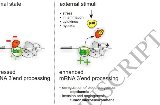
expression when needed
98. This is achieved through complex, mutually exclusive binding of 8
suppressive and stimulatory RNA binding proteins (RBPs), and is regulated by activation of 9
p38 MAPK (Figure 3)
99. After phosphorylation by p38 MAPK, inhibitory RBPs (FBP2, FBP3) 10
can no longer bind to the processing sites in the F2 pre-mRNA, allowing 3’-end processing to 11
proceed. Thus, virtually all types of ‘environmental’ conditions that lead to activation of p38 12
MAPK
100, 101can induce F2 expression.
13
Inflammatory conditions are known to trigger F2 expression
102-107. Consistently, the mechanism 14
described here was found to account for the induction of
F2 expression under inflammatory15
conditions, including septicemia
99, 108. While this may contribute to the initial onset and 16
undesirable propagation of hemostatic perturbances during septicemia, such mechanisms 17
may also play a compensatory role
3. After an initial hypercoagulable state, septicemia is often 18
followed by a hemorrhagic phase, in part due to consumption of procoagulant components
109. 19
Such conditions of increased turnover and demand require mechanisms to restore the 20
hemostatic balance and stockpile hemostatic components
110. 21
In addition to the critical function in hemostasis, the role of thrombin in angiogenesis
11122
suggests that regulatory mechanisms have evolved a sensor for low oxygen pressure. This 23
could explain why F2 is overexpressed due to ischemic events
112or in the tumor micromilieu
99. 24
Consistent with its role in oxygen pressure sensing
100, 101, activation of p38 MAPK also drives 25
F2 overexpression in the tumor microenvironment. This activates protease-activated receptors
26
(PARs) that induce genes with a role in angiogenesis and tumor dissemination
99. 27
Thus, regulated 3′-end processing emerged as an important mechanism of gene regulation in 28
the control of the hemostatic system. While such mechanisms are desirable under 29
physiological conditions (to replenish the amount of blood coagulation factors under high 30
turnover, see above), they can be ‘hijacked’ under pathological conditions (such as 31
inflammation or cancer), thereby leading to a thrombophilic state
108, 113. Since prothrombin is 32
expressed in a wide variety of organs and cells
108, this type of regulation may become relevant 33
to numerous other thrombin-mediated diseases
113. However, it also appears that tissue- 34
specific mechanisms can be used to selectively target deleterious prothrombin expression 35
without altering essential prothrombin expression in the liver
108. 36
ACCEPTED MANUSCRIPT
Downloaded from https://academic.oup.com/cardiovascres/advance-article/doi/10.1093/cvr/cvad046/7082876 by Universite de Bordeaux user on 17 April 2023
Targeted interference with cleavage and polyadenylation is increasingly perceived as an 1
important therapeutic means. This involves either redirection of aberrant RNA processing 2
(through ASOs, U1snRNP interference or trans-splicing) or the elimination of faulty 3
transcripts
89to prevent the fatal consequences of aberrant 3’-end processing
114, 115. 4
Perturbations of 3’-end processing can, for example, act as nongenomic oncogenic drivers of 5
tumorigenesis
115, but they also play important roles in inflammatory conditions
116. Deciphering 6
the underlying mechanisms is of paramount importance for establishing targets with 7
therapeutic selectivity and specificity.
8
RNA-protein interactome studies
117and transcriptome-wide profiling of polyadenylation
118are 9
thus central to defining new therapeutic targets, their specificity and downstream 10
consequences
119. Since most miRNA binding sites are localized in the 3’-UTR, when and 11
where a pre-mRNA is polyadenylated has a critical impact on the regulatory properties of the 12
resulting mRNA molecule (see below). A significant proportion of genetic variants in 3’-UTRs, 13
often dismissed as ‘non-functional’ polymorphisms, are therefore likely to disrupt important 14
regulatory mechanisms, ultimately leading to pathologies including a dysbalanced hemostatic 15
system
89. This is supported by the thrombophilia variants discovered in the F2 gene. However, 16
this also extends to other coagulation factor 3’-UTR variants that affect, for example, miRNA 17
regulation
120, 121. 18
19
Role of microRNAs in the hemostatic system
20
MicroRNAs (miRNAs) are small single-stranded non-coding RNAs (17-25 nucleotides in 21
length) that post-transcriptionally down-regulate target gene expression by RNA silencing
122. 22
After transcription, miRNAs are processed in the nucleus by the microprocessor complex 23
consisting of Drosha and DGCR8 to produce a pre-miRNA
123. After export to the cytoplasm 24
and further processing by Dicer
124, the mature miRNA duplex is incorporated into the RNA- 25
induced silencing complex (RISC)
125. This complex is guided by miRNA base pairing to a target 26
gene mRNA resulting in translational inhibition and/or transcript degradation
126. Generally, 27
miRNAs target mRNAs via the 3'-UTR. In a few cases, miRNAs can also carry out their 28
inhibitory function by binding to the coding region or the 5’-UTR of target mRNAs
127. 29
Over 2600 human miRNAs have been identified
128, regulating the majority of human genes
129. 30
Thus almost every biological process is modulated through miRNAs
130. Although miRNAs 31
generally fine-tune gene expression
131, they can also function as master regulators
132. For 32
example, multiple miRNAs can cooperatively silence a single gene to gain regulatory 33
specificity, with the targeting of particular network hub genes enabling the regulation of entire 34
pathways
133. In addition, a single miRNA can target multiple genes, allowing broad regulation 35
of molecular networks
127. Perturbations of miRNA expression are observed in most disorders, 36
ACCEPTED MANUSCRIPT
Downloaded from https://academic.oup.com/cardiovascres/advance-article/doi/10.1093/cvr/cvad046/7082876 by Universite de Bordeaux user on 17 April 2023
with some of them even causally contributing to the development and progression of 1
disease
130. 2
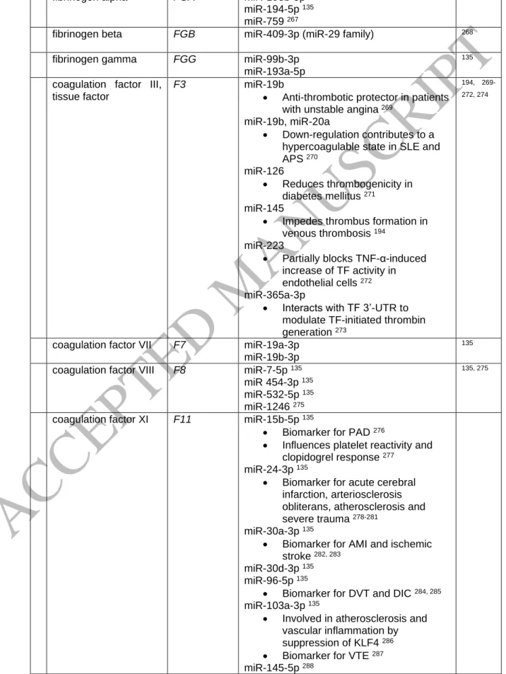
A growing number of studies document a contribution of miRNAs to the regulation of 3
hemostatic
134-138and thrombotic
121, 135, 137-140functions. miRNAs directly regulate multiple 4
hemostatic factors through interactions with the 3'-UTR (Table 1). Additionally, miRNAs can 5
tune hemostatic factors indirectly, for example fibrinogen via interleukin-6-mediated 6
signaling
141, factor IX by repressing NMD
142, plasminogen activator inhibitor 1 (PAI-1) via 7
SMAD2 signaling
143and CXCL12 to reduce inflammatory response and thrombosis, altering 8
the expression of multiple factors including TF, PAI-1 and VWF
144. 9
Further evidence implicating miRNAs in the hemostatic system comes from the important roles 10
that miRNAs play in the development of bleeding disorders and thrombosis. Blood miRNA 11
levels are associated with hemostatic perturbations, suggesting their potential use as 12
prognostic or diagnostic tools in VTE
145and beyond
146. These include aberrant coagulation in 13
sepsis
147, venous thromboembolism
140, 148-158, trauma-induced coagulopathy
159, 14
atherosclerosis
160-164, coronary artery disease
165-167, ischemic stroke
168, 169and autoimmune 15
inflammatory conditions such as systemic lupus erythematosus (SLE)
170-172. 16
Recently, using an unbiased systematic search based on a biophysical miRNA interaction 17
study coupled to high-throughput sequencing, the Atlas of the Hemostatic miRNA Targetome 18
was released
135. This screening identified more than 1500 miRNA/3'-UTR interactions with 19
potential function in the hemostatic system from nearly 4500 miRNA/3'-UTR biophysical 20
interactions
135. A proof-of-concept, rigorous filtering combined with loss-of-function studies 21
(limited to 96 of the 1500 miRNA/3’-UTR interactions with a potential function) identified dozens 22
of miRNAs targeting 27 hemostasis-associated gene 3′-UTRs globally or in a gene-specific 23
manner (Figure 4). This highlights the global importance of miRNAs in controlling the 24
hemostatic system and suggests that many more functional miRNAs will be discovered in this 25
system.
26
The unbiased view on miRNAs regulating the hemostatic system also sheds light on hitherto 27
functionally poorly characterized connections between different physiological systems and 28
diseases. These include the link between tumor formation and hemostatic perturbations
135, or 29
the intricate relationship between the hemostatic system and inflammatory processes (Table 30
1). For example, miR-181 family members that target the 3'-UTR of F11 mRNA
135are involved 31
in several aspects of hemostasis, including vascular inflammation
152, 173-175and platelet 32
activation
176. Another example is miR-24 which controls the expression of VWF
177. Here, 33
hyperglycemia-induced repression of miR-24 increases VWF expression and secretion in 34
diabetes mellitus, linking metabolic dysfunction to a miRNA-mediated mechanism of 35
hemostatic deregulation.
36
ACCEPTED MANUSCRIPT
Downloaded from https://academic.oup.com/cardiovascres/advance-article/doi/10.1093/cvr/cvad046/7082876 by Universite de Bordeaux user on 17 April 2023
On the other hand, polymorphisms affecting miRNA binding sites in hemostatic genes can be 1
associated with disease. For example, deletion of the miR-759 binding site of
FGA is2
associated with susceptibility to chronic thromboembolic pulmonary hypertension
178, and SNPs 3
in the 3′-UTR of the
F2, F8 and F11 genes are associated with increased activity levels of4
these hemostatic components
120, 179-182. 5
The importance of miRNAs in hemostasis is further corroborated by their role in platelet 6
biology
136. Here miRNAs modulate the expression of target mRNAs important for hemostatic 7
and thrombotic function
183-187. For example, miRNA levels are altered in platelets from patients 8
with essential thrombocythemia and this in turn is associated with elevated platelet counts and 9
an increased risk of thromboembolic events
188. Additionally altered miRNA expression is often 10
observed in atherosclerotic plaques
189(and refs therein).
11
In light of the functional importance of miRNAs in the hemostatic system
135and the increasingly 12
recognized role of miRNA therapeutics
190currently conquering the cardiovascular system
191, it 13
is tempting to turn this knowledge into new therapeutics (see targeting section below). In 14
support of this, miRNA treatment has been demonstrated to result in therapeutic response in 15
thrombosis and hemostasis. In murine models of venous thrombosis, overexpression of 16
miRNAs contributes to thrombus resolution
193, reduces thrombogenesis
194, enhances 17
endothelial progenitor cell migration and tubulogenic activity
195, angiogenesis and thrombosis 18
recanalization
196. Furthermore, the use of antagomirs (i.e., molecules that silence miRNAs) 19
has been shown to block miR-19b-3p-mediated silencing of
SERPINC1 (antithrombin),20
resulting in increased antithrombin expression and activity in vivo
135. This documents the in- 21
principle druggability of the hemostatic system in a miRNA-directed manner and opens 22
opportunities to target other hemostatic components such as coagulation FXI
138. 23
24
Other means of posttranscriptional regulation of the hemostatic system
25
RNA binding proteins beyond their function in the biogenesis of mRNAs
26
In addition to co- and posttranscriptional processing, much of the fate of RNAs from synthesis 27
to decay depends on RNA-binding proteins
197. RBPs regulate RNA localization, transport, 28
translation, stabilization and degradation of bound RNA molecules. In fact, much of the rapid 29
adjustment of gene expression in inflammation and the immune system
30-32is executed via 30
modulation of RNA stability and decay. The same is likely to apply to the hemostatic system 31
as well, and the adaptation of prothrombin expression (Figure 3) may be a prototype for 32
analogous occurrences
198. It is interesting to note that even in apparently non-polar cells such 33
as hepatocytes, the major source of most hemostatic components, localization of transcripts 34
and thus protein output critically depends on UTR-RBP interactions
199. This suggests that 35
dynamic changes of 5’ and 3’-UTR structures of mRNAs, due to the use of alternative 36
transcription start sites and alternative splicing/polyadenylation, may have a critical impact on 37
ACCEPTED MANUSCRIPT
Downloaded from https://academic.oup.com/cardiovascres/advance-article/doi/10.1093/cvr/cvad046/7082876 by Universite de Bordeaux user on 17 April 2023
protein output and ultimately function. This is corroborated, for example, by the role of 5’-UTR 1
variants that alter upstream open reading frames in cardiovascular disorders (CVD)
200. 2
3
RNA modification and networks of competitive RNA-RBP binding
4
As soon as the nascent RNA molecules emerge from the RNA polymerase during transcription, 5
they are instantly decorated with RBPs. While this ensures that co-transcriptional processing 6
takes place effectively and at the right position, RBP loading also prevents the hybridization of 7
the nascent RNA molecule with the DNA strand. This helps to avoid the formation of reactive 8
RNA:DNA hybrids (so called R-loops)
201, 202, which can lead to genomic instability
203, 204. Most 9
importantly, binding of RBPs and non-coding RNAs to (pre-)mRNAs can occur in a complex, 10
sometimes mutually exclusive manner, thereby determining the posttranscriptional fate of 11
mRNAs selectively
84or in a global manner
205, 206. This is supported by the observation that the 12
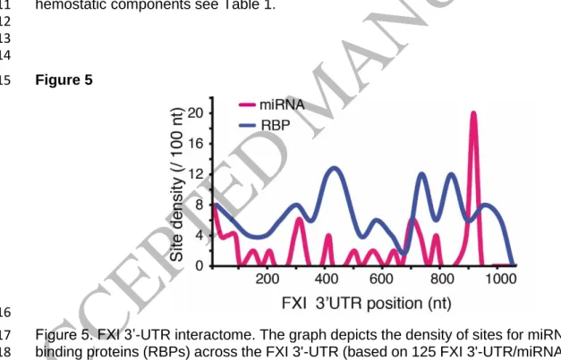
density of RBP and miRNA binding to the UTRs of coagulation factor mRNAs is very high
138, 13
and that numerous RBP and miRNA binding sites are in close proximity (Figure 5).
14
Hence, there must be mechanisms that coordinate the binding of such molecules. Although 15
not yet studied in great detail, it is likely that modifications of both RNAs
207and RBPs
208can 16
result in remodeling of the 3’-UTR-RBP architecture and thereby change the fate of RNAs 17
encoding coagulation factors under inflammatory conditions. In support of this notion, 18
posttranslational modifications of RBPs have been shown to change the fate of mRNAs 19
encoding central hemostatic components (Figure 3)
99. But also variations in N
6- 20
methyladenosine (m
6A), the most prevalent RNA modification with a wide biological impact
209,21
210
, have been documented in various RNA transcripts in vascular tissues of septic rats
211. 22
Additionally, there is growing evidence that m
6A modification is closely related to the 23
development and progression of CVD, including cardiac hypertrophy, heart failure, ischemic 24
heart disease and pulmonary hypertension
212, 213. It is tempting to explore if therapeutic 25
modulation of the cellular m
6A machinery (for example in COVID-19
214) might be useful in 26
preserving vascular integrity and function in sepsis and/or CVD. Interestingly, the fat mass and 27
obesity-associated protein (FTO), one of the few m
6A erasers, has emerged as an important 28
pharmaceutical target in many pathophysiological conditions
209. As many more RNA 29
modifications are currently being discovered
215, this holds great potential for systematically 30
uncovering their importance in human diseases and defining novel therapeutic avenues.
31 32
Long non-coding RNAs and circRNAs
33
Despite the unexpectedly small number of protein-coding genes identified by the human 34
genome project, RNA sequencing has shown that up to 85% of the human genome is 35
transcribed
216. This led to the identification of a large number of non-coding RNA molecules 36
with regulatory functions
217. In contrast to small non-coding RNAs (such a miRNAs, snoRNAs 37
ACCEPTED MANUSCRIPT
Downloaded from https://academic.oup.com/cardiovascres/advance-article/doi/10.1093/cvr/cvad046/7082876 by Universite de Bordeaux user on 17 April 2023
or piRNAs), long-noncoding (lnc)RNAs are around 200 nucleotides or more
218and often 1
undergo alternative splicing, which further expands their repertoire. LncRNAs can bind to DNA, 2
mRNAs, miRNAs and proteins depending on sequence and secondary structure, thereby 3
modulating gene expression under physiological and pathological conditions
219. Their modes 4
of action include epigenetic, transcriptional and post-transcriptional mechanisms. Accordingly, 5
this new class of ncRNAs is increasingly taking center stage in the modulation of the 6
cardiovascular system. As an example, lncRNA H19 is involved in the pathogenesis of 7
atherosclerosis
220. The expression of lncRNA H19 is significantly increased in patients with 8
ischemic stroke compared to healthy controls
221. Genome-wide association studies have 9
identified SNPs in the lncRNA ANRIL associated with CVD, such as coronary atherosclerosis 10
and cardiac infarction
222, 223, while variants in lncRNA ZFAS1 are associated with susceptibility 11
to ischemic stroke
224. Recently, a transcriptome wide association study on VTE also revealed 12
further lncRNA hits (RP11-747H7.3, RP4-737E23.2)
225, corroborating their function in CVD.
13
Unlike miRNAs or proteins, lncRNA function cannot currently be simply inferred from sequence 14
or structure, and the diversity of lncRNAs described to date precludes simple 15
generalizations
219. In the context of the hemostatic system, this hitherto poorly explored area 16
deserves attention. This is also supported by the role lncRNAs have in platelets
226, 227, although17
their role is still under active investigation. In analogy to the central regulatory function of non-
18
coding RNAs in the immune system and because of the resulting therapeutic implications
228, 19
it will be important to better understand the pathophysiological dimension of this class of 20
regulators in thrombosis and its connection to inflammation.
21
Circular RNAs (circRNAs) are another class of endogenous non-coding regulatory 22
biomolecules. They are prevalent and arise from a non-canonical splicing event called 23
‘backsplicing’
229. They exert important biological functions by acting as miRNA or protein 24
sponges, by regulating protein function or by being translated
230. As such, circRNAs regulate 25
a plethora of biological functions including ROS formation and cardiovascular metabolic 26
inflammation
231. Accordingly, perturbations of these process(es) can become pathogenic and 27
result in CVD. For example, a haplotype on 9p21 that protects against coronary artery disease 28
has been shown to be associated with the abundance of circRNA ANRIL, which in turn 29
regulates ribosomal RNA maturation, conferring atheroprotection
232. Accordingly,
circANRIL30
has been proposed as a potential therapeutic target for the treatment of atherosclerosis. The 31
in-principle therapeutic utility of circRNA is also supported by recent preclinical observations 32
demonstrating their use, for example, to attenuate cell apoptosis in cerebral ischemia- 33
reperfusion
233. Finally, circulating circRNA may have diagnostic potential and serve as 34
biomarkers for acute ischemic stroke
234and even help distinguish different etiologies (i.e., 35
atherothrombotic, cardiothrombotic vs undetermined stroke)
235. 36
37
ACCEPTED MANUSCRIPT
Downloaded from https://academic.oup.com/cardiovascres/advance-article/doi/10.1093/cvr/cvad046/7082876 by Universite de Bordeaux user on 17 April 2023
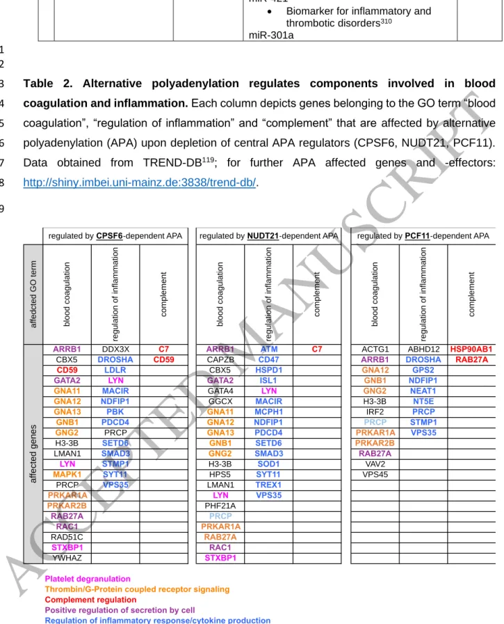
What comes next? Alternative polyadenylation and 3’-UTR diversity as central
1
regulatory hubs
2
Much of the posttranscriptional regulation of the hemostatic system depends on players that 3
determine the fate of RNAs encoding the respective hemostatic components. The different 4
layers of regulation are largely inter-dependent, as alternative splicing and polyadenylation are 5
coupled to each other
84and thereby determine not only the final open reading frame, but also 6
the 3’-UTR sequence and hence the susceptibility of the mature mRNA to posttranscriptional 7
control by RBPs and ncRNAs.
8
Since much of the posttranscriptional regulation of gene expression takes place at the level of 9
the 3’-UTR, to which RBPs and ncRNAs are abundantly recruited, the 3’-UTR architecture has 10
an important regulatory function (Figure 6)
84. Diversification of the transcriptome at the 3’-end 11
by alternative polyadenylation (APA) has recently emerged as a pervasive and evolutionarily 12
conserved layer of gene expression control
236(Figure 1), which affects more than 70% of all 13
genes. APA considerably expands the diversity of the transcriptome 3’-end, affecting protein 14
output, isoform composition and protein localization
237. 15
APA is globally regulated in various conditions, including developmental and adaptive 16
programs
89. It is thus likely that APA also tunes the hemostatic system, as exemplified by 17
alternative processing of TF and TFPI, where alternative splicing also generates different 3’- 18
UTRs (Figure 2). In addition, a recent large scale RNAi screen based on the depletion of more 19
than 170 putative APA regulators revealed how individual regulators affect the APA 20
landscape
115, including the resulting impact on gene ontologies
119. Several significantly 21
enriched GO terms suggest a critical function of UTR structures in inflammatory processes and 22
innate and adaptive immunity
119. APA affects key components broadly involved in inflammation 23
and blood coagulation (Table 2). This is consistent with findings that APA is a critical 24
component in the control of inflammatory processes
116, 238, 239(including COVID-19
240), that 25
typically result in shorter mRNA isoforms (Figure 6).
26
Strikingly, several hemostatic components have alternative transcripts that differ not only in 27
their exon composition but also in their 3’-UTR structure (see NCBI Ref seq). These include 28
essential components of the protein C pathway (i.e., protein C and protein S) with established 29
functions at the interface of coagulation and inflammation
241. For the protein C cofactor protein 30
S, 3'-UTR dynamics are already documented
119, which appear to be regulated by specific 31
RBPs (RNPS1) or other components (CDKN2D). This points to a regulatory function of APA 32
at the interface of the hemostatic and the immune system. Due to the pervasive regulatory 33
function of APA in various processes
119(with perturbations leading to numerous diseases
89), 34
it is plausible that much of this diversity in the hemostatic system is regulated in response to 35
inflammatory signals. This is illustrated by inflammation-triggered alternative processing of the 36
FGG mRNA242
, resulting in gamma prime (γ') fibrinogen
74. γ' fibrinogen is the fibrinogen fraction 37
ACCEPTED MANUSCRIPT
Downloaded from https://academic.oup.com/cardiovascres/advance-article/doi/10.1093/cvr/cvad046/7082876 by Universite de Bordeaux user on 17 April 2023
that contains the γ' chain, which arises when the
FGG mRNA is polyadenylated at an1
alternative polyadenylation signal, resulting in a polypeptide with a unique 20-amino acid 2
extension encoded by intron 9
74. Thanks to the strongly negatively charged C-terminus of the 3
γ' chain, fibrinogen γ' can bind with high affinity to thrombin exosite II, decreasing thrombin 4
activity on several substrates (antithrombin I activity)
243. As a consequence, low γ' fibrinogen 5
levels have been associated with an increased risk of venous thrombosis
74, 244, while a potential 6
role in CVD
245and ischemic stroke
246is under debate
247. This highlights how seemingly subtle 7
changes through alterations of APA and 3’-UTR diversity can have most significant functional 8
effects in the hemostatic system. It also serves as an example illustrating the complex 9
interdependency of posttranscriptional processing of RNA molecules and hence functional 10
output.
11
Interrogating system-wide posttranscriptional gene regulation
38, 39and transcriptome 3’-end 12
diversity
118, 119, combined with unbiased RNA interactome studies
117, 135and strategies to 13
disentangle the functional significance of genomic perturbations in non-coding elements
248, 14
therefore holds great potential to unravel novel layers of coupling of the hemostatic system 15
with inflammatory processes. This could also open entirely new therapeutic perspectives
89to 16
combat medical threats centering around thromboinflammation such as sepsis, which is still 17
the leading cause of death in the Western world and in critically ill patients worldwide
1. 18
19
Targeting post-transcriptional regulation of the hemostatic system
20
The multiple layers of posttranscriptional control of gene expression offer various opportunities 21
and targets for therapeutic intervention. For example, RNA-based therapeutics can be used 22
not only to re-direct splicing
80and polyadenylation
249, but also to silence an mRNA or to prevent 23
its interaction with other RNAs or RBPs
250, 251. 24
Compared to ‘conventional’ small therapeutic molecules, RNA-based therapeutics such as 25
ASOs, siRNAs and miRNAs offer the advantage of being able to act on ‘non-druggable’ targets 26
(i.e., proteins that lack enzymatic function or whose conformation is inaccessible to traditional 27
drug molecules), as they can be designed to affect virtually any gene of interest
192. 28
ASOs are relatively short, chemically modified single-stranded nucleic acids that selectively 29
pair to specific regions of mRNA resulting in endonucleolytic cleavage and degradation
250. 30
Currently, more than 60 ASO therapies are in or have completed phase I/II trials, with a 31
substantial number of antithrombotic ASO therapeutics currently under development
138. 32
The recent introduction of ASOs down-regulating FXI expression exemplifies the potential of 33
such therapeutics to modulate the hemostatic system via post-transcriptional mechanisms
34. 34
This phase II study in patients undergoing knee surgery revealed that the FXI-targeting ASO 35
effectively protects patients against venous thrombosis with a relatively limited risk of bleeding.
36
However, this proof-of-concept trial was too small to assess the effect on other thrombotic end 37
ACCEPTED MANUSCRIPT
Downloaded from https://academic.oup.com/cardiovascres/advance-article/doi/10.1093/cvr/cvad046/7082876 by Universite de Bordeaux user on 17 April 2023
points. Other genes that are being explored as potential targets for antithrombotic therapy 1
using silencing ASOs are FII, FVII, FXII, prekallikrein, plasmin activator inhibitor, 2
thrombopoetin and FMO3
138. A possible concern is that changes in platelet counts were 3
observed in non-human primates treated with ASOs
252, which has been attributed to peripheral 4
clearance
253and could potentially impact hemostasis.
5
miRNA therapeutics represent another highly versatile therapeutic means in the context of the 6
hemostatic system
138. MiRNA mimics may be employed to silence pro-coagulant genes to treat 7
thrombosis (or alternatively, anticoagulant genes to treat bleeding). Conversely, antagomirs or 8
target site blockers can be used to relieve silencing of anticoagulant genes to treat thrombosis.
9
Moreover, some miRNAs target several hemostatic components at the same time (Figure 4), 10
and silencing of such miRNAs can be intentionally used to control several hemostatic 11
components. On the other hand, undesired pleiotropy is one of the conceptual downsides of 12
therapeutic miRNA targeting.
13
MiRNA therapeutics are currently at an early stage of development and not yet applicable in 14
the clinical setting
254. In preclinical studies, several miRNA mimics and antagomirs have been 15
shown to reduce thrombus formation
138or increase the antithrombin activity in vivo
135. One of 16
the biggest challenges in the clinical development of miRNA-based therapeutics is the 17
identification of key miRNA candidates and targets, their specificity and effect size. There is 18
currently a relatively small number of experimentally validated miRNA:mRNA interactions, 19
making knowledge of the miRNA targetome in the hemostatic system a major trove for future 20
targeted therapeutics
135. 21
ASOs and most siRNAs exhibit perfect complementary to their targets, which usually results 22
in degradation of the target mRNA
255. In contrast, partial base-pairing of miRNAs prevents the 23
cleavage activity of RISC, predominately causing translational repression, and only in some 24
cases deadenylation, decapping and finally mRNA degradation
256. Although the proportion of 25
mRNA target degradation varies widely
257, a number of targets are almost exclusively 26
repressed at the level of translation
258. How much each mechanism contributes to down- 27
regulation depends on characteristics, such as seed-flanking nucleotides, of the individual 28
miRNA–mRNA pair
259. 29
In the context of the hemostatic system, it is interesting to note that miRNA regulation of 30
transcripts encoding secretory proteins results almost exclusively in translational repression, 31
because miRNA translational repression is stronger for mRNAs translated at the endoplasmic- 32
reticulum compared to free cytosolic ribosomes
258. Thus, miRNA-mediated therapeutic 33
targeting without degradation of the target mRNAs preserves physiological cell intrinsic 34
regulatory mechanisms carried out by 3’-UTRs and their binding partners (such as RBPs, 35
miRNAs, lncRNAs, circRNA or miRNA sponges). This allows for ‘compensatory’ on-demand 36
ACCEPTED MANUSCRIPT
Downloaded from https://academic.oup.com/cardiovascres/advance-article/doi/10.1093/cvr/cvad046/7082876 by Universite de Bordeaux user on 17 April 2023
adjustments of protein output even in the presence of the miRNA therapeutic, and thus may 1
represent a conceptual advantage of miRNA therapeutics over ASO-based approaches
138. 2
While RNA therapeutic approaches have been used in the development of new drugs and 3
clinical trials are underway
260, there are still concerns and challenges to be overcome. These 4
include, but are not limited to, off-target effects
261, triggering innate immune responses
262, 5
stability of the therapeutic RNA molecule and design of optimal delivery systems for disease- 6
specific release with minimal toxicity
190. 7
Finally, there are increasingly strategies to modulate other facets of the RNA biogenesis. This 8
concerns the targeted interference with splicing
80or with cleavage and polyadenylation
249, 9
involving either redirection of aberrant RNA processing (through ASOs, U1snRNP interference 10
or trans-splicing) or the elimination of aberrant transcripts
79, 89. The characterization of the 11
transcriptome dynamics thus becomes the next milestone to exploit the untapped therapeutic 12
opportunities arising from the increasingly available RNA therapeutics.
13 14
Summary
15
Besides transcriptional control, posttranscriptional regulation of gene expression is taking 16
center stage in the modulation of the hemostatic system. The highly regulated use of 17
alternative transcription start sites, exons and polyadenylation sites makes the transcriptome 18
highly dynamic in time, space and in response to pathological processes. Additional 19
posttranscriptional regulation by non-coding RNAs, RNA-binding proteins and RNA 20
modification mechanisms further modulate the functional output of numerous biological 21
processes, including the hemostatic system. Many of these regulatory principles also play an 22
important functional role in tuning the immune system
28-32, suggesting conserved regulatory 23
links between both systems. It will be critical to characterize these links to identify rational 24
targets for the emerging repertoire of RNA therapeutics to effectively combat the dangerous 25
alliance of the hemostatic and the immune system.
26 27
Acknowledgments:
28
The authors would like to express their gratitude to current and former members of the 29
Danckwardt and the Castoldi lab. Work in the Danckwardt lab is kindly supported by the 30
German Research Foundation Priority Program SPP 1935, German Research Foundation 31
grants DA 1189/2-1 and DA 1189/5-1, Germany Research Foundation Major Research 32
Instrumentation Program INST 371/33-1, the German Research Foundation graduate school 33
GRK 1591, Federal Ministry of Education and Research (BMBF01EO1003), German Society 34
of Clinical and Laboratory Medicine and the Hella Bühler Award for Cancer Research. Work in 35
the labs of S.D. and E.C. is supported by the EU Horizon 2020 Innovative training network 36
ACCEPTED MANUSCRIPT
Downloaded from https://academic.oup.com/cardiovascres/advance-article/doi/10.1093/cvr/cvad046/7082876 by Universite de Bordeaux user on 17 April 2023