HAL Id: hal-00895662
https://hal.archives-ouvertes.fr/hal-00895662
Submitted on 1 Jan 2007
HAL
is a multi-disciplinary open access archive for the deposit and dissemination of sci- entific research documents, whether they are pub- lished or not. The documents may come from teaching and research institutions in France or abroad, or from public or private research centers.
L’archive ouverte pluridisciplinaire
HAL, estdestinée au dépôt et à la diffusion de documents scientifiques de niveau recherche, publiés ou non, émanant des établissements d’enseignement et de recherche français ou étrangers, des laboratoires publics ou privés.
and DSC to elucidate the crystallisation properties and polymorphism of triglycerides in milk fat globules
Christelle Lopez, Claudie Bourgaux, Pierre Lesieur, Michel Ollivon
To cite this version:
Christelle Lopez, Claudie Bourgaux, Pierre Lesieur, Michel Ollivon. Coupling of time-resolved syn-
chrotron X-ray diffraction and DSC to elucidate the crystallisation properties and polymorphism of
triglycerides in milk fat globules. Le Lait, INRA Editions, 2007, 87 (4-5), pp.459-480. �hal-00895662�
Original article
Coupling of time-resolved synchrotron X-ray di ff raction and DSC to elucidate
the crystallisation properties and polymorphism of triglycerides in milk fat globules
Christelle L
opez
a,b*, Claudie Bourgaux
b,c, Pierre Lesieur
c,d,Michel O
llivon
baUMR1253 Science et Technologie du Lait et de l’Œuf, INRA-Agrocampus Rennes, 65 rue de Saint-Brieuc, 35042 Rennes Cedex, France
bUMR8612 CNRS Université Paris-Sud, Physico-Chimie des Systèmes Polyphasés, 92296 Châtenay-Malabry, France
cLaboratoire pour l’Utilisation du Rayonnement Électromagnétique (LURE), 91898 Orsay, France
dPhysico-Chimie des colloïdes, Université Henri Poincaré, UMR7567, BP 239, 54506 Vandœuvre-lès-Nancy, France
Abstract –Crystallisation properties and polymorphism of triacylglycerols (TG) in natural milk fat globules are studied with an apparatus permitting the coupling of time-resolved synchrotron X-ray diffraction (XRD) and high-sensitivity differential scanning calorimetry (DSC) for cooling rates 0.1<Rcooling<1000◦C·min−1. We showed that the fat phase of dairy products displays a complex polymorphism. For the first time, six different types of crystals were identified, several of them in coexistence, and their time- and temperature-dependent evolutions were quantitatively monitored.
They correspond to lamellar structures with double-chain length (2L: 40.5–48 Å) and triple-chain length (3L: 54–72 Å) organisations of TG. Depending on the cooling rate, at least five crystalline subcell species were observed at wide angles:αand sub-α, twoβ’ and oneβ. All these crystalline structures coexist with a liquid phase even at low temperatures. Thermal events recorded by DSC were related to the structural information on the organisation of TG obtained by XRD. Moreover, we showed that crystallisation in fat globules induces a higher disorder and a smaller size of crystals than in bulk, i.e. in anhydrous milk fat. Furthermore, XRD permitted the identification of different crystallisation behaviour in natural milk fat globules with different sizes, which could be implicated in the manufacture of dairy products involving tempering periods in the technological process (e.g.
butter, ice cream, whipped products). The increased knowledge of milk fat crystallisation in water- dispersed and highly complex food products might have a certain value for technical applications as well as for the development of new processes and dairy products.
crystal/phase transition/triacylglycerol/emulsion droplet/fat globule size 摘
摘摘要要要–时时时间间间分分分辨辨辨的的的X射射射线线线衍衍衍射射射和和和差差差示示示量量量热热热扫扫扫描描描法法法研研研究究究乳乳乳脂脂脂肪肪肪球球球中中中三三三酸酸酸甘甘甘油油油酯酯酯的的的晶晶晶体体体性性性质质质 和
和和多多多晶晶晶性性性。。。本文采用时间分辨的X射线衍射仪(XRD)和高灵敏度的差示量热扫描仪(DSC) 在冷却速率为0.1<Rcooling<1000◦C·min−1时,研究了天然乳脂肪球中三酸甘油酯(TG)晶体 特性和多晶性。实验证明乳制品中的脂肪相具有复杂的多晶性。首次鉴定出6种不同的晶 型,他们中大多数是共存的,并且定量地测定了这六种晶型的时间和温度之间的变化关系。
他们对应的层状结构是具有双倍链长(2L: 40.5–48 Å)和三倍链长(3L: 54–72 Å)的TG组织。
* Corresponding author (通讯作者): Christelle.Lopez@rennes.inra.fr Article published by EDP Sciences and available at http://www.lelait-journal.org or http://dx.doi.org/10.1051/lait:2007018
根据冷却速率,在一个很宽的范围内至少观察到5个亚晶胞结构,分别为α、亚-α、2个β 和1个β型结构。即使在较低的温度下,这些晶型结构也是与液态相共存的。DSC的热分析 结果与XRD观察到的TG组织结构特性是一致的。此外,我们还证明了脂肪球中的脂肪晶 体与游离脂肪(如无水脂肪)相比,前者处于高度的无序状态,并且晶体的尺寸较小。根据 XRD分析证明了不同大小的天然乳脂肪球具有不同的晶体性质,这种性质有可能与乳制品 (如奶油、冰淇淋、搅打乳制品)加工过程中所涉及到老化工艺有直接的关系。了解乳脂肪 晶体在水分散的和复杂的食品体系中的结晶特性将有助于解决生产实际问题和开发新的加 工技术及新的乳制品。
晶 晶
晶体体体/相相相转转转移移移/三三三酸酸酸甘甘甘油油油酯酯酯/乳乳乳状状状液液液滴滴滴/脂脂脂肪肪肪球球球大大大小小小
Résumé – Couplage de la DSC et de la diffraction des rayons X résolue en temps en utili- sant le rayonnement synchrotron pour élucider les propriétés de cristallisation et le polymor- phisme des triglycérides dans les globules gras du lait. Les propriétés de cristallisation et le polymorphisme des triglycérides (TG) dans les globules gras naturels du lait sont étudiés avec un appareil permettant le couplage de la diffraction des rayons X (DRX) résolue en temps et de la microcalorimétrie différentielle (DSC), pour des vitesses de refroidissement comprises entre 0,1 et 1000◦C·min−1. Nous avons montré que la phase lipidique des produits laitiers possède un polymor- phisme complexe. Pour la première fois, six types différents de cristaux ont été identifiés, plusieurs d’entre eux étant en coexistence, et leurs évolutions ont été caractérisées de manière quantitative, en fonction du temps et de la température. Elles correspondent à des structures lamellaires avec des structures à deux longueurs de chaîne (2L : 40,5–48 Å) et à trois longueurs de chaîne (3L : 54–72 Å). En fonction des vitesses de refroidissement, au moins cinq sous-cellules cristallines ont été identifiées aux grands angles :α, sub-α, deuxβetβ. Toutes ces structures cristallines coexistent avec une phase liquide, même à basse température. Les évènements thermiques enregistrés par DSC ont été reliés aux informations structurales de l’organisation des TG obtenues par DRX. Nous avons montré également que la cristallisation dans les globules gras induit un plus grand désordre et une plus petite taille des cristaux qu’en milieu anhydre. De plus, la DRX a permis d’identifier différents comportements de cristallisation dans les globules gras de différentes tailles, qui pourraient inter- venir lors de la fabrication de produits laitiers nécessitant des périodes de tempérage (par exemple le beurre, la crème glacée, les produits foisonnés). L’augmentation des connaissances sur la cris- tallisation de la matière grasse du lait dans les systèmes dispersés et dans les produits alimentaires complexes contribuera aux innovations technologiques ainsi qu’au développement de nouveaux produits.
cristal/transition de phase/triglycéride/gouttelette d’émulsion/taille des globules gras
1. INTRODUCTION
Triacylglycerols (TG), which are the main constituents of natural fats, deter- mine the physical and thermal properties of high-fat products. Moreover, the fat phase of food products is sometimes partially crystallised at room and storage (4–7◦C) temperatures, as is the case for milk and dairy products.
Milk fat is an essential nutriment con- sumed in many food products, in which it is frequently found dispersed as small droplets called milk fat globules, e.g. in milk, cream and cheeses. Crystallisation of
TG in milk fat globules is of prime im- portance because it affects many properties such as (i) the rheological properties, (ii) the resistance of fat globules to disruption and then to coalescence, (iii) the suscepti- bility of globules to churning, (iv) the sta- bility of whipped cream and (v) the consis- tency and mouth feel of high-fat products.
Thus, it is important to understand better the physical properties of fat globules, e.g.
their thermal and crystallographic proper- ties, for industrial applications and to im- prove the quality of food products.
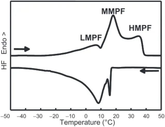
Milk fat globules have a diame- ter ranging from 0.02 to 15 μm with
−50 −40 −30 −20 −10 0 10 20 30 40 50
Temperature (°C)
HF Endo>
LMPF MMPF
HMPF
Figure 1.Thermal properties of anhydrous milk fat. Crystallisation and melting curves recorded upon cooling at 2 ◦C·min−1 and subsequent heating at 2 ◦C·min−1 using differential scanning calorimetry (DSC). LMPF: low melting point fraction; MMPF: middle melting point fraction;
HMPF: high melting point fraction.
a volume-weighted diameter of about 4μm [9]. Natural milk fat globules, which are enveloped by a biological membrane (the milk fat globule membrane, MFGM), are mainly composed of TG (∼98% of milk lipids), which are esters of fatty acids and glycerol. More than 400 fatty acids have been identified in milk, with a wide diversity of chain lengths, num- ber of unsaturation, branching and position on the glycerol. More than 200 individ- ual molecular species of even-numbered TG have been quantified [4]. Furthermore, it is well known that milk fat compo- sition changes with cow feeding, season and stage of lactation. Thus, milk fat is undoubtedly the most complex fat found in nature. The consequence of this TG and fatty acid composition is that anhy- drous milk fat (AMF), which is the fat isolated from butter, has a broad melting range from −40 ◦C to +40 ◦C (Fig. 1) and no true melting point, like pure com- pounds. Therefore, at intermediate temper- atures and at equilibrium (e.g. room and fridge temperatures) milk fat is a mix- ture of crystals and oil. Moreover, this
fat-composition-related complexity is dra- matically enhanced by the existence of a polymorphism of monotropic type for each TG [22, 25]. As observed for most of the lipids, each TG of milk fat can exhibit several crystalline forms, the occurrence of which strongly depends on its thermal history. Each polymorphic form is charac- terised by its own melting point. Further- more, the fact that TG mixtures also ex- hibit multiple melting points, depending on their composition, renders the overall melt- ing behaviour of milk fat even more com- plex.
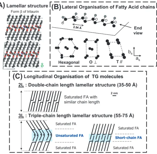
TG polymorphism relates to the abil- ity of molecules to arrange themselves within a crystal lattice in a number of different ways of lateral packing of the fatty acid chains and of longitudinal stack- ing of molecules in lamellar structures (Fig. 2) [6]. Thus, pure TG and mix- tures of TG can adopt several crystalline arrangements. Transitions between these polymorphic forms are mostly irreversible;
monotropism implies that they are only possible via a liquid and when leading to the formation of more stable species [25].
O⊥⊥ T //
Hexagonal
as bs
2.54 Å
2.54 Å End
view
(B)
Lateral Organisation of Fatty Acid chainsFormβof trilaurin
δ
(C)
Saturated FA Unsaturated FA
Saturated FA
Saturated FA with similar chain length
Short-chain FA Saturated FA
Saturated FA
3L : Triple-chain length lamellar structure (55-75 Å) 2L : Double-chain length lamellar structure (35-50 Å)
2 nm
LongitudinalOrganisation of TG moleculesOrganisation of TG molecules
(A)
Lamellar structureFigure 2.Main types of TG packings. (A)Lamellar structure formed by triacylglycerol (TG) molecules in the solid state: example of theβ form of trilaurin.(B)Top: the stable conforma- tion of the hydrocarbon chains of saturated fatty acid (FA) is a planar zigzag shown here as a 3D view along its main axis; bottom: three main types of chain lateral packings (only carbon atoms are drawn, hydrogen atoms are not drawn): hexagonal, orthorhombic perpendicular (O⊥) and triclinic parallel (T//) subcells to whichα,β’ andβpolymorphic forms correspond.(C)Two main types of TG chain longitudinal stacking (fatty acids are drawn as straight lines).
The three main polymorphic forms fre- quently observed for the lateral pack- ing of fatty acid chains correspond to different subcells that have been described in detail [22, 25]: hexagonal (αform), or- thorhombic perpendicular (β’ form) and triclinic parallel (β form) (Fig. 2B). The density, enthalpy of fusion, melting point and stability increase in the orderα,β’ and
β, according to the monotropic character of the polymorphism. In the α form, the packing of chains is not very tight and the chains have considerable rotational free- dom, whereas in the β form, the chains are very densely packed. TG crystals are made by the stacking of TG molecule lay- ers, the thickness of which depends on the length and unsaturation of the fatty
acid chains, and their angle of tilt with re- spect to the basal planes formed by the methyl end groups of the TG (Fig. 2C). The longitudinal organisations of TG in lamel- lar structures are primarily related to the number of chains stacked one on top of the other in the main crystalline cell. For TG, this number frequently takes a value of 2 or 3 and corresponds to the stacking of dou- ble (2L) or triple (3L) chain length lamellar structures, leading to distances in the range 35–50 Å and 55–75 Å, respectively [6, 25].
Roughly, 3L forms are usually related to low-melting, long-chain monounsaturated and/or mixed long- and short-chain TG, whereas 2L forms are generated mostly by similar long-chain, high-melting, trisat- urated TG [25].
Crystallisation in milk fat globules has been investigated by different techniques.
Freeze-fracture electron microscopy has made possible the study of fats and food systems such as milk, cream and but- ter [24, 26]. Polarised light microscopy permitted the classification of fat glob- ules into four main types as a function of the crystal habit [14, 28]. These stud- ies gave an insight into the orientation and the size of the crystals at a micro- scopic level, but little information exists at a molecular level on the organisation of TG molecules in the crystals formed within fat globules. The techniques most frequently used for the study of the thermal and crys- tallographic properties of TG are differen- tial scanning calorimetry (DSC) and X-ray diffraction (XRD), respectively.
DSC studies have given an insight into the thermodynamics of fat phase transi- tion in bulk and in emulsions. The authors that studied anhydrous milk fat by DSC observed that it crystallises and melts in several steps (Fig. 1). A typical melting curve of AMF shows three endothermic peaks, corresponding to low melting point (LMP), medium melting point (MMP) and high melting point (HMP) fractions [27].
These peaks correspond to large groups of
TG that melt separately and behave like solid solutions. The occurrence of numer- ous thermal transitions during DSC exper- iments, the partial overlapping of the melt- ing peaks, their respective enthalpies and temperatures depend strongly on the ther- mal treatments (e.g. heating and cooling rates, tempering) and on the entire thermal history of the sample [1, 22]. The complex DSC recordings are difficult to interpret.
Thus, DSC experiments need to be coupled with other techniques.
XRD is an essential tool for elucidat- ing the molecular packing of molecules in the solid state and polymorphism of pure TG and complex fats, since it pro- vides structural information on the organ- isation of molecules, and it complements DSC. X-rays are the ideal direct probe to determine the internal structure of crys- tals, since they provide information on the repetitive patterns of the electron density of the array of atoms. The atoms that consti- tute the TG molecules inside a perfect crys- tal are arranged periodically in planes at a repetitive distance, d (Fig. 3). As a beam of X-rays comes to the crystal at an an- gle,θ, the X-rays of wavelengthλare re- flected by the planes at that same angleθ.
The optical path length required to produce constructive interference is nλ, with inte- gral values of n, providing specific crys- tal symmetry allows it. The equation that determines the angle at which constructive interference occurs is known as Bragg’s law: 2dsinθ=nλ[5,25]. The actual diffrac- tion measurement during an experiment is the one formed between the incident beam and the reflected beam, which is denoted by 2θ. It is often convenient to use the scattering vector q instead of the scatter- ing angle 2θsince the former is indepen- dent of the X-ray wavelength. They are re- lated as follows: q = (4π/nλ) sinθ. Thus, q=2π/d.
The two levels of organisation, e.g. the lateral packing of the fatty acid chains and the longitudinal stacking of TG molecules
2 d sin θθ = n λ
2θ
d X-rays
θ
λ ~ 1.5 Å
θ
Figure 3.Demonstration of Bragg’s law of diffraction: 2dsinθ=nλ(d in Å is the repetitive distance between the planes arranged periodically;θin degrees is the angle of incidence of X-ray relative to the crystalline plane;λin Å is the X-ray wavelength; the integral values of n are required to produce constructive interference nλ).
in lamellae formed in the solid state (Fig. 2), are easily identifiable from XRD patterns recorded at wide and small angles, respectively. The thickness of the lamel- lar structures, which can be measured by XRD at small angles (0◦ < θ < 5◦), corresponds to TG longitudinal stacking called TG long spacings. From the mea- surement of d (Å) and with knowledge of the fatty acid composition (chain length and unsaturation), it is possible to deduce if the stackings correspond to 2L or 3L or- ganisations (Fig. 2C). The cross-sectional packings of the aliphatic chains are charac- terised by specific short spacings, indepen- dent of chain length and observable at wide angles in the range 8.5◦ < θ<13◦. Short spacings are widely used for identifying the various crystalline subcells character- ising the polymorphic forms (Fig. 2B). A single line around 4.15 Å characterises the αform (hexagonal subcell), and a strong line at 4.6 Å among other sharp lines iden- tifies theβform (triclinic parallel subcell), while theβ form (orthorhombic perpen- dicular subcell) frequently shows associa-
tion of two lines, among which one is about 4.2 Å and the other around 3.8 Å.
Recent use of synchrotron radiation, which provides X-ray flux 103to 106times more intense than that generated by usual X-ray sources, permits recordings to be performed within a few seconds or mil- liseconds. Thus, direct continuous record- ings can be performed as a function of time (XRDt) or temperature (XRDT).
Moreover, synchrotron radiation permits the study of the organisation of TG in water-dispersed systems such as emulsions and food products and to quantitatively monitor phase changes within emulsion droplets. This is especially interesting to relate the textural and rheological proper- ties of milk fat, and of high-fat food prod- ucts, to the thermal and crystallographic properties of TG. Therefore, the function of milk fat in many food products can- not be understood without knowing both its composition and physical properties, as well as those of their respective dependen- cies.
Determination of the thermal and crys- tallographic properties of TG molecules in the dispersed state, e.g. in milk fat glob- ules, is much more challenging than for fat in bulk, e.g. anhydrous milk fat, since the presence of numerous compounds (e.g.
casein micelles, minerals, lactose and wa- ter) not involved in the TG organisation will necessarily decrease the intensity of the signal recorded by both DSC and XRD techniques, by a simple dilution effect.
Overcoming the difficulties associated with milk fat globule examination requires pow- erful X-ray sources such as those available at synchrotron laboratories.
The objective of this paper is to pro- vide an overview of the possibilities of investigation offered by the coupling of high-sensitivity DSC and time-resolved synchrotron radiation XRD. Selected ex- amples illustrate the study of the thermal and crystallographic properties of milk fat within the globules, the evolution of organ- isation of milk fat TG in the solid state and their polymorphism as a function of tem- perature and time.
2. MATERIALS AND METHODS 2.1. Samples
Concentrated creams (fat content=400 to 800 g·kg−1) were obtained by industrial skimming of whole milk. Anhydrous milk fat (AMF) was extracted from the creams as detailed in Lopez et al. [15]. Natural milk fat globules with different sizes were selected from the same raw whole milk by using a novel microfiltration process [21].
2.2. Methods
2.2.1. Coupled XRD and DSC measurements
Experiments were performed using a technique allowing the coupling of time- resolved synchrotron X-ray diffraction as
a function of temperature (XRDT) or time (XRDt) and high-sensitivity differential scanning calorimetry (DSC) in the same apparatus from the same sample. This ap- paratus, called Microcalix, was recently described in Ollivon et al. [23].
This set-up was used on the D22 and D24 benches of the DCI synchrotron of LURE (Laboratoire pour l’utilisation du rayonnement électromagnétique, Orsay, France). On the D22 bench (λ=1.5498 Å), the XRD data were recorded simulta- neously at small and wide angles with two position-sensitive gas linear detec- tors (1024 and 512 channels), which were placed about 1 770 mm and 300 mm from the sample, respectively (Fig. 4). On the D24 bench (λ=1.489 Å), a single detector can be placed at 300 mm or 900 mm (q= 0–0.55 Å−1). The small-angle detector al- lows the characterisation of the longitudi- nal organisation of TG molecules, whereas the wide-angle detector permits the identi- fication of the chain packing (Fig. 2). The channels of the detectors were calibrated to express the XRD data in scattering vector q (q= 4πsinθ/λ; q in Å−1,θin degrees is the angle of incidence of the X-ray rela- tive to the crystalline plane,λin Å is the X-ray wavelength) [5]. The crystalline 2L βform of high-purity tristearin was used as a reference for the wide-angle channel to scattering vector q calibration of the detec- tor (4.59,3.85,3.70±0.01 Å) [22]. Silver behenate, which is characterised by a long spacing of 58.380±0.001 Å, was used to calibrate the small-angle detector [3]. The calorimeter coupled to XRD was calibrated with lauric acid.
XRD data and DSC measurements were synchronously collected versus time by a single microcomputer. A Visual Basic (Microsoft, Redmond, USA) program was specially written to ensure simultaneous display of the small-angle XRD, wide- angle XRD and DSC results during their collection (Fig. 4A).
Calorimeter Cell
X-Rays
Sample
Small-angle detector Wide-angle
detector
(A)
(B)
T C C T C C
Counting CountingElect.Elect. nVmeter
nVmeter T Ctrl
T Ctrl
LD2LD2
LinearLineardetector 1detector 1
Sample Sample Ref Ref..
X X--raysrays
(synchrotron) (synchrotron)
MCD SAXD WAXD
1770 mm
2
)
) CalorimeterT C C T C C
Counting CountingElect.Elect. nVmeter
nVmeter T Ctrl
T Ctrl
LD2LD2
LinearLineardetector 1detector 1
Sample Sample Ref Ref
X X--raysrays
Small Angles (synchrotron)
(synchrotron)
q = 0 – 0.45 Å-1
MCD SAXD WAXD
300 mm
2θ
)
)Wide Angles q = 1.1 – 2.1 Å-1
Figure 4. Experimental set-up of the microcalorimeter in the time-resolved synchrotron X-ray diffraction environment.(A)Schematic representation: the cell is positioned with the capillary con- taining the sample perpendicular to the beam in such a way that the diffraction patterns are recorded in the vertical plane by one or two one-dimensional proportional detectors (LD) at small and wide angles. Counting electronic (Counting Elect.), nanovoltmeter (nVmeter) and temperature controller (T Ctrl) are monitored by a single computer. The temperature-controlled cryostat (TCC) is kept at constant temperature (e.g. 6◦C).(B)Set-up on the D22 bench of the synchrotron (LURE, Orsay, France).
XRD patterns were recorded by trans- mission through Lindeman glass capillar- ies (GLAS, Muller, Berlin, Germany), with 0.01 mm of wall thickness and diame- ter Φ = 1.40 ±0.10 mm. These capil- laries were specially designed for X-ray studies since they allow minimum atten-
uation of the beam and parasitic scatter- ing. Furthermore, they are characterised by poor thermal conductivity and thus guaran- tee minimum thermal losses. Samples were prepared by loading the capillaries with about 20μL of cream or melted AMF us- ing a syringe and a thin Teflon capillary.
The samples in the capillary were heated to 60 ◦C for 5 min to ensure that all crystals and nuclei were melted. Then, the samples were cooled with cooling rates in the range 0.15 ◦C·min−1 Rcooling 1000 ◦C·min−1. The most rapid Rcooling
were obtained by rapid introduction of the capillary into the calorimeter pre-cooled to the temperature, e.g.−8◦C or 4◦C. Tem- pering in isothermal conditions was also performed, e.g. at−8◦C, 4◦C and 20◦C.
Then, XRD patterns were recorded as a function of time and upon subsequent cool- ing or heating (in general at 2◦C·min−1).
The samples of concentrated fat globules were not cooled at T <−8◦C to avoid ice formation.
Each XRD pattern recorded as a func- tion of time (crystallisation and melting experiments) was analysed using Peak- fit software (Jandel scientific, Erkrath, Germany). XRD peaks were fitted by the Gaussian-Lorentzian (sum) equation, to determine the position of the maximum, maximal intensity and area under each XRD peak [11].
2.2.2. DSC experiments
Thermal analyses were conducted by DSC, using a DSC-7 calorimeter (Perkin Elmer, St Quentin en Yvelines, France) equipped with an Intracooler II and run- ning under Pyris software and a TA Q 1000 calorimeter (TA Instruments, New Castle, DE). About 5 to 10 mg of cream were weighed in a hermetic aluminium pan of 50 μL. An empty pan was used as refer- ence.
2.2.3. Fat globule size measurements The fat globule size distributions were measured by laser light scattering using a Mastersizer 2000 (Malvern Instruments,
Malvern, UK). From the size distribu- tion, the average volume-weighted diam- eter, d43 = Σnid4i/Σnid3i (where ni is the number of fat globules in a size class of diameter di), was calculated by the instru- ment software.
3. RESULTS AND DISCUSSION
The organisation of TG molecules in the solid state (e.g. in fat crystals) was investigated in milk fat globules, by vary- ing the cooling rates. The crystallisa- tion properties of TG in milk fat glob- ules were studied upon slow cooling (0.15 ◦C·min−1), at intermediate cooling rates (0.5 < Rcooling ≤ 3 ◦C·min−1) and after quenching (∼1000◦C·min−1), to char- acterise the most unstable crystalline struc- tures and their reorganisation as a function of time.
The examination of TG polymorphism in milk fat globules is especially difficult since (i) both small- and wide-angle XRD should be considered at the same time and compared to determine the evolution of each of the species as a function of time;
(ii) the X-rays diffracted by each of the crystalline structures are reduced to the proportion of the fraction considered; (iii) the whole XRD signal is largely absorbed by the surrounding water and its solutes (e.g. casein micelles); and (iv) the peak broadening results from the crystallisation constraints in dispersed systems.
3.1. Crystallisation in milk fat globules during slow cooling:
Rcooling <0.5◦C·min−1
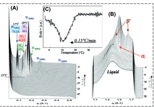
The crystals formed in milk fat glob- ules upon slow cooling (0.15 ◦C·min−1) and their melting behaviour (2◦C·min−1) were considered. For the first time to the authors’ knowledge, the XRD experiments performed at small angles showed that
Figure 5.Structural evolution, expressed in scattering vector q (Å−1), of triacylglycerols dispersed in milk fat globules during cooling from 60◦C to−8◦C at 0.15◦C·min−1. Three-dimensional plots of the XRD patterns recorded (A) at small angles and (B) at wide angles. (C) Differential scanning calorimetry curve recorded simultaneously.
crystallisation in fat globules occurs suc- cessively, producing four types of crys- tals [11]. The lamellae formed upon slow cooling correspond to two 2L structures (46.5 and 40 Å) and to two 3L structures (71.3 and 65 Å) (Fig. 5A). Nucleation oc- curs in theαform, then theα+β poly- morphic forms coexist until the end of the cooling (Fig. 5B). The DSC crystallisa- tion curve recorded simultaneously shows a single exothermic peak (Fig. 5C). The 4 types of crystals start to form within a 10 ◦C range, from about 22◦C, prevent- ing separation of overlapped peaks by DSC recording. Before the formation of crys- tals in milk fat globules, characterised by diffraction peaks, both small- and wide- angle XRD patterns show scattering peaks, respectively, centred at 0.28 Å−1(22.44 Å) and 1.38 Å−1 (4.55 Å), corresponding to
the X-ray signature of TG in their liquid state, as described by Larsson [7].
A quantitative analysis of the XRD data was performed to determine the rel- ative proportion of the 5 different phases, 4 solids and 1 liquid, formed in milk fat globules upon slow cooling and their evo- lution as a function of temperature. It is generally accepted that the sum of integrals of the whole diffraction peaks of a phase is roughly proportional to its abundance in a material [5]. The total diffracted inten- sity is not affected by the effect of crystal perfection, crystal size or the polymorphic form, since X-ray scattering is only propor- tional to the number of atoms present in the organised system. Thus, we determined the areas for each XRD peak recorded at small angles during cooling, using Peakfit soft- ware (Fig. 6 left). The relative abundance
of each of the phases was obtained by summing the areas of each of the small- angle XRD peaks corresponding to the same crystalline structure (e.g. the 5 areas of peaks corresponding to the 5 orders of the 3L1 structure). Figure 6 right shows the evolution of the relative proportions of the 5 phases as a function of tempera- ture during cooling of milk fat globules at 0.15◦C·min−1. The successive occurrences and growths of the 4 different solid phases at the expense of the liquid or of one of the phases, newly formed but metastable, are clearly evidenced.
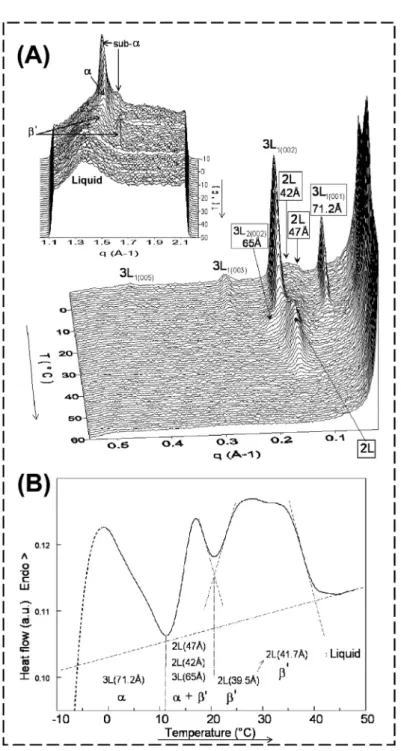
After cooling, the samples were heated from −8 ◦C to 60 ◦C at 2 ◦C·min−1 to study the melting behaviour of TG in milk fat globules (Fig. 7). Figure 7A shows the XRD patterns recorded at small and wide (insert) angles during heating. The deter- mination of the evolution of maximal in- tensity of each small-angle XRD peak as a function of temperature (Fig. 6B) permits one to relate the structural and the ther- mal events (Fig. 6C). The decrease in in- tensity of the XRD peaks recorded at small angles corresponds to the melting of the corresponding crystalline structures. From about 12◦C, a new 2Lβ’ (40 Å) structure formed during the heating, corresponding to the increase in intensity (Fig. 7B) of the peak observed at small angles (Fig. 7A). At wide angles, the XRD patterns showα→ β’→liquid transitions (Fig. 7A). This re- crystallisation of TG during heating with a polymorphic transition of monotropic type (α → β’) showed that fat crystals formed during slow cooling of milk fat globules were still metastable. The DSC melting curve showed 3 endotherms that corre- spond to the melting of the crystalline structures, as indicated in Figure 7C. TG dispersed in milk fat globules were in their liquid state for T>37◦C.
The crystallisation behaviour of AMF in similar conditions was completely differ- ent [13], showing the influence of the dis- persion state.
3.2. Crystallisation in milk fat globules for cooling rates 0.5Rcooling3◦C·min−1 At intermediate cooling rates, the first crystalline structures formed in milk fat globules are 2L structures (2L1 = 47 Å and 2L2 = 42 Å). Then, the succes- sive formation of 5 sharp XRD peaks corresponds to the crystallisation of TG molecules in a 3L (71.2 Å) structure (re- sults not shown in this paper; see [13]). The 3 lamellar structures that are successively formed upon cooling have a hexagonal packing of the fatty acid chains (αform).
At −8 ◦C, the reversible formation of a pseudo-orthorhombic perpendicular pack- ing of the acylglycerol chains, called sub- α, was observed. This α → sub-αtransi- tion that occurs at low temperature has also been observed in AMF but is favoured in the dispersed state [16].
The melting behaviour was studied upon heating at 2 ◦C·min−1. Figure 8A shows the 3D plot of the small- and wide- (insert) angle XRD patterns. Numerous structural rearrangements were observed before final melting of TG molecules in milk fat globules, showing that cooling at intermediate rates leads to the formation of unstable crystalline varieties. Roughly, the melting of the α 3L (71.2 Å) struc- ture is first observed and some TG trans- form into a new 3L (65 Å) structure that melts rapidly, then the melting of theα2L (47 and 42 Å) structures is observed. In the 16 ◦C T 24 ◦C domain, XRD patterns show a transition and the forma- tion of a new diffraction line, correspond- ing to aβ’ 2L (39 Å) structure, before fi- nal melting of TG for T>40◦C. Thisβ’
2L (39 Å) structure that crystallises during heating is probably formed by the reorgan- isation of the TG initially incorporated in the other lamellar structures formed during the cooling process. At wide angles, we ob- served the following transitions upon heat- ing: sub-α↔ α→ β→liquid (Fig. 8A,
Liquide3L1 (005)
3L2 (002)
2L2 2L1 3L2 3L1 (003)
3L1 (002) 3L1
-8°C Figure6.AnalysisofX-raydiffraction(XRD)patternallowingthedeterminationoftherelativeproportionofthecrystallinestructuresasafunction oftemperature.Left:exampleofPeakfitanalysisperformedonasmall-angleXRDpatternrecordedat−8◦Cattheendofcoolingofmilkfatglobules from60◦Cto−8◦Cat0.15◦C·min−1(Fig.5).Theareaunderthepeakscorrespondingtothesamelamellarstructurehavethesamecolour.Right: decompositionofthesmall-angleXRDpatternsintothefoursolidphasesandtheliquidone,asevaluatedfrompeakareasusingPeakfitanalysis.Phase contentsareexpressedasapercentageofthetotalfat.
Figure7.Meltingbehaviouroftriacylglycerolsinmilkfatglobulesuponheatingat2◦C·min−1afterslowcoolingat0.15◦C·min−1.(A)Three- dimensionalplotsobtainedfromtheX-raydiffractionpatternsrecordedatsmallandwide(insert)anglesasafunctionoftemperature.(B)Evolutionof theintensitiesofthesmall-angleX-raydiffractionpeaks,recordedduringtheheating(theintensitieshavebeenexpressedasapercentageofthepeak maximum).(C)Meltingcurverecordedsimultaneouslybydifferentialscanningcalorimetry.
insert). The evolution of maximal intensi- ties of each XRD peak recorded at both small and wide angles during heating of cream allowed us to delimit the domains of existence of the crystals and to relate the thermal events recorded by DSC to the structural information recorded by XRD (Fig. 8B) [14].
3.3. Crystals formed after quenching:
Rcooling ∼1000◦C·min−1
The most unstable crystalline structures formed by TG molecules in natural milk fat globules and in AMF were investigated af- ter quenching from 60◦C to−8◦C [10,12].
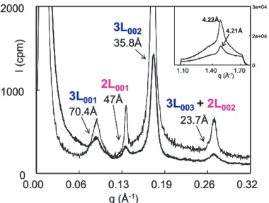
We identified the formation of 2 lamellar structures from the liquid: 3L (70.4 Å) and a transient 2L (47 Å) with a lateral organ- isation of the fatty acid chains correspond- ing to the most unstable polymorph, i.e. the α form (hexagonal subcell) (Fig. 9). The acquisition of XRD patterns as a function of time after quenching permitted the ob- servation that theα2L (47 Å) structure was very unstable since it disappeared during a 20-min isothermal recording.
The crystallographic properties of milk fat globules were also investigated after quenching from 60 ◦C to 4 ◦C, which is the temperature of storage of dairy prod- ucts, and followed as a function of time in isothermal conditions at 4 ◦C [15]. After such a rapid cooling, the main crystalline structure formed corresponded to anα3L (70.5 Å). As for quenching at lower tem- peratures (e.g.−8◦C), theα2L (47 Å) was also characterised, but it disappeared more rapidly after quenching at 4◦C in compar- ison with−8◦C.
The rapid cooling of milk fat from 60◦C to low temperatures, e.g.−8 ◦C or 4 ◦C, induces a brusque liquid → solid phase transition. The amount of the liq- uid phase at the temperature of quenching can favour the structural rearrangement of TG molecules. Phase transitions occurred
as a function of time, in isothermal condi- tions, with changes in both the polymor- phic forms and the longitudinal organisa- tion of TG molecules. These polymorphic transitions, e.g.α→β→ β, were related to successive increases in the density of fat globules during storage at 4◦C [15].
Furthermore, we observed that the phase transitions were delayed in fat glob- ules in comparison with AMF [15].
3.4. Crystals formed after tempering in isothermal conditions
The crystals formed in milk fat globules after tempering at different temperatures corresponding to the storage and consump- tion of dairy products were characterised.
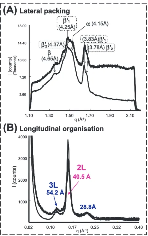
The structures formed after storage for t>5 days at 4◦C were identified (Fig. 10).
They correspond to 2 lamellar organisa- tions: mainly a 2L (40.5 Å) and a 3L (54.2 Å) structure (Fig. 10A). The lat- eral packing of the fatty acid chains corre- sponds to the coexistence of 4 crystalline packings: one α form, two β forms and traces of the β form (Fig. 10B). Similar structures were characterised in milk fat globules and AMF [15]. The absence of evolution of the thickness of the lamel- lae upon subsequent heating and the ab- sence of recrystallisation were in favour of the polymorphic stability of the crys- tals formed after this tempering period (re- sults not shown in this paper, see [15]). The endothermic peaks recorded by DSC upon heating were related to the successive melt- ing of the crystalline structures formed dur- ing the tempering period: the 3L structure, then the 2L structure [15].
This work also evidenced the formation of crystals during storage of milk fat glob- ules (creams) for t>20 h at room temper- ature (e.g. T ∼20◦C): 2L (40.5–41.1 Å) lamellar structure [8]. The melting of this 2L structure corresponds to a broad en- dothermic peak recorded by DSC [18].
Upon subsequent slow cooling of the fat
Figure 8.Structural evolution, expressed in scattering vector q (Å−1), of triacylglycerols dispersed in milk fat globules during heating at 2◦C·min−1after cooling at 1◦C·min−1. (A) Three-dimensional plots of the XRD patterns recorded at small and wide (insert) angles. (B) Differential scanning calorimetry curve recorded simultaneously.
3L
0013L
0023L
003+ 2L
0022L
0010.00 0.06 0.13 0.19 0.26 0.32 q (Å-1)
0 1000 2000
I (cpm)
70.4Å 47Å
35.8Å
23.7Å
1.10 1.40 1.70
q (Å-1) 0 2e+04 3e+04
4.22Å 4.21Å
3L
0013L
0023L
003+ 2L
0022L
0010.00 0.06 0.13 0.19 0.26 0.32 q (Å-1)
0 1000 2000
I (cpm)
70.4Å 47Å
35.8Å
23.7Å
1.10 1.40 1.70
q (Å-1) 0 2e+04 3e+04 4.22Å
0.00 0.06 0.13 0.19 0.26 0.32 q (Å-1)
0 1000 2000
I (cpm)
70.4Å 47Å
35.8Å
23.7Å
1.10 1.40 1.70
q (Å-1) 0 2e+04 3e+04
4.22Å 4.21Å
Figure 9.Small- and wide- (insert) angle X-ray diffraction patterns recorded at−8◦C after quench- ing of cream (thick line) and anhydrous milk fat (thin line) from 60◦C.
globules tempered at 20 ◦C, the amount of crystals in this 2L form increases and two additional types of 3L structures are formed (3L1 type: 68.5–70.7 Å; 3L2type:
61.2–64.2 Å), the characteristics of which depend on the size of the milk fat glob- ules [20].
The crystalline structures formed after a tempering period, mainly the 2L (40–41 Å) structure, correspond to stable polymor- phic species and are characterised by a higher final melting point [18].
3.5. Influence of milk fat globule size on their crystallisation properties
The thermal and crystallographic prop- erties of creams were analysed as a func- tion of temperature and time with the whole droplet size distribution of natural milk fat globules [10,11,14,15]. In this part of the work, we focused on the influence of fat globule size on TG crystallisation and polymorphism.
Natural milk fat globules (i.e. covered by the MFGM) of smaller size (∼1–3μm) and larger size (∼5–7μm) were selected from the same initial milk (fat globules
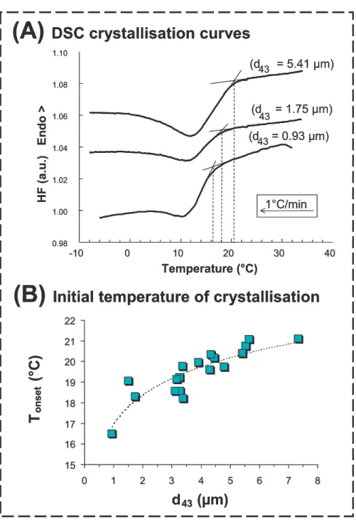
∼4 μm) by using a novel microfiltration process [21]. Figure 11A shows that the DSC crystallisation curves recorded for creams containing natural milk fat glob- ules with different sizes correspond to a single exothermic peak and that the ini- tial temperature of crystallisation (Tonset) is delayed for smaller fat globules. Fig- ure 11B shows the decrease in Tonset as a function of the decrease in natural milk fat globule size [20]. Lopez et al. [14]
prepared emulsions by homogenising milk fat with β-lactoglobulin and using differ- ent pressures to obtain significantly differ- ent average droplet sizes. The latter authors showed that reducing the size of the emul- sion droplets induces a higher supercool- ing and that the Tonset is displaced toward lower temperatures. The delay in crystalli- sation observed for natural milk fat glob- ules and emulsion droplets was indepen- dent of the composition of the milk fat and
0.02 0.10 0.17 0.25 0.32 0.40 q (Å-1)
1000 2000 3000 4000
I (counts)
54.2 Å
40.5 Å
28.8Å
2L 3L
1.10 1.30 1.50 1.70 1.90 2.10
q (Å 3.60
7.20 10.80 14.40 18.00
(Thousands)I (counts)
(4.65 (4.65ÅÅ))
(4.37 (4.37ÅÅ))
(4.25
(4.25ÅÅ)) (4.15Å(4.15Å))
(3.83 (3.83ÅÅ))
(3.783.78ÅÅ)
β β
α α
β β ' β β '
22'
11
'
2 2
β β β β
11-1)
(B) Longitudinal organisation
(A) Lateral packing
0.02 0.10 0.17 0.25 0.32 0.40
q (Å-1) 1000
2000 3000 4000
I (counts)
54.2 Å
40.5 Å
28.8Å
2L 3L
1.10 1.30 1.50 1.70 1.90 2.10
q (Å 3.60
7.20 10.80 14.40 18.00
(Thousands)I (counts)
(4.65 (4.65ÅÅ))
(4.37 (4.37ÅÅ))
(4.25
(4.25ÅÅ)) (4.15Å(4.15Å))
(3.83 (3.83ÅÅ))
(3.783.78ÅÅ)
β β
α α
β β ' β β '
22'
11
'
2 2
β β β β
11-1)
-1)
(B) Longitudinal organisation
(A) Lateral packing
Figure 10.X-ray diffraction patterns recorded at wide angles (A) and small angles (B) after storage of cream (thick line) and anhydrous milk fat (thin line) for 5 days at 4◦C.
(A) DSC crystallisation curves
(B) Initial temperature of crystallisation
1515 16 16 17 17 1818 1919 20 20 2121 2222
0
0 11 22 33 44 55 66 77 88
d
43(µm) T
onset(°C)
-
-1010 00 1010 2020 3030 4040
Temperature (°C)
0.98 1.00 1.02 1.04 1.06 1.08 1.10
HF (a.u.) Endo> (d
43 = 1.75 µm) (d43 = 5.41 µm)
(d43= 0.93 µm)
1°C/min
Figure 11. Crystallisation properties of natural milk fat globules as a function of their size.
(A)Differential scanning calorimetry (DSC) curves recorded upon cooling of milk fat globules at 1◦C·min−1. The size of small fat globules is indicated in the figure.(B)Evolution of the initial temperature of crystallisation as a function of natural milk fat globule diameter (d43) calculated from the DSC crystallisation curves.
the surface of droplets (MFGM vs. milk proteins) and was related to the size of dispersed fat particles. This delay in crys- tallisation can be explained by the the- ory of heterogeneous nucleation in dis- persed systems, e.g. initiated by catalytic impurities. In emulsions such as creams, at least one nucleus must be formed in each droplet to achieve full crystallisation, and the time needed to obtain the nucleus is inversely proportional to globule volume.
Consequently, a lower temperature due to a higher supercooling is needed to induce crystallisation in a dispersed system with smaller fat globules since more catalytic impurities are required [29].
Lopez et al. [14] showed that what- ever the size of fat globules, crystallisation mainly occurs in the α 3L (72 Å) upon cooling at 1 ◦C·min−1. However, the de- crease in the fat globule size induces (i) the formation of less 2L structures and (ii) a decrease in small-angle XRD peak maxi- mum intensity correlated with an increase in peak width. This was interpreted as re- sulting from defaults in the organisation of TG molecules in the crystals, either di- rectly due to the curvature of the surface from which crystals are supposed to grow, or indirectly to the faster relaxation that can also induce the formation of crystals of smaller size.
This work showed that there are no ma- jor differences in the type of crystalline structures formed by TG, after eliminating thermal history. However, the level of or- ganisation of TG in the crystals and/or the size of the crystals can differ as a function of the size of the fat globules. As a sum- mary, the smaller the fat globule size, the larger the supercooling, the faster the crys- tals’ growth and the larger the crystal dis- organisation inside the globules.
Differences in the crystallisation prop- erties were observed after tempering milk fat globules of different sizes at 20 ◦C and subsequent cooling at 0.5◦C·min−1to
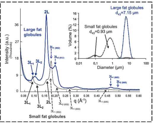
−8 ◦C (Fig. 12). The smaller fat globules
contained much more 3L structures and less 2L structures than larger ones [20].
Moreover, the crystals formed in the larger fat globules (d43 = 7.15μm) melted upon heating but did not evolve from a polymor- phic point of view, whereas the crystals formed in the smaller fat globules (d43 = 0.93 μm) were metastable and reorgan- ised to form more stable species before fi- nal melting of TG. These differences could have interesting applications in the manu- facture of dairy products in which temper- ing periods are involved in the technologi- cal process (e.g. butter, whipped cream, ice cream).
3.6. Compound crystals are formed in milk fat globules
The studies performed on milk fat glob- ule crystallisation showed that it occurs successively, producing 1, 2, 3 or 4 dif- ferent varieties depending on the cooling rate and that only small-angle XRD exper- iments permit the characterisation of these different types of crystals [10, 11, 14, 15].
We showed that upon cooling the first lon- gitudinal organisations of TG molecules dispersed in fat globules correspond to 2L lamellar structures with long spacings of 40–42 and 46–48 Å. These organisa- tions may correspond to crystallisation of high-melting point TG, with saturated and similar chain length fatty acids (e.g. PPS, PPP, MPP; P: palmitic acid, S: stearic acid, M: myristic acid). Then, crystallisation of 3L structures occurs: 3L1 = 70–72 Å and 3L2 = 65 Å. They may correspond to crystallisation of TG molecules with un- saturated fatty acids, or to TG with fatty acid chains of different lengths (e.g. BuPP, BuPO, PPO; Bu: butyric acid, O: oleic acid). Recently, Lopez et al. [19] showed that the 2L structures are formed by the stearin fraction, whereas the 3L structures are formed by the olein fraction, after dry
Large fat globules
Small fat globules
0.05 0.10 0.15 0.20 0.25 0.30 0.35 0.40 0.45 0.50 0.55 0.60
q (Å-1)
(Thousands)Intensity(a.u.)
3L1 3L2 2L
3L1 (002)
3L2 (002)
3L1 (005)
3L2 (005)
3L1 3L2
2L
3L1 (002)
3L2 (002)
3L1 (003)
3L1 (005) 0
9 18 27 36
0 2 4 6 8 10 12 14 16
0,01 0,1 1 10 100
Small fat globules d43=0.93 µm
Large fat globules d43=7.15 µm
Volume (%)
Diameter (µm)
0 2 4 6 8 10 12 14 16
0,01 0,1 1 10 100
0 2 4 6 8 10 12 14 16
0,01 0,1 1 10 100
Small fat globules d43=0.93 µm
Large fat globules d43=7.15 µm
Volume (%)
Diameter (µm)
0.05 0.10 0.15 0.20 0.25 0.30 0.35 0.40 0.45 0.50 0.55 0.60
q (Å-1)
(Thousands)Intensity(a.u.)
0.05 0.10 0.15 0.20 0.25 0.30 0.35 0.40 0.45 0.50 0.55 0.60
q (Å-1)
()
0.05 0.10 0.15 0.20 0.25 0.30 0.35 0.40 0.45 0.50 0.55 0.60
q (Å-1)
()
3L1 3L2 2L
3L1 (002)
3L2 (002)
3L1 (005)
3L2 (005)
3L1 3L2
2L
3L1 (002)
3L2 (002)
3L1 (003)
3L1 (005) 0
9 18 27 36
0 2 4 6 8 10 12 14 16
0,01 0,1 1 10 100
Small fat globules d43=0.93 µm
Large fat globules d43=7.15 µm
Volume (%)
Diameter (µm)
0 2 4 6 8 10 12 14 16
0,01 0,1 1 10 100
0 2 4 6 8 10 12 14 16
0,01 0,1 1 10 100
d43=0.93 µm
d43=7.15 µm
Volume (%)
(µm)
Figure 12.Comparison of the small-angle X-ray diffraction patterns recorded at−8◦C with small and large natural milk fat globules that were initially stabilised at 20◦C for 24 h and subsequently cooled at 0.5◦C·min−1down to−8◦C. Insert shows the size distribution of milk fat globules.
fractionation from the same initial batch of milk fat, performed at 21◦C.
The comparison of the small number of crystal types formed (4 longitudinal organisations and 5 polymorphic forms identified) with the large number of TG molecules present in milk fat (200 TG species were identified) provides evidence that each of the varieties corresponds in fact to compound crystals. The forma- tion of compound crystals, also called mixed crystals as they contain different TG molecular species in the crystal lattice, is not surprising in a natural fat with a wide compositional range of TG molecules, such as milk fat.
4. CONCLUSION
The use of high-flux X-ray sources (syn- chrotron) permitted the study of the poly- morphism and phase transitions displayed as a function of temperature and time by the complex mixture of TG dispersed in milk fat globules. XRD experiments per- formed at both small and wide angles allowed the identification on a molecu- lar scale of the TG longitudinal stack- ing and the lateral packing of fatty acid chains in lamellar structures, respectively.
Thanks to the resolution obtained with the various set-ups used in these studies, the small-angle XRD analysis performed for the first time on milk fat in the dispersed
state was essential for the identification of the longitudinal organisation of TG molecules in the lamellar structures and their time- and temperature-induced tran- sitions. Earlier studies concerning milk fat polymorphism were based on the descrip- tion of XRD peaks corresponding to the lateral packing of TG, and permitted only the identification of the crystalline sub- cells. Furthermore, we showed that differ- ent types of crystals (1 to 4) and several polymorphic forms (1 to 5) coexist in milk fat globules at low temperatures, together with a liquid phase. The coupling of XRD with DSC recordings permitted us to re- late the structural transitions to the ther- mal behaviour of milk fat globules. These tools allowed, for the first time to our knowledge, the identification and the char- acterisation of crystalline structures within water-dispersed and highly complex, natu- ral or processed food products as well as their transitions (several studies on cheese and ice cream are under way). Moreover, the comparison of the crystallisation prop- erties in the dispersed state and in bulk pro- vides information on the influence of the interface on crystallisation and its compo- sition (Lopez et al., in preparation). This work was performed on bovine milk fat globules but similar work is under way for other species: dromedary [17], goat [2], ewe and buffalo.
Acknowledgements: The authors gratefully acknowledge the French dairy board ARILAIT Recherches (Paris, France) for supporting this research and all the members of the steering committee on fat research. They also thank M.C. Michalski, N. Leconte and V. Briard (UMR STLO, INRA, Rennes, France) for col- laborating in the studies on the crystallisation properties of natural milk fat globules as a func- tion of their size.
REFERENCES
[1] Ali M.A.R., Dimick P.S., Thermal analysis of palm mid-fraction, cocoa butter and milk
fat blends by differential scanning calorime- try, J. Am. Oil Chem. Soc. 71 (1994) 299–
302.
[2] Amara-Dali W., Karray N., Lesieur P., Ollivon M., Goat milk fat: Thermal and structural behavior. 1. Crystalline forms ob- tained by slow cooling, J. Agric. Food Chem.
53 (2005) 10018–10025.
[3] Blanton T.N., Barnes C.L., Lelental M., Preparation of silver behenate coatings to provide low- to mid-angle diffraction cali- bration, J. Appl. Cryst. 33 (2000) 172–173.
[4] Gresti J., Burgaut M., Maniongui C., Bezard J., Composition of molecular species of tria- cylglycerols in bovine milk fat, J. Dairy Sci.
76 (1993) 1850–1869.
[5] Guinier A., Théorie et technique de la cristal- lographie, 3rd edn., Dunod, Paris, 1964.
[6] Hagemann J.W., Thermal behaviour and polymorphism of acylglycerides, in:
Garti N., Sato K. (Eds.), Crystallisation and polymorphism of fats and fatty acids, Marcel Dekker, Inc., New-York, 1988, pp. 9–95.
[7] Larsson K., Molecular arrangement in glyc- erides, Fette Seifen Anstrichm. 74 (1972) 136–142.
[8] Lopez C., Contribution à l’étude de la cris- tallisation des triacylglycerols: application aux émulsions laitières, Ph.D. thesis, Univ.
Paris VI, 2001.
[9] Lopez C., Focus on the supramolecular structure of milk fat in dairy products, Reprod. Nutr. Dev. 45 (2005) 497–511.
[10] Lopez C., Lesieur P., Keller G., Ollivon M., Thermal and structural behavior of milk fat:
1. Unstable species of cream, J. Colloid Interface Sci. 229 (2000) 62–71.
[11] Lopez C., Lesieur P., Bourgaux C., Keller G., Ollivon M., Thermal and structural behavior of milk fat: 2. Crystalline forms obtained by slow cooling of cream, J. Colloid Interface Sci. 240 (2001) 150–161.
[12] Lopez C., Lavigne F., Lesieur P., Bourgaux C., Ollivon M., Thermal and structural be- havior of milk fat: 1. Unstable species of an- hydrous milk fat., J. Dairy Sci. 84 (2001) 756–766.
[13] Lopez C., Lavigne F., Lesieur P., Keller G., Ollivon M., Thermal and structural behavior of anhydrous milk fat: 2. Crystalline forms obtained by slow cooling, J. Dairy Sci. 84 (2001) 2402–2412.
[14] Lopez C., Bourgaux C., Lesieur P., Bernadou S., Keller G., Ollivon M., Thermal and structural behavior of milk fat:
3. Influence of cooling rate and droplet size
on cream crystallisation, J. Colloid Interface Sci. 254 (2002) 64–78.
[15] Lopez C., Bourgaux C., Lesieur P., Ollivon M., Crystalline structures formed in cream and anhydrous milk fat at 4 ◦C, Lait 82 (2002) 317–335.
[16] Lopez C., Lesieur P., Bourgaux C., Ollivon M., Thermal and structural behavior of anhy- drous milk fat: 3. Influence of cooling rate, J.
Dairy Sci. 88 (2005) 511–526.
[17] Lopez C., Karray N., Lesieur P., Ollivon M., Crystallisation and melting properties of dromedary milk fat globules studied by X-ray diffraction and differential scanning calorimetry. Comparison with anhydrous dromedary milk fat, Eur. J. Lipid Sci.
Technol. 107 (2005) 673–683.
[18] Lopez C., Briard-Bion V., Camier B., Gassi J.-Y., Milk fat thermal properties and solid fat content in Emmental cheese: a differential scanning calorimetry study, J. Dairy Sci. 89 (2006) 2894–2910.
[19] Lopez C., Bourgaux C., Lesieur P., Riaublanc A., Ollivon M., Milk fat and primary fractions obtained by dry frac- tionation. 1. Chemical composition and crystallisation properties, Chem. Phys.
Lipids 144 (2006) 17–33.
[20] Michalski M.C., Ollivon M., Briard V.,
Leconte N., Lopez C., Native fat globules