HAL Id: tel-00865651
https://tel.archives-ouvertes.fr/tel-00865651
Submitted on 24 Sep 2013
HAL is a multi-disciplinary open access archive for the deposit and dissemination of sci- entific research documents, whether they are pub- lished or not. The documents may come from teaching and research institutions in France or abroad, or from public or private research centers.
L’archive ouverte pluridisciplinaire HAL, est destinée au dépôt et à la diffusion de documents scientifiques de niveau recherche, publiés ou non, émanant des établissements d’enseignement et de recherche français ou étrangers, des laboratoires publics ou privés.
as indicator of environmental conditions : A pioneer study on the influence of volcanic gases on the cuticle
structure in extant plants
Antonello Bartiromo
To cite this version:
Antonello Bartiromo. The cuticle micromorphology of extant and fossil plants as indicator of environ- mental conditions : A pioneer study on the influence of volcanic gases on the cuticle structure in extant plants. Paleontology. Université Claude Bernard - Lyon I, 2012. English. �NNT : 2012LYO10017�.
�tel-00865651�
Università degli Studi di Napoli “Federico II”
Scuola di Dottorato in Scienze della Terra
Dottorato di Ricerca in Analisi dei Sistemi Ambientali
“XXIV Ciclo”
Tesi preparata in cotutela con:
Université Claude Bernard Lyon 1
Ecole doctorale E2M2 Evolution Ecosystèmes Microbiologie Modélisation Thèse de Doctorat en Paléonvironnements et Évolution
Thèse en Biologie et Science de la Terre
The cuticle micromorphology of extant and fossil plants as indicator of environmental conditions.
A pioneer study on the influence of volcanic gases on the cuticle structure in extant plants
Bartiromo Antonello 2011
RELATORI/DIRECTEURS DE RECHERCHE:
Dott.ssa Guerriero Giulia Maître de conférences Guignard Gaëtan
COORDINATORE DEL DOTTORATO: Prof. Barattolo Filippo CORRELATORE:Dott.ssa Barone Lumaga Maria Rosaria
COMMISSIONE/JURY: Raschi Antonio, Prat Daniel, Guignard Gaëtan, Guida Marco DATA D’ESAME/DATE DE SOUTENANCE: 14/02/2012
The cuticle micromorphology of extant and fossil plants as indicator of environmental conditions.
A pioneer study on the influence of volcanic gases on the cuticle structure in extant plants
Antonello Bartiromo
antonello.bartiromo@gmail.com
Alla mia Terra, che non merita tutto questo … specie e di tutti gli ecosistemi della Terra.
Niles Eldredge, 2000
INDEX
Riassunto 7
Résumé 9
Abstract 11
Keywords 13
Laboratories 13
General Introduction 14
CHAP. I Influence of volcanic gases on the epidermis of Pinus halepensis Mill. in Campi Flegrei, Southern Italy: A possible tool detecting volcanism in present and past floras
17
1.1. Introduction 17
1.2. Material and methods 20
1.3. Results 24
1.3.1. Sulphur measures 24
1.3.2. Scanning electron microscopy observations 24
1.3.3. Transmission electron microscopy observations 27
1.4. Discussion 30
1.4.1. Environmental response of Pinus halepensis to volcanism 31
1.4.1.1. Epicuticular and epistomatal wax 31
Stomatal aperture 31
Volcanic toxic compounds and wax alterations 33
Other sulphur considerations 35
Wettability of leaf surface 36
Microenvironment 37
1.4.1.2. Cuticular membrane (CM) + cell wall (CW) 38
Pinus halepensis cuticle type 38
CM + CW thickness 38
CM + CW development 40
CM and CW ageing 40
Calcium-oxalate deposits 41
1.4.2. Potential application for extant and fossil material 42
CHAP. II
The cuticle micromorphology of in situ Erica arborea L.
exposed to long-term volcanic gases in Phlegrean Fields, Campania, Italy
46
2.1. Introduction 46
2.2. Material and methods 48
2.2.1. Plant material and sites description 48
2.2.2. Gas vent 49
2.2.3. SEM, TEM and EDS preparations 50
2.2.4. Gas concentration measurements in air and soil 51
2.2.5. Statistical analysis 52
2.3. Results 52
2.3.1. Energy diffractive X-ray analysis with SEM 52
2.3.2. Scanning electron microscopy observations 53
2.3.3. Trasmission electron microscope observations 55
2.4. Discussion 58
2.4.1. Chemical and SEM considerations 58
2.4.2. TEM considerations 60
CHAP. III An Early Cretaceous flora from Cusano Mutri, Benevento,
southern Italy 63
3.1. Introduction 63
3.2. Geological setting 63
3.2.1. Stratigraphy 65
3.3. Material and methods 68
3.4. Systematic palaeontology 69
3.5. Taphonomic and palaeoecological remarks 88
3.5.1. Taphonomy 88
3.5.2. Palaeoecology of the Cusano Mutri sedimentary basin 89
3.5.3. Xeromorphic adaptations of the plants 90
3.5.4. Palaeoclimate and floral comparison 91
CHAP. IV Plant remains from the Early Cretaceous Fossil-Lagerstätte of
Pietraroja, Southern Italy, Benevento 93
4.4.1. Introduction 93
4.2. Geological setting 94
4.3. Material and methods 96
4.4. Systematic Palaeontology 97
4.5. Taphonomic and palaeoecological implications 111
4.5.1. Taphonomy 111
4.5.2. Palaeoecology of the sedimentary basin 114
4.5.3. Palaeoclimate and comparison with other Albian florae 114
General Conclusions 117
Acknowledgments / Ringraziamenti 120
References 121
Photos 155
RIASSUNTO
Lo strato acellulare che ricopre le parti aeree delle piante vascolari superiori è chiamato cuticola. Quest‟ultima funge da barriera protettiva ed è un efficace rilevatore d‟inquinamento ambientale. Lo studio delle cuticole vegetali e in particolare degli apparati stomatici delle conifere, è estesamente utilizzato quale strumento di analisi ai fini della comprensione delle caratteristiche ecologiche e paleoecologiche. E‟ interessante notare che, sebbene siano numerosi gli studi inerenti le cuticole delle piante, poco o niente è stato fatto relativamente agli effetti prodotti sulle cuticole delle piante da parte dei gas vulcanici.
La Campania, con la presenza di numerose località caratterizzate da emissioni di gas di origine vulcanica (Pisciarelli, Solfatara, complesso del Somma-Vesuvio, ecc.), consente di effettuare studi di questo tipo.
L‟obiettivo di questa ricerca è quello di contribuire a individuare le potenzialità delle conifere e delle angiosperme (attuali e fossili) quali indicatori ecologici utili per la comprensione delle variazioni dei parametri ambientali. A tale scopo sono state compiute osservazioni macroscopiche e microscopiche di piante vascolari in relazione all‟influenza di fattori ambientali quali: aerosol vulcanici, intensità luminosa, disponibilità di acqua e salinità. Nel corso della ricerca sono state campionate numerose aree e sono state utilizzate apparecchiature quali: microscopio ottico, SEM, TEM e EDS. La statistica è stata utilizzata per l‟analisi delle caratteristiche micromorfologiche.
Le osservazioni condotte su piante attuali hanno consentito di studiare, per la prima volta, gli effetti dei gas vulcanici sull‟ultrastruttura delle cuticole della conifera Pinus halepensis [pino d‟Aleppo; siti di raccolta: Pisciarelli (presenza di gas vulcanici) e Cigliano (assenza gas vulcanici)] e dell‟angiosperma Erica arborea [siti di raccolta: Solfatara di Pozzuoli e Pisciarelli (presenza di gas vulcanici per entrambi) e Cigliano (assenza gas vulcanici)].
Le osservazioni al TEM effettuate su cuticole di P. halepensis, influenzate e non da gas vulcanici, hanno evidenziato che lo spessore totale di CM (cuticola) + CW (parete primaria) non subisce sostanziali variazioni di spessore. In particolare, la cuticola degli aghi influenzati da gas vulcanici mostra (a forti ingrandimenti: TEM) un aumento dei depositi di ossalato di calcio e un riarrangiamento delle fibrille che si dispongono parallelamente alla superficie. Le osservazioni condotte al SEM e al TEM su aghi di P.
halepensis attuali hanno permesso altresì di realizzare una chiave dicotomica che consente di identificare possibili alterazioni (dovute alla presenza di gas potenzialmente tossici come quelli vulcanici) anche in cuticole di pino sub-fossili o fossili.
Le osservazioni condotte su E. arborea, hanno permesso di costatare che gli spessori totali delle cuticole, “fumigated and not fumigated”, sono diversi. Comunque, in presenza di gas vulcanici lo strato esterno A2 subisce un sensibile incremento di spessore. Quest‟ultimo aumenta quando la concentrazione di CO2 in atmosfera è elevata, mentre non subisce sostanziali variazioni quando la quantità di CO2 al suolo varia drasticamente. Ciò conferma che la cuticola è il mediatore principale negli scambi gassosi tra ambiente interno ed esterno.
Per entrambe le specie attuali studiate non è stata riscontrata presenza di zolfo nella cuticola, nella parete cellulare o nel citoplasma. Ciò conferma l‟ipotesi che gli scambi gassosi avvengono essenzialmente attraverso gli apparati stomatici e che lo zolfo in eccesso è metabolizzato nelle foglie.
Relativamente ai macroresti vegetali fossili, sono state studiate le cuticole rinvenute nei Fossil-Lagertätten cretacici di Cusano Mutri (Aptiano superiore) e Pietraroja (Albiano inferiore). Il primo sito fossilifero ha consentito di: 1) identificare svariati taxa riconducibili alle conifere; 2) descrivere una nuova specie di conifera caratterizzata dalla presenza di caratteri xeromorfici: Frenelopsis cusanensis Bartiromo et al.; 3) rinvenire, per la prima volta al di fuori dei confini spagnoli, un‟angiosperma ancestrale: Montsechia vidalii. Lo studio tassonomico condotto sulle cuticole cretaciche di Cusano Mutri e Pietraroja ha permesso di descrivere entità tipiche della Provincia Euro-Siniana. Lo studio sedimentologico e sistematico denota un clima tropicale-subtropicale piuttosto arido. E‟
interessante notare come per il sito di Cusano Mutri, il rinvenimento di abbondanti
“fusain” sulle superfici di strato sia la prova che incendi naturali, frequentemente innescati da fulmini, interessavano le terre emerse.
Questo studio (almeno per quanto riguarda le piante attuali) può essere considerato pionieristico proprio perché per la prima volta sono state studiate le variazioni dell‟ultrastruttura della cuticola in presenza di gas vulcanici.
RÉSUMÉ
La couche qui recouvre les parties aériennes des plantes vasculaires supérieures est appelée cuticule. Cette dernière agit comme une barrière protectrice et est un détecteur efficace de la pollution de l'environnement. L‟étude de la cuticule des plantes, en particulier des appareils stomatiques des conifères, est largement utilisée comme un outil d‟analyse pour comprendre les caractéristiques écologiques et paléoécologiques. Il est intéressant de noter que, bien que les études sur la cuticule des plantes soient nombreuses, peu ou rien n‟a été réalisé sur les effets sur la cuticule des plantes par les gaz volcaniques. La Campanie, avec ses nombreux endroits caractérisés par des émissions de gaz d'origine volcanique (Pisciarelli, Solfatara, complexe du Somma-Vésuve), permet d‟effectuer ce type d‟études.
L‟objectif de cette recherche est donc de contribuer à individualiser les potentialités des conifères et angiospermes (actuelles et fossiles) comme indicateurs écologiques dans la reconnaissance des variations de paramètres environnementaux. Pour cela, des observations macroscopiques et microscopiques de plantes vasculaires ont été effectuées par rapport à l'influence des facteurs environnementaux tels les aérosols volcaniques, l'intensité de la lumière, disponibilité d‟eau et la salinité. Au cours de la recherche un certain nombre de localités ont été échantillonnées et on a utilisé des équipements comme le microscope optique, le MEB, le MET et l‟EDS. La statistique a été largement mise à contribution, avec l‟intervalle de confiance portant sur 30 mesures.
Les observations effectuées sur les plantes actuelles ont permis d‟étudier, pour la première fois, les effets des gaz volcaniques sur l‟ultrastructure des cuticules du conifère Pinus halepensis [le pin d‟Alep, sites de récolte: Pisciarelli (fumigé) et Cigliano (non fumigé)] et de l‟angiosperme Erica arborea [la bruyère arborescente, sites de récolte: Solfatara et Pisciarelli (fumigé) et Cigliano (non fumigé)].
Les observations conduites au TEM sur les cuticules de P. halepensis, influencé et non influencé par les gaz volcaniques, ont montré que l'épaisseur totale de la CM (cuticule) + CW (paroi pectocellulosique) ne subit pas de variations significatives d'épaisseur. En particulier, la cuticule des aiguilles influencée par les gaz volcaniques montre (à fort grossissement TEM) une accumulation d‟oxalate de calcium ainsi qu‟un réarrangement des fibrilles disposées parallèlement à la surface. Les observations SEM et TEM sur des aiguilles de P. halepensis actuelles ont permis également de réaliser une clé dichotomique permettant d‟identifier les altérations possibles (dues à la présence de gaz potentiellement toxiques comme les gaz volcaniques) des cuticules de pins sub-fossiles ou fossiles.
Les observations conduites sur E. arborea ont permis de constater que les épaisseurs totales des cuticules, influencées ou non par les gaz, sont significativement différentes. En présence de gaz volcaniques la couche externe A2 subit un sensible accroissement d‟épaisseur. Cette dernière augmente quand la concentration en CO2 en atmosphère est élevée, alors qu‟elle ne subit pas de variations substantielles quand la quantité de CO2 au sol varie de manière drastique. Ceci démontre que la cuticule est le médiateur principal dans les échanges entre l‟environnement interne et externe.
Grâce à des analyses EDS, pour les deux espèces actuelles étudiés il n‟a pas été trouvé de présence de soufre dans la cuticule, dans la paroi cellulaire ou dans le cytoplasme. Ceci confirme que la cuticule est le principale médiateur des échanges gazeux entre l‟environnement interne et externe.
Par rapport aux macro-restes végétaux fossiles, les cuticules du Fossil-Lagertätten du Crétacé de Cusano Mutri (Aptien sup.) et de Pietraroja (Albien inf.) ont été étudiées. Le première site fossilifère a permis 1) d‟identifier plusieurs taxa appartenant aux conifères; 2) de décrire une nouvelle espèce de conifère caractérisée par la présence de caractères xéromorphiques: Frenelopsis cusanensis Bartiromo et al.; 3) de trouver, pour la première fois à l‟extérieur de l‟Espagne, une angiosperme ancestrale: Montsechia vidalii. L‟étude taxonomique conduite sur des cuticules du Crétacé de Cusano Mutri et Pietraroja a permis de décrire entités typiques de la Province Euro-Sinienne. L‟étude sédimentologique et systématique montre une climat tropical-subtropical plutôt sec. Il est intéressant de noter, comme pour le site de Cusano Mutri, la présence des abondants fusains sur les surfaces des couches rocheuses, montrant les incendies naturels fréquemment amorcés par des éclairs intéressant les terres émergées.
Cette étude (au moins en ce qui concerne les plantes actuelles) peut être considérée comme pionnière car, pour la première fois, a été étudié les variations de l'ultrastructure de la cuticule en présence de gaz volcaniques.
ABSTRACT
The leaves of many tracheophytes are covered with a cuticle, an extracellular membrane covering aerial organs of plants. The gas exchanges between the plant and the surrounding atmosphere are mediated by the cuticle; its acts as the main barrier to air pollutants. The study of the plant cuticle, in particular the stomatal apparatuses of conifer, is largely used as a tool analysis revealing ecological and paleoecological features. It is worth noting that little is known about the long-term response of micromorphology of natural vegetation to volcanic toxic gases. Fortunately, Campania Region with its numerous volcanic localities (Pisciarelli, Solfatara, complexe du Somma-Vésuve) represents a natural laboratory allowing experiments involving plant-volcano interactions.
The object of this research is to study the conifer and angiosperms potentialities (extant and fossil) as ecological indicators useful in the identification of the environmental parameters variations. That is why, macroscopical and microscopical observations in vascular plants in relation to various environmental factors (volcanic gases, light intensity, water availability and salinity), have been analysed. A number of localities have been sampled and SEM, TEM and EDS equipments have been used together with statistic.
Observations made on extant plants allowed for the first time, the study of the effects of volcanic gases on the cuticle ultrastructure of Pinus halepensis [Aleppo pine; Pisciarelli (fumigated) and Cigliano (not fumigated) localities] and Erica arborea [tree heather;
Solfatara, Pisciarelli (fumigated) and Cigliano (not fumigated) localities].
TEM observations on P. halepensis cuticles fumigated or not by volcanic gases revealed insignificant thickness variations of the cell wall plus cuticle among current- and first-year- old needles of both fumigated and not fumigated trees. In particular, the needle cuticles experiencing chronic fumigation display (TEM) a calcium oxalate accumulation.
Moreover, in respect to the cell surface, fibrils are parallel disposed. SEM and TEM observations allowed an identification key enabling distinction between not fumigated and fumigated material with 9 characters, providing a good tool detecting the influence of volcanism for extant and fossil plants.
In specimens of E. arborea fumigated or not by volcanic gases, the total thickness of cuticles varies significantly. In plant experiencing chronic fumigation the A2 layer records an increase of its thickness. Within three localities, a good correlation between the atmospheric CO2 concentration and the thickness variation of A2 layer has been found.
This fact confirms that the cuticle is the main mediator between the plant and the atmosphere.
As for fossil plants, the cuticles of Cretaceous Fossil-Lagertätten of Cusano Mutri (Late Aptian) and Pietraroja (Lower Albian) have been studied. In the former: 1) numerous taxa belonging to conifers have been identified; 2) the new species Frenelopsis cusanensis Bartiromo et al. bearing xeromorphic features has been described; 3) the occurrence of Montsechia vidalii is recorded for the first time outside of Spain. Taxonomical studies carried out on Cretaceous cuticles from Cusano Mutri and Pietraroja allowed the description of typical Euro-Sinian fossil plants. Sedimentological and taxonomical studies suggest semi-arid or arid conditions in a subtropical or tropical climate. It is worth noting as for Cusano Mutri locality, evidence of wildfire (fusain) suggests a periodic combination of arid periods, high temperatures and lightning strikes.
This study (at least for extant plants) can be considered pioneering, because, for the first time, the relationships between cuticle ultrastructure variations and volcanic gases have been studied.
Parole chiave:
Campi Flegrei, gas vulcanici, Pinus halepensis, Erica arborea, epidermide, ultrastruttura della cuticola, idrogeno solforato (H2S), piante fossili, Cretacico inferiore, Sud Italia, Cusano Mutri, Pietraroja, Cheirolepidiaceae, Frenelopsis cusanensis sp. nov., paleoecologia, Provincia Euro-Siniana.
Mots clés:
Campi Flegrei, gaz volcaniques, Pinus halepensis, Erica arborea, épiderme, ultrastructure de la cuticule, hydrogéné sulfuré (H2S), plant fossiles, Crétacée inferieur, Italie du Sud, Cusano Mutri, Pietraroja, Cheirolepidiaceae, Frenelopsis cusanensis sp. nov., paléoécologie, Province Euro-Sinien.
Keywords:
Campi Flegrei, volcanic gases, Pinus halepensis, Erica arborea, epidermis, cuticle ultrastructure, hydrogen sulphide (H2S), fossil plants, Early Cretaceous, Southern Italy, Cusano Mutri, Pietraroja, Cheirolepidiaceae, Frenelopsis cusanensis sp. nov., Palaeoecology, Euro-Sinian Province.
Laboratori dove la tesi è stata preparata/L'intitulé et l'adresse de l'unité ou du laboratoire où la thèse a été préparée:
Dipartimento di Scienze della Terra, Università degli Studi di Napoli “Federico II”, Largo San Marcellino, 10, 80138 Napoli, Italia.
Université de Lyon, F-69622, Lyon, France; Université Lyon 1, Villeurbanne; CNRS, UMR 5276 Laboratoire de Géologie de Lyon, Herbiers de l‟Université Claude-Bernard Lyon 1.
Dipartimento delle Scienze Biologiche, Università degli Studi di Napoli “Federico II”, Via Mezzocannone, 8, 80134 Napoli, Italia.
Orto Botanico, Università degli Studi di Napoli “Federico II”, Via Foria, 239, 80139 Napoli, Italia.
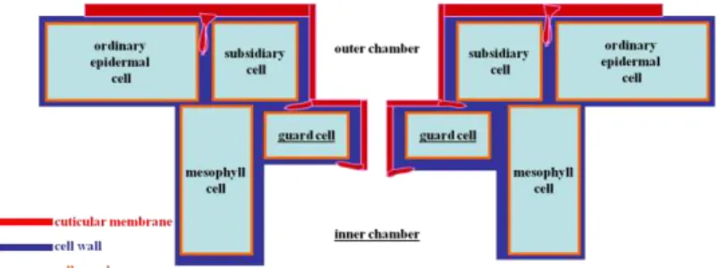
Fig. 1. Scheme showing the location of cuticle (= cuticular membrane in red) within the epidermis and in a stomatal apparatus context.
GENERAL INTRODUCTION
The leaves, fruits, and primary stems of higher plants are covered by a cuticular membrane (CM), or cuticle, that occupies approximately the outer 0.1-10 m of the aerial plant surface (Stark and Tian, 2006). On leaves the cuticle is present in both adaxial and abaxial surfaces. The CM occupies the outer surfaces of epidermal cell walls and is impregnated with an extracellular matrix (Domínguez et al., 2011). The cuticle has often been called the
“skin” of the primary parts of higher plants and has a very long history on the palaeobiological timescale (Riederer, 2006). With exception of fossil pollen and spores, cuticles represent the most widespread unaltered fossil plant remains and are known from the Devonian to the recent (Taylor et al., 1989). For this reason paleobotanists always played an important role in the study of plant cuticles (Kerp, 1990). The earliest references to fossil cuticles are by Göppert (1841–1846), while Brongniart (1834) gave the name
“cuticula” to a superficial membrane isolated from the cabbage leaf epidermis by retting in water. Later, Norris and Bukovac (1968) defined the limits of the cuticle as “all of the layers that can be separated from the underlying cellulose cell wall”.
Cuticular membrane is a translucent film of polymeric lipids and soluble waxes (Jeffree, 2006). The cuticle is usually
thicker above the anticlinal epidermal cell walls (CW), often forming pegs or span- drels by penetrating deeply between the anticlinal walls of adjacent epidermal cells (Jef- free, 2006). The CM bears an imprint (a “ghost”) of the epi-
dermal cell pattern of the plant organs on which it was formed (Fig. 1), which may survive as the only remaining fossil evidence of multicellular structure of the earliest land plants (Edwards et al., 1996).
In most plants the CM is not structurally or chemically homogeneous but composed of a number of layers, each of which is defined by its position and chemical constitution (Holloway, 1982). As Taylor et al. (1989) claimed, the cuticle consists of various layers which can be delimited based on the substances (e.g. waxes) embedded within the polymeric matrix and various structural features (e.g. lamellae and fibrillae). Generally
speaking, TEM revealed that the cuticle of the cells of the upper epidermis is made of an outer stratum named cuticle proper (CP) and an inner one called cuticular layer (CL).
According to the international terminology, the former (CP) can be constituted by an A1 (lamellate) or/and an A2 (uniformly electron dense with areas of lacunae) layers; the latter (CL) is constituted by a reticulate (B = B1) layer (see: Holloway, 1982; Archangelsky, 1991; Jeffree, 2006), in some rare cases an innermost B2 granular layer is present.
The outer surface of cuticle can be coated with epicuticular waxes which confer water repellency (Adam, 1963). Cuticular wax plays pivotal physiological and ecological roles in the interactions between plants and their abiotic and biotic environments, respectively (Jetter et al., 2006).
The cuticle performs numerous functions such as: transpiration control; control of loss and uptake of polar solutes; control of the exchange of gases and vapours; interface for biotic interactions and so on. However, the primary function of the cuticle is a permeability barrier against water vapour loss from tissues (Schreiber et al., 1996). For these and other reasons (see: Huttunen, 1984; Jeffree, 1986; Archangelsky et al., 1995; Garrec, 1996) the cuticle can be considered as an “external skeleton” as it represents the interface between the plant and the atmosphere (McElwain and Chaloner, 1996). As cuticle forms the interface between plants and atmospheric environment, it is the first point of contact between plants and air pollutants and it presents an effective barrier to pollutant entry.
During recent years considerable progress has been made for investigating this more external part of plants and its relations with external environment (Holloway, 1982; Hill and Dilcher, 1990; McElwain and Chaloner, 1996; Newrath, 2006; Jeffree, 2006; Shepherd and Griffiths, 2006). However, few studies have been carried out in the study of plant- volcanic gases interaction.
This doctoral thesis represents a contribution to the study of extant and fossil plant cuticles by means of optical and electronic (SEM and TEM) microscopy as well as EDS analyses.
In particular, this research is pioneer in the study of ultrasctructure of plant cuticle submitted to the influence of volcanic gases representing the first part of this thesis.
Similar researches are highly advisable in the Campania Region (Ischia island, Phlegrean Fields, Roccamonfina and Somma–Vesuvio complex) where volcanic and not volcanic gas emissions, give great opportunities in the analysis of plant–volcano interaction. However, Campania Region represents also a “virgin reservoir” for the study of Cretaceous plants that exactly make the object of the second part of this thesis (Fig. 2).
Fig. 2. Showing the location of the studied sites in Campania Region (the red asterisks). The dotted circle indicates the area occupied by the volcanic area of Phlegrean Fields.
Organisms of the past, in the same ways as those of the present, became adapted to their environments. The distributions of plant and animal species, as well as community characteristics, are strongly in-
fluenced by climate (Wing and Greenwood, 1993). As a matter of fact, thick cuticles and sunken sto- mata of fossil leaves also suggest lack of available water (Stewart and Rothwell, 1999).
Therefore, the “ability” of cuticle to register the environmental con- ditions is herein used as “trait d‟union” between the first (extant
plants) and second (fossil plants) part of this research as detector of environmental conditions.
For extant plants, a representative of conifers (Pinus halepensis Mill.) and angiosperms (Erica arborea L.) long-term fumigated by toxic volcanic gases have been studied. In Pisciarelli area P. halepensis is the only conifer growing near the vent. In Pisciarelli and Solfatara localities, E. arborea represents the only xeromorphic species capable to grow in the volcanic plume.
The present study, carried out over three years, rises from the collaboration between the Université Claude Bernard Lyon–1 and the Università degli Studi di Napoli Federico II and has as main goals the following:
1) analyze microscopical (essentially) and macroscopically features of the extant species Pinus halepensis Mill. and Erica arborea L. submitted to the influence of volcanic gases;
2) use experiences and observations make in the first part of the project to carried out in–depth studies of Cretaceous plants from Campania Region as to improve the knowledge of these “virgin” fossil site.
For every chapter a dedicated introduction and a “material and methods” section are proposed, because different techniques have been used for extant and fossil plant cuticles analyzed and for every site studied.
CHAPTER I
Influence of volcanic gases on the epidermis of Pinus halepensis Mill. in Campi Flegrei, Southern Italy: A possible tool detecting volcanism in present and past floras
1.1. Introduction
Over the geological history of the planet, among chronic environmental stress factors advocated as killing agents (Visscher et al., 2004), changes in atmospheric chemistry had world-wide dramatic effects on plant life in land (e.g. Visscher et al., 1996; Meyer and Kump, 2008). For instance, among chemical contaminants that could have disrupted end- Permian biota, volcanogenic SO2 (Visscher et al., 2004) and biological H2S (euxinia mechanism: Kump et al., 2005; Berner and Ward, 2006) gases are favoured to explain extinction. In particular, volcanism subsequently played a role in both maintaining and perturbing the atmosphere chemistry and physics, with important implications in terms of the evolution of life (Mather, 2008). The development of large igneous province (LIP) and continental flood basalt province (CFBP) (Courtillot and Renne, 2003; Jerram et al., 2005;
Keller, 2008; Bryan et al., 2010) commonly coincides with mass extinction events (Wignall, 2001, 2005; Rampino, 2010; Whiteside et al., 2010) and results in the release of significant volumes of gases, such as CO2, H2S and SO2 into the atmosphere (Beerling and Berner, 2002; Berner and Beerling, 2007; Hori et al., 2007). It is widely recognized that volcanic sulfur dioxide (SO2) and hydrogen sulfide (H2S) emissions are significant sources of sulfur release to the atmosphere (Bates et al., 1992; Berner and Berner, 1996).
Gases emitted by volcanoes represent both a factor inhibiting vegetation development (Whittaker et al., 1989) and could have been responsible of the decline of vegetation during periods of global-scale volcanism (Bond et al., 2010; Visscher et al., 2004;
Whiteside et al., 2010; McElwain and Punyasena, 2007). In particular, H2S is often thought to be a phytotoxin, being harmful to the growth and development of plants (Lisjak et al., 2010) especially when the quantities are higher than plant necessity (Thompson and Kats, 1978; Lorenzini and Nali, 2005). Moreover, atmospheric pollutants produced by volcanic activity and OAEs, such as SO2 and H2S, are said to be absorbed via the cuticle as well as the stomata (Haworth and McElwain, 2008).
Plants exposed to poisonous volcanic gases may show signs of diseases to total defoliation and death (e.g. Dickson, 1965; Clarkson and Clarkson, 1994; Delmelle et al., 2002). However, plant damages are related to both gas concentration (Delmelle, 2003) and its persistence (Grattan et al., 1998) in the atmosphere. Under severe pollution conditions, the direct phytotoxic effects of gaseous pollutants as well as long-term effects of acid washout (Grattan and Pyatt, 1994) can even be considered as potential environmental mutagens disturbing plant growth and community structure (Visscher et al., 1996). As a matter of fact, as Visscher et al. (2004) pointed out, variation in structure and composition of leaf cuticles is a potential source of botanical evidence on mutational effects of environmental stress factors.
Therefore, leaves in natural environments are subjected to a range of physical processes which may damage their surfaces, leading to alterations in the structure and integrity of the cuticle, and consequently changes in the physical properties of the leaf surfaces (van Gardingen et al., 1991).
To this end, numerous articles have been published in relation to the effects and interactions of the volcanic activity products (e.g. tephra or ash fall) on both fossil (e.g.
Kovar-Eder et al., 2001; García Massini and Jacobs, 2011) and extant plants (Winner and Mooney, 1980b; Cook et al., 1981; Seymour et al., 1983; Dale et al., 2005). Moreover, in extant plants the concentration of chemical elements in the leaves (Notcutt and Davies, 1989; Martin et al., 2009a and b) and the analysis of the log (Baillie and Munro, 1988;
Battipaglia et al., 2007) together with field studies led to significant advances in understanding the composition and dispersion of volcanic emissions at source (e.g.
Kempter et al., 1996; Delmelle et al., 2002), including major “gas species” (Costa et al., 2005; Chiodini, 2008; Chiodini et al., 2010a).
Leaves of plants act as passive and active collectors for natural (e.g. Martin et al., 2009a) and anthropogenic (e.g. Bačić et al., 1999) airborne pollutants (e.g. gas, aerosols and dusts) and are more sensitive to air quality than other plant organs (e.g. roots) (Landolt et al., 1989; Casseles, 1998; Kabata-Pendias, 2001); the gas exchanges between the plant and the surrounding atmosphere are mediated by the cuticle; this non-living (Riederer, 2006) thin (<0.1-10 m thick in extant plants) and heterogeneous membrane (van Gardingen et al., 1991) covers the epidermis of the aerial part of many tracheophytes (Guignard et al., 2004) and consists of a polymer matrix (cutin), polysaccharides and associated solvent-soluble lipids which are synthesised by the epidermal cells and deposited on their outer wall (Kirkwood, 1999; Riederer and Schreiber, 2001). The outer
surface of the cuticle is coated with epicuticular waxes, a general term (Jeffree, 2006) designating very long chain hydrocarbons found embedded within the cuticle and also in the crystalline epicuticular wax layer (Bird and Gray, 2003). The main function ascribed to waxes is to limit the diffusional flow of water and solutes across the cuticle (Heredia and Dominguez, 2009), providing protection for the leaf cells (Turunen and Huttunen, 1990) and acting as the main barrier to air pollutants (e.g. Jeffree, 1986). The composition and amount of waxes in the cuticle have been shown to vary depending to environmental conditions of the plant (Baker, 1982; Bird and Gray, 2003) and according to many authors air pollution seems to increase the rate of wax tubules degradation (e.g. Huttunen and Laine, 1983; Riding and Percy, 1985; Berg, 1987; Turunen and Huttunen, 1990, 1991, Huttunen, 1994). In particular, wax load and structure can be used as an indicator of pollution level (Hansell and Oppenheimer, 2006; Holroyd et al., 2002). The epicuticular wax of pine needles undergoes an ageing procedure during the needle lifetime (Turunen and Huttunen, 1996; Bačić et al., 1999) and is disturbed by polluted air (Huttunen, 1994).
The literature is replete with references to structural changes in epicuticular waxes following exposure to air pollutants (see Turunen and Huttunen, 1990), and as a matter of fact, the erosion of epicuticular waxes is a relevant factor of the multiple forest decline syndrome (Turunen and Huttunen, 1990).
Few paleobotanical works have been achieved on cuticular characters related to volcanic stress. Archangelsky et al. (1995) and Villar de Seoane (2001) studied Early Cretaceous plants from Patagonia (recovered in Baqueró and Springhill Formations, respectively) demonstrating that the volcanic ash fall played an important role in the formation of xeromorphic structures. As Haworth and McElwain (2008) claimed, the effect of toxic atmospheric gases and volcanic dust would explain xeromorphic features of Pseudofrenelopsis parceramosa (Fontaine) Watson from the Early Cretaceous of England.
Moreover, the relationship between ultrastructural characteristics of cuticle and the environment is still poorly understood for extinct as well as extant plants (Guignard et al., 2001) and cuticular ultrastructure data are not numerous for fossil conifers (e.g. Guignard et al., 1998; Villar de Seoane, 1998; Yang et al., 2009) and seem to be still lacking for some species belonging to the genus Pinus (Jeffree, 2006).
However, to date, no studies have been carried out relatively to the response of the ultrastructural features of plant cuticle exposed to the persistent volcanic gases. Conifers are well suited for studies of pollutant levels because they are evergreen and often have long-lived foliage. Usually the needles have a life cycle of several years (Hellström, 2003).
Therefore, the protective role of the epicuticular waxes is particularly important for conifers that have to ensure their investment in leaf tissue for several years (Chabot and Chabot, 1977). In the volcanic area of Pisciarelli (Campi Flegrei, Southern Italy) the gymnosperm Pinus halepensis Mill. (Aleppo pine) is the only conifer growing adjacent to the fumaroles, and much of the surrounding vegetation (under study) displays indications of damage caused by toxic gases. P. halepensis is the most abundant pine species in the western Mediterranean Basin, where it occupies 2.5 million ha (Quézel, 2000) and it is considered as an opportunistic species (Nathan and Ne‟eman, 2000) which is able to regenerate either in the absence or as a result of fire. In addition, P. halepensis has an elevated resistance to drought (Boddi et al., 2002), so much so that Emberger (1930) identifies it as being semiarid, and Oppenheimer (1968) considers it as the most arid- tolerant of all the Pinus species. As a matter of fact, the present study aims to assess the cuticular response of this conifer at a prolonged exposition to the volcanic gases using both SEM and TEM approaches. Moreover, to our knowledge, this is the first study that tests the cuticle ultrastructure behaviour during two subsequent years (current- and first-year-old needles) in response to the fumigation of volcanic gases containing H2S.
In particular, this research aimed to investigate: 1) response of plants to volcanic gases through different aspects: epicuticular and epistomatal waxes and ultrastructural features of the cuticle; 2) potential implications of the conifer cuticle response across environmental stress periods during the geological past; 3) a new method detecting the influence of volcanism for extant and fossil plants.
1.2. Material and methods
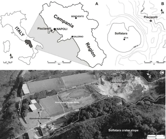
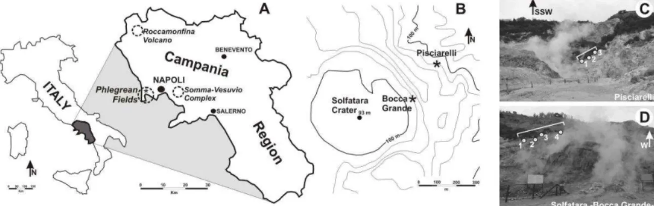
The material was collected from two localities in the Phlegrean Fields (Campi Flegrei, Campania Region), an active caldera which spans the last 50000 years (Scandone et al., 2010), characterized by significant recent ground deformation (Morhange et al., 2006) and considered as one of the most dangerous volcanic areas in the world (Chiodini et al., 2010a). In particular, pine needles were recovered from the famous fumaroles field in Pisciarelli locality (40°49‟48.88‟‟N, 14°08‟46.95‟‟E) about 1 km SE of the Solfatara volcano, both characterised by volcanic gas emissions (Fig. 1A,B). Control sample of needles were collected from a volcanic quiescent area (Cigliano: 40°50‟46.46‟‟N, 14°07‟36.31‟‟E) about 2.5 km from Pisciarelli and characterised by the absence of volcanic gas emissions and the presence of clean air. Both localities are characterised by the same
Fig. 1. (A) Location of the Pisciarelli area in the Campania Region. (B) Sketch map showing the location of Solfatara crater and Pisciarelli localities. (C) Close up view of Pisciarelli area with the main fumarole.
Dotted line indicates the area of diffuse degassing.
soil features (Di Gennaro and Terribile, 1999; Di Gennaro, 2002) and sun exposition and are far away from traffic and industries.
The temperature reaches about 97°C at the Pisciarelli fumaroles (Chiodini et al., 2010a) and the analysis of gaseous compositions (Caliro et al., 2007) revealed that the main component of the fumaroles is H2O followed by CO2, H2S, N2, H2, CH4, He, Ar, and CO.
The absence of acidic gases (SO2, HCl, and HF) can be also noted (Chiodini et al., 2010a).
Natural high atmospheric concentration of sulphur gas may occur locally in areas with volcanic and geothermic activity (De Kok et al., 2007). As a matter of fact, at Pisciarelli volcanic vent, the high H2S air concentration (equal to ca 600 m/mol) at source (Chiodini et al., 2010a) is also testified by both typical smell (the odour threshold is >0.02 m l-1: De Kok et al., 2007) of rotten eggs in the surrounding air and by indirect corrosive action of this gas -well visible on the iron objects- which caused people to abandon some buildings in the area (Fig. 1C). Nevertheless, this extreme environment is inhabited by very few
angiosperm species providing xeromorphic features (e.g. Erica arborea) and the boiling water of fumaroles retains the cyanidialean alga Galdieria phlegrea (Pinto et al., 2007).
Distal volcanic impacts have shown that plants are generally less sensitive to eruptions outside the growing season (Zobel and Antos, 1997; Hotes et al., 2004) and, as Payne and Blackford (2008) pointed out, in winter, plants are senescent and higher rainfall may serve to remove rapidly volcanic pollutants. In case of Pinus halepensis, an evergreen plant permanently fumigated by volcanic gases, retaining leaves for over a year, these “ground noises” do not exist.
The trees present diffuse damages along the North sides of the crown, while needles show symptoms consisting in leaf-tip non-specific discoloration which gradually increasing shootward (terminology from Baskin et al., 2010). Current- and first-year-old needles were collected from branches at heights over 1.5 m from three trees (15-20 years old) at each site (Pisciarelli and Cigliano). Needles were carefully handled to avoid damaging the epicuticular waxes. Following Reed‟s remarks (1982) and also Crang and Klomparens‟ ones (1988) about possible changes in epicuticular wax structures occurring during sample preparation, in order to limit any chemical or physical damages, especially for preserving and dehydrating samples for wax morphology studies (e.g. Turunen and Huttunen, 1991; Tuomisto and Neuvonen, 1993), needles were air-dried for 1-week at mild room temperatures. Among several hundreds of pine needles collected at each site (fumigated and not fumigated), 60 were selected for scanning electron microscope (SEM), then 16 were carefully selected for transmission electron microscope (TEM). Stomata were observed on 15 current- and first-year-old needles for each site. Analysis was performed within two weeks from sampling. Taxonomical identification of García Álvarez et al.
(2009) approach has been used. Light microscope observations were made using a Leitz microscope.
In order to quantify the quality of epicuticular and epistomatal waxes, SEM observations were carried out. Untreated needle sections of approximately 5 mm in length - obtained from the middle of each needle- were mounted on stubs using double-sided adhesive tape; both abaxial and adaxial surfaces have been studied. Part of specimens were sputter-coated with gold using an AGAR Auto Sputter Coater, while the specimens for energy diffractive x-ray (EDX) analyses were coated with carbon in a Emitech K450, observed and photographed with JEOL JSM-5310 SEM adapted with an Energy Diffractive X-ray Oxford Inca X-act at the CISAG (Centro Interdipartimentale di servizi per analisi Geomineralogiche) in the Dipartimento di Scienze della Terra, Università degli
Studi di Napoli “Federico II”. The operative conditions were as follows: 25-30 KV accelerating voltage, 100 A emission current, 15 m spot size, 20 mm microscope work distance and 1 min spectra collection time. To quantify the wax change in stomatal chamber, the Nicolotti et al. (2005) needle damage classes have been used as a criterion for the level of crystalline wax degradation.
Samples for TEM were dropped in paraformaldehyde solution mixed in a phosphate- sodium buffer for 3 weeks using Lugardon's technique (1971), washed and postfixed in a 1% osmium tetroxide solution mixed in a phosphate–sodium buffer for 24 hours.
Dehydrated in graded ethanol series during 48 h, the samples were dropped in propylene oxide with an increasing percentage of Epon resin for 24 h. Transferred into pure Epon resin during 24 hours, they were embedded in fresh Epon resin using flat moulds. The preparations were subsequently treated for polymerization at 56 °C for 3 days. Ultrathin (60-70 nm) sections were sectioned with a diamond knife, using a Reichert Ultracut microtome. Ultrathin sections were placed on uncoated 300 Mesh copper grids and stained manually both with a methanol solution of 7% uranyl acetate for 15 min and an aqueous lead-citrate solution for 20 min, then observed and photographed with a Philips CM 120 TEM at 80 kV, in the Centre de technologie des microstructures (CT ) of Lyon-1 University, Villeurbanne, France. Totally 16 pieces of material were embedded in Epon resin blocks. 90 uncoated mesh copper grids were prepared (80 as transversal sections, i.e.
perpendicular to the leaf length; 10 as longitudinal sections, i.e. parallel to the leaf length).
In order to detect the presence of sulphur in the cuticle, 40 measurements (i.e. 10 measures for each current- and first-year-old needles, fumigated and not fumigated by volcanic gases) with EDS microanalysis were carried out on different parts of the cuticle and also on the cytoplasm remnants of the epidermal cells. The sulphur analysis was performed on 25 coated 300 Mesh grids with transmission electron microscope JEOL 1200EX coupled to a microanalysis system EDS Si(Li) 30mm² NORAN VOYAGER III with an acceleration voltage = 80 kV and a spectres acquisition time = 60 s.
All the quantitative TEM measurements were made with tools in the ImageJ program (Abramoff et al. 2004). The terminology of Holloway (1982) and Archangelsky (1991) was used for the ultrastructural analysis. All specimens and SEM stubs are housed in the Dipartimento di Scienze della Terra, Largo San Marcellino, 10, Napoli, Italy. The resin blocks and TEM negatives are stored in the Lyon-1 University, Villeurbanne, France.
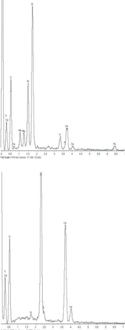
Fig. 2. Two different qualitative SEM X-ray diffractograms. A) Example of volcanic dust spectrum. B) Example of CaSO4 crystals spectrum. Current-year- old needle. Abaxial side. Pisciarelli.
1.3. Results
1.3.1. Sulphur measures
Energy diffractive X-ray analysis with the SEM, carried out on needles undergone chronic fumigation, showed two different graphics displaying typical volcanic products coming from Pisciarelli area (Valentino and Stanzione, 2004):
the leaves were covered with thin volcanic dust coming from the main fumarole of Pisciarelli (Fig.
2A); they also displayed numerous CaSO4 crystals (Fig. 2B).
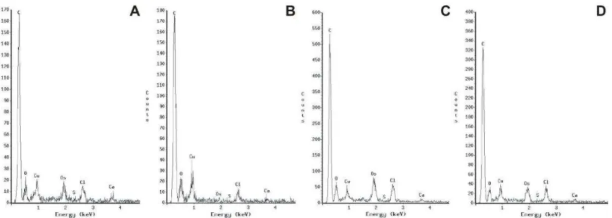
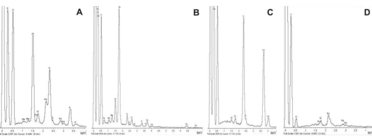
EDS microanalyses, within fumigated and not fumigated needles, concerned 4 sets of 10 measurements on outer, middle and inner part of cuticle + cytoplasm remnants of the epidermal cells. The elements are largely homogeneous (Fig.
3A-D) and the absence of sulphur is clear in all analysed parts. Extraneous elements detected by means of TEM X-ray analyses are Cu, Os and Cl derived from: the TEM-grids, chemical treatments and polyvinyl formal (FORMVAR, the most widely film for TEM grids) respectively. However, TEM electron energy-loss spectroscopy has proved that the cation calcium can be found in cuticle and cytoplasm of epidermal cells, especially in correspondence of low electron-density areas related to calcium-oxalate (CaC2O4) deposit.
1.3.2. Scanning electron microscopy observations
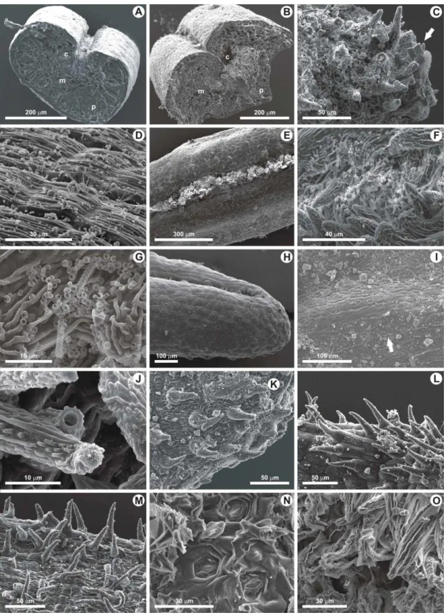
Stomatal apparatuses together with epicuticular and epistomatal waxes of fumigated and not fumigated needles have been observed (Plate I).
Fig. 3. Cuticle qualitative TEM X-ray diffractograms made on 70 nanometers sections, in three parts of the cuticle (outer, middle and inner parts) plus cell cytoplasm remnants, showing four examples among the elements content of: not fumigated, current- (A) and first-year-old (B) needles; fumigated, current (C) and first-year-old (D) needles.
Current- and first-year-old needles which did not experience chronic fumigation, retain quite well-preserved wax tubes in both abaxial and adaxial needle sides (Plate 1, Figs. 3,4).
The abaxial side of not fumigated current- and first-year-old needles displays the borders of the epidermal cells making folds between them (Plate I, Fig. 1).
The abaxial sides, and to a lesser degree the adaxial ones, of current- and first-year-old needles experiencing chronic fumigation, have tubular wax crystals which are converted into scale-like crystalloid formations on epicuticular surface (Plate I, Fig. 2) owing to the wide wax fusion phenomena. Rare crystalloid structures have been preserved underneath the epicuticular crusts (Plate I, Fig. 7, arrows). Epistomatal chambers show eroded and fused crystalloids wax, clearly visible in transverse sections (Plate I, Figs. 7,8) as a top sheet distinct to waxes embedded in the cuticle. Crusts were not usually seen in samples from Cigliano (not fumigated), where, apart from some scattered particles and granules the surface was quite smooth, even relatively clean (Plate I, Fig. 1). Crusts appear on most of the samples from Pisciarelli and are clearly visible in the area which surrounds stomatal apparatuses.
The abaxial side of the needles fumigated by volcanic gases presents the most part of stomatal apparatuses affected by crystalloids wax erosion and fusion phenomena in both epistomatal chamber and cuticle surface (Plate I, Figs. 5,7,8). On the other hand, the adaxial surface is less damaged and crystal tubules, even if eroded, can be seen (Plate I, Fig. 9). For not fumigated needles epicuticular waxes are more or less well-preserved in both epistomatal chamber and cuticle surface (Plate I, Figs. 1,3,4,6).
The majority of the stomatal apparatuses in abaxial side of current- and first-year-old needles from Pisciarelli (= fumigated) have fused tubules inside the epistomatal chambers
Plate I. Pinus halepensis, SEM of abaxial and adaxial cuticles, for to both trees experiencing and not chronic fumigation by volcanic gases. Stomatal class refers to Nicolotti et al. (2005) classification in which: class 0 = stomatal structure displaying no sign of alteration, class 1 = for slight sign of alteration, class 2 = for moderate sign of alteration, class 3 = for severe alterations.
1. Abaxial side of not fumigated current-year-old needle showing crystalline wax structure around epistomatal rim and along the contact zones of epidermal cells. Photo MC3lon1. 2. Abaxial side of fumigated current-year-old needle showing heavily melted epicuticular waxes partially occluding epistomatal chambers. Numerous volcanic dust particles obstruct stomatal apparatuses. Photo PP1.2long4. 3. Transversal section of not fumigated abaxial side of current-year-old needle showing a stomatal apparatus with well-preserved crystalline wax in epistomatal chamber (Class 0). Photo MC3s.t.1.1. 4. Detail of Fig. 3, showing completely uninfluenced wax crystal in stomatal antechamber (Class 0). Photo MC3s.t.1.2. 5. Transversal section of fumigated abaxial side of current-year-old needle showing a stomatal apparatus displaying severe alterations with the formation of wax granules and a volcanic dust particle that totally obstruct the epistomatal chamber; the network of microtubules is almost all melted (Class 3). Photo PP1.2s.t.3. 6. Abaxial side of not fumigated first-year-old needle showing slightly influenced wax crystals in epistomatal chamber (Class 1). Photo MC2.3long4. 7.
Transversal section of adaxial side of fumigated current-year-old needle showing a stomatal apparatus with its epistomatal chamber almost filled with a wax clump, except a small area in the right part in which the wax tubes are visible (Class 2). The arrows show crystalloids structures underneath the epicuticular crusts. Photo PP.2.1s.t.2. 8. Transversal section of fumigated first-year-old adaxial side of needle showing a rather flattened and deformed stomatal apparatus lacking in wax tubes in the epistomatal chamber, except for the base (arrow) in which the wax is heavily fused (Class 2-3). Photo PP2.2s.t.2. 9. Outer view of fumigated adaxial side of current-year-old needle showing stomatal aperture with more or less dense tuft of tubular wax structures in the upper part of epistomatal chamber. Mycelia fungi are also visible (arrow) (Class 1). Photo PP1.1long5.
forming a flat and solid wax plug or amorphous crusts above the pore which was completely or partially occluding the stomata in most cases (Plate I, Fig. 7). Instead, current- and first-year-old needles that do not experience chronic fumigation, regardless of the age of the needles sampled, present most of their stomatal apertures not occluded, thereby remaining almost completely open, apart from some minor fusing tubules (Plate I, Fig. 3). On the abaxial side of current- and first-year-old needles experiencing chronic fumigation, crystalloid wax tubules rarely persisted and they occupy limited portion only in epistomatal chambers (Plate I, Fig. 7 top right). It was noticed that in current-year-old needles from Pisciarelli the wax degradation was beginning very early and it increases.
Frequently, in abaxial side of some Pisciarelli stomatal apparatuses, amorphous wax and/or particles of other material -30-40 m width- filled the stomatal aperture, therefore blocking the direct view into the chamber (Plate I, Figs. 5,7).
In abaxial side of not fumigated needles, the rim of stomatal apparatuses retains crystalloid wax, whereas in fumigated needles the wax tubes are absent (Plate I, Figs. 1,2).
Frequently, the abaxial side of needles experiencing chronic fumigation presents deformed stomatal apparatuses so losing the typical funnel-like cavity (Plate 1, Fig. 8).
Stomatal damages are common and may include collapses (Plate I, Fig. 8), depression, degradation of guard cells (Plate 1, Fig. 2), and occlusion with wax clumps (Plate I, Figs.
2,7). Decay of the epistomatal chambers and empty cavities in stomatal chambers can also be noted (Plate I, Figs. 5,8).
On both abaxial and adaxial surfaces of fumigated needles, a great amount of dust occurred (Plate I, Figs. 2,9), while on the needles not fumigated by volcanic gases non- reactive dusts only mechanically disturb the wax structures (Plate I, Fig. 1). Moreover, most of Pisciarelli needles are affected by fungal infection (Plate I, Fig. 9).
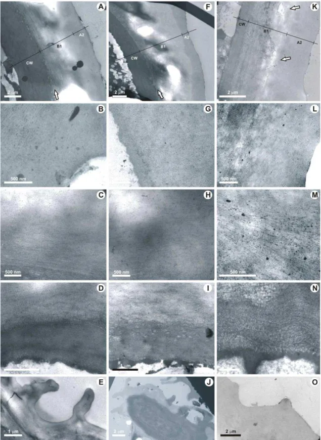
1.3.3. Transmission electron microscopy observations
The ultrastructure (cuticle and cell wall) of abaxial epidermal cells from fully grown needles (current- and first-year-old needles) of both localities have been studied in details.
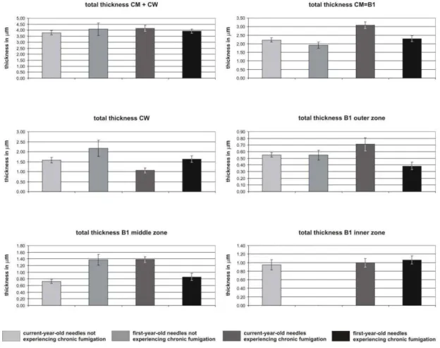
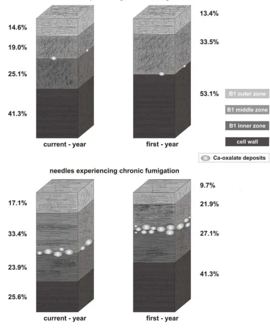
Cuticular membranes (CM) of all ordinary epidermal cell are composed of a fibrillar layer B (CL) in which an outer, middle and inner zones can be distinguished (Plate II). The cuticle proper (CP = A) is absent. Differences between the cuticular structures are given below. All the data given below are the means based on 30 measurements, the percentages of each component of the cuticle and of cell wall are also given (Table1).
In the current-year-old not fumigated needles (Plate II, Figs. 1-5) the total thickness of B1 + cell wall (CW) is 3.80 m (Plate II, Figs. 1,2). 58.7% is composed of the cuticular membrane, which may be further divided into three fibrillous zones: B1 outer (14.6%; 0.56 m), B1 middle (19%; 0.72 m) and B1 inner (25.1%; 0.95 m). In the first-year-old needles (Plate II, Figs. 6-11) the total thickness of CM + CW is 4.09 m (Plate II, Figs. 6- 7). 46.9% is composed of the cuticular membrane, which may be further divided into an outer fibrillous zone B1 (13.4%; 0.55 m), and middle zone (33.5%; 1.37 m). The B1 inner zone is lacking in this case.
In the current-year-old fumigated needles (Plate II, Figs. 12-16) the total thickness of CM + CW is 4.14 m (Plate II, Figs. 12,13). 74.4% of the CM + CW is composed of the cuticular membrane, which may be further divided into three fibrillate zones: B1 outer (17.1%; 0.71 m), B1 middle (33.4%; 1.38 m) and B1 inner (23.9%; 0.99 m).
Table 1. Statistical values, made with 30 measurements for cuticular membrane (CM) and cell wall (CW) of the epidermal cells. Note: the cuticular membrane CM is made up with cuticular layer CL (= B1 outer, middle and inner layers). All the measurements are in m. min-max = minimum and maximum values observed; % = percentage of each detailed part of the cuticle and cell wall; st-d = standard deviation; var = variance.
not fumigated (current year) not fumigated (first year)
mean min-max % st-d var mean min-max % st-d var Total CM + CW 3.80 3.03-5.01 100 0.53 0.28 4.09 2.64-7.58 100 1.47 2.17 CM = B1 2.23 1.48-2.79 58.7 0.35 0.12 1.92 1.17-2.88 46.9 0.49 0.24 B1 outer 0.56 0.29-0.69 14.6 0.09 0.01 0.55 0.34-1.14 13.4 0.21 0.04 B1 middle 0.72 0.39-1.15 19.0 0.18 0.03 1.37 0.52-2.17 33.5 0.46 0.21
B1 inner 0.95 0.39-1.60 25.1 0.34 0.12 0
CW 1.57 0.87-2.32 41.3 0.37 0.14 2.17 0.84-5.02 53.1 1.12 1.26
fumigated (current year) fumigated (first year)
Total CM + CW 4.14 3.40-5.86 100 0.70 0.49 3.92 3.25-5.52 100 0.50 0.25 CM = B1 3.08 2.27-4.29 74.4 0.56 0.31 2.30 1.93-3.46 58.7 0.48 0.23 B1 outer 0.71 0.37-1.27 17.1 0.28 0.08 0.38 0.22-0.80 9.7 0.16 0.03 B1 middle 1.38 0.94-1.94 33.4 0.24 0.06 0.86 0.44-1.60 21.9 0.29 0.08 B1 inner 0.99 0.36-1.53 23.9 0.28 0.08 1.06 0.40-1.61 27.1 0.27 0.07 CW 1.06 0.66-2.40 25.6 0.37 0.14 1.62 0.22-2.15 41.3 0.52 0.27
In the first-year-old needles (Plate II, Figs. 17-21) the total thickness of CM + CW is 3.92 m thick (Plate II, Figs. 17,18). 58.7% of the CM + CW is composed of the cuticular membrane, which may be further divided into the fibrillous zones: B1 outer (9.7%; 0.38
m), B1 middle (21.9%; 0.86 m) and B1 inner (27.1%; 1.06 m).
Plate II. Pinus halepensis, TEM of fumigated and not fumigated needles. All photographs were taken from ordinary epidermal cells in transversal sections, except a longitudinal section of photograph 11. The cuticle is made up with B1 fibrillous layer (= CL, cuticular layer) divided in three zones: o = outer zone, m = middle zone and i = inner zone.
1-5. Not fumigated current-year-old cuticle. 1. General view of cuticle and cell wall. Photo GGAB0018.
2. Magnification of cuticle and cell wall showing also the three B1 zones. Photo GGAB0007. 3. Detail of B1 outer zone showing slightly dense fibrils more or less random orientated. The upper part of middle zone just appears in the bottom of the photo, more densely stained. Photo GGAB0013. 4. Detail of B1 middle zone of cuticle showing more dense and crowded fibrils randomly orientated and in variable densities. Photo GGAB0012. 5. Detail of B1 inner zone showing less dense and the random disposition of fibrils. Note the lower part of middle zone just at the top right of the photo. Photo GGAB0011.
6-11. Not fumigated first-year-old cuticle. 6. General view of cuticle and cell wall. Photo GGAB0012. 7.
Magnification of cuticle and cell wall: the lack of B1 inner zone can be noted. Photo GGAB0006. 8. B1 outer less dense zone showing a prevalent random orientation of fibrils. Photo GGAB0008. 9. B1 middle zone more dense in parallel fibrils (especially visible downwards) and randomly orientated (upwards). Photo GGAB0008. 10. B1 base part of middle zone showing fibrils oblique or parallel to the cell wall. Photo GGAB0006. 11. B1 other middle zone of cuticle showing fibrils perpendicularly orientated, the upper part of cell wall being at the bottom of the photo. Photo GGAB0009. 12-16. Fumigated current-year-old cuticle. 12.
General view of cuticle and cell wall with numerous calcium oxalate crystal deposits between middle and inner zones of the cuticle. Photo GGAB0017. 13. Magnification of cuticle and cell wall showing the three B1 zones and a big amount of calcium oxalate crystals deposit between the B1 middle and B1 inner zones. Photo GGAB0028. 14. B1 outer zone of cuticle showing a prevalent parallel orientation of fibrils and quite numerous granules more or less aligned tending to simulate the A1 polylamellate layer of the cuticle proper A. Photo GGAB0042. 15. B1 middle zone more dense in fibrils showing fibrils orientated parallel. Photo GGAB0019. 16. B1 inner zone of cuticle less dense showing a parallel disposition of fibrils, above the upper part of cell wall at the bottom of the photo. Photo GGAB0020. 17-21. Fumigated first-year-old cuticle. 17.
General view of cuticle and cell wall with numerous and extensive calcium oxalate crystal deposits, especially at the anticlinal wall location between the two cells. Photo GGAB0040. 18. Magnification of cuticle and cell wall showing the three B1 zones and calcium oxalate crystal deposits. Photo GGAB0016. 19.
B1 outer zone of cuticle showing the parallel orientation of fibrils tending to form dense clusters (arrow) and quite numerous granules at the top part tending to simulate a cuticle proper “A”. Photo GGAB0027. 20. B1 middle zone with more dens and thicker fibrils showing fibrils more or less parallel orientated tending to form clusters. Photo GGAB0010. 21. B1 inner zone showing the parallel disposition of fibrils, above the upper part of cell wall at the bottom of the photo. Photo GGAB0044. CW = cell wall, o = outer, m = middle, i = inner. All the microphotographs are presented with the same orientation, the top of each print corresponding to the upper side of the cuticle, the bottom corresponding to the lower side of the cuticle.
For both fumigated and not fumigated needles a decrease of CM thickness between current- and first-year-old needles, has been observed.
A tendency to a reduction in the fibrillar structure followed by an increase of the granular component has been noted between not fumigated and fumigated needles. This feature is visible for all B1 zones but especially for B1 middle zone (Plate II, Figs.
4,9,15,20). Moreover, it is worth noting the disposition of fibrils: they are more or less chaotic in the three B1 zones of not fumigated needles, while they are much more parallel to the cell surface in the B1 outer (Plate II, Figs. 14,19) and inner (Plate II, Figs. 16,21) zones of influenced material. The cuticle of current- and first-year-old symptomatic needles, in respect to control needles, exhibits a much higher number of more or less rounded shaped areas of low electron-density; these areas represent calcium oxalate deposits, usually located between the middle and inner B1 zones (Plate II, Figs.
12,13,17,18).
1.4. Discussion
The present study based on 30 measurements is the most precise statistic analysis so far done for extant plant cuticles. Similar statistic analyses were provided for fossil plants as Pachypteris gradinarui from the Early Jurassic of Romania (Guignard et al., 2004) and the