HAL Id: tel-00772913
https://tel.archives-ouvertes.fr/tel-00772913
Submitted on 11 Jan 2013
HAL is a multi-disciplinary open access archive for the deposit and dissemination of sci- entific research documents, whether they are pub- lished or not. The documents may come from teaching and research institutions in France or abroad, or from public or private research centers.
L’archive ouverte pluridisciplinaire HAL, est destinée au dépôt et à la diffusion de documents scientifiques de niveau recherche, publiés ou non, émanant des établissements d’enseignement et de recherche français ou étrangers, des laboratoires publics ou privés.
Sélection d’anticorps recombinants dirigés contre des matériaux inorganiques pour des applications en
nanosciences
Purvi Jain
To cite this version:
Purvi Jain. Sélection d’anticorps recombinants dirigés contre des matériaux inorganiques pour des applications en nanosciences. Autre [cond-mat.other]. Université de Grenoble, 2012. Français. �NNT : 2012GRENY040�. �tel-00772913�
Université Joseph Fourier / Université Pierre Mendès France /
THÈSE
Pour obtenir le grade de
DOCTEUR DE L’UNIVERSITÉ DE GRENOBLE
Spécialité : Physique pour les sciences du vivant
Arrêté ministériel : 7 août 2006
Présentée par
Purvi Jain
Thèse dirigée par Dr Avraham Halperin codirigée par Dr Clément Nizak
Laboratoire Interdisciplinaire de Physique, LIPhy École Doctorale de physique
Sélection d’anticorps recombinants dirigés contre des matériaux inorganiques pour
des applications en nanosciences
Thèse soutenue publiquement le 27/09/12, devant le jury composé de :
Prof Franz Bruckert
Professeur INP Grenoble, Président
Prof Pierre Cosson
Professeur, Centre Médical Universitaire de Genève, Rapporteur
Prof Phillipe Minard
Professeur, Université Paris Sud 11, Rapporteur
Dr Erik Dujardin
Directeur de recherches CNRS, CEMES Toulouse, Examinateur Dr Avraham Halperin
Directeur de recherches CNRS, LIPhy Grenoble, Directeur de thèse Dr Clément Nizak
Chargé de recherches CNRS, LIPhy Grenoble, Co-directeur de thèse
Acknowledgment
Thanking people in just few words makes these pages the hardest and most incomplete part of this thesis. First of all, I would like to thank my thesis supervisor, Clément Nizak to give me opportunity to work on such an interesting project. The project presented interesting questions during entire three years, which kept me amazed. Most of the questions attempted to answer in this thesis are new with no prior knowledge. Thus experience of finding new results which sometimes contradict my earlier assumptions was truly thrilling part of this thesis. Major contribution to maintain this voyage as fun goes to my supervisor, Clément Nizak. He continuously guided me and never allowed me to feel lost in the subject. He not only guided me scientifically but also helped me in dealing with frustrations caused by inevitable failure of experiments and helped me seeing light at the other end of the tunnel.
He has so many things to be learned as a scientist as well as a human being. Working with him, made my PhD training an unforgettable learning platform.
Thank you to all jury members for your time and attention to review my work and also for constructive criticism.
I would take opportunity to thank nanoscience foundation for funding my project. I would also like to thank to the collaborators of different projects of this thesis – S.Moutel, F.Perez at Institute Curie, Paris and Benoit Dubertret at ESCPI, Paris.
It was amazing to work with talented peers working on very diverse areas at BIOP (LiPhy). Your suggestions, comments and criticisms along the way have been most helpful. In particular Jan Bednar, Avraham Helperin and Anand Shushi always provided their extended and constructive comments on my work.
I would like to offer special thanks to Noël and Mathilde, whose constant efforts make the laboratory very organised place to work. Your efforts with everything from ordering chemicals to occasional helping during experiment are very much appreciated. Special thanks to Jessi, without her I might be probably lost in dealing with administrative work. She is an (very) essential part of the lab in making any foreign students feel home.
Thanks to many people in the lab, who created an atmosphere which makes the laboratory a joy to work. Thanks to people at third floor for watching cricket world cup 2011 with great enthusiasm and cheering with me for glorious victory of India. Edith for being my translator in the lab as well as outside the lab, for all pause de café, giving me training for French bisous, and being my first gateway to learn French culture and Grenoble mountains, above all a good friend, list will go long.
Thanks to Darja for being coolest colleague on the planet earth (I doubted her origin from earth in first few encounters). She has magic tricks to lighten up the environment. Thanks to Marc Joyeux for providing me quiet place in his office for thesis writing and creating nice musical environment.
Thanks to Pierre-yves for his bottomless bag of incredible questions counterbalanced by Levan’s calmness, Olivier Vincent for solving my physics related doubts, Michael for his friendship and lending his ears whenever needed. Luca, Jawani and Valentina for giving Italian touch to the
environment and also occasionally to our taste buds, Madhu for being new Indian entry and source of 4 pm Indian sweets, Catherine Quilliet for occasional chick talks, Prem Prabhakaran for nano talks, and Nathalie for her cat talks.
This section would be incomplete without thanking very talented colleagues I met during my stay at Indian Institute of Science (IISc), Bangalore. A special thanks to Amartya Sanyal, his positive attitude towards research is infectious and it really helped me to shape my interest in research at the very beginning of my career. Richa Tyagi, who set up an example to balance research life, by doing excel in science and never missing any opportune moment of fun which life offers. And also by pursuing personal interests without any excuse of busy research schedule. I feel lucky to meet you all as colleagues and now friends.
Life is rarely complete without friends, who directly or otherwise help achieve one’s goals. My friends must be brave, for they put up with me. I cannot thank them enough, and will only name them here - Shubhra for being great buddy, Vivien as coolest flatmate, who believed that I do some science fiction sort of research in the lab, it gave me funny but great feeling when I was back at home.
Friends whom I met in Grenoble – Tarun, Mahesh, Nirmal, Pallavi, Krutika, and Nivs, life would not be same here without all those parties, midnight b’day cakes, movies, and shopping together.
Tarun’s ice cream break helped me a lot to melt all sorts of worries. Special thanks to Sylvana, François, Jeremy, Lucky and Jack (Lucky and Jack being THE cats!) for their unconditional love and affection, I call them extended French family.
Thanks to papa, for setting an example of how to love one’s job passionately. Thanks to mummy for much needed dose of encouraging words. Your lessons about being calm in hardest situations, and accepting all challenges with smile on the face, actually paid off during this thesis. I have never appeared in any exam from my school days to the college without talking to mummy-papa. PhD was for sure was toughest of all, It would have been impossible for me to complete it without your constant support. My elder sister actually share equal credit for all my degrees up till now, she was left with no choice than being awake all night, specially for my mathematics and physics tests. Now I am studying something out of her expertise, so she cannot help me directly, but thanks for helping me able to reach this level and also for spoiling me a bit with lots of pocket money during my student life. Having elder sister like you is one of the best things of my life. Thanks for your blessings nani (grand-mother), I will miss you throughout my life.
Very special thanks to Rohan, knowing you as a friend then boy friend and now as fiance is the next best thing happened to me. Ever since I met you, you are laughing on all my problems and actually you make them appear small, then I start wondering why I was worried about them. At the same time, even on small success, you make me feel like great achiever. Sometimes I am able to accomplish tasks just because you believe in my strength. It was your joyful spirit, which provided boost to enjoy my research work more.
An interesting project, nice supervisor, loving family, and trusted friends have always kept me motivated during this voyage of my career.
Sélection d’anticorps recombinants dirigés contre des matériaux inorganiques pour des applications nanoscientifiques
Résumé
Les matériaux inorganiques ont des propriétés uniques à l'échelle nanométrique. Ces propriétés ont généré beaucoup d'intérêt pour fabriquer des nouveaux matériaux utilisant des nano-objets comme unité de construction. Nous avons suivi une approche biomimétique pour la fabrication de dispositifs à base de nanoparticules afin d'améliorer les méthodes actuelles de fabrication top-down et bottom-up. Certaines protéines naturelles se lient en effet spécifiquement à des matériaux inorganiques, et déclenchent notamment la croissance de cristaux inorganiques. Une première étape dans cette approche biomimétique est de comprendre comment des protéines se lient spécifiquement à des nanomatériaux inorganiques. Nous avons exploré ce mécanisme de reconnaissance en sélectionnant des anticorps (les protéines de notre système immunitaire spécialisées dans les interactions avec de nombreuses cibles) contre des matériaux inorganiques par la méthode combinatoire biotechnologique appelée "phage display". Cette technique permet d'obtenir la séquence génétique codante des anticorps sélectionnés se liant à leur cible à partir d'une banque aléatoire d'anticorps. L'analyse statistique des séquences des anticorps sélectionnés fournit de nouvelles informations sur les interactions protéines/matériaux inorganiques. Notre principale conclusion est l'identification de l'acide aminé arginine en tant que contributeur majeur dans les interactions protéine/or. L'ingénierie génétique des anticorps permet de fonctionnaliser ces nouvelles sondes de matériaux inorganiques en vue de leur utilisation pour des applications dans le domaine des nanomatériaux. Les anticorps recombinants sélectionnés et leurs dérivés fonctionnalisés peuvent être exprimés par sécrétion à l'aide d'un hôte eucaryote (Dictyostelium discoideum) mis au point au cours de cette thèse.
Mots-clés: biomimétisme, nanoparticules, anticorps, Phage display, or, arginine, Dictyostelium discoideum.
Selection of recombinant antibodies against inorganic materials for applications in nanosciences
Abstract
Inorganic materials have unique properties at the nanometer scale. These properties have generated a lot of interest among researchers to fabricate novel materials using nano objects as building units. In this PhD thesis, we have attempted to mimick nature in the fabrication of nanoparticle based devices in order to improve upon current top-down and bottom-up nanomaterial fabrication methods. Proteins can specifically bind inorganic materials and trigger crystal growth and thus are considered as the main building units for a biomimetic approach of fabrication. The first step towards mimicking nature is to explore how proteins bind specifically to nanomaterials. We have explored this recognition mechanism by selecting antibodies (the protein binders of our immune system) against inorganic nanomaterials using the combinatorial biotechnology method of phage display.
This technique provides us with the genetic sequence of selected antibodies from a random antibody library exposed against a target. Statistical analysis of selected antibody sequences provides new information on proteins/inorganics interactions. Our main finding in this regard is the identification of the amino acid arginine as a major contributor to protein/gold interactions. Additional functionality to these new binders of inorganic materials is obtained by antibody engineering, allowing for their value added use in nanomaterial science applications. Selected recombinant antibodies and their engineered derivatives along with other recombinant protein can be expressed and secreted using a eukaryotic expression platform (Dictyostelium discoideum) developed during this thesis.
Keywords: biomimetics, nanoparticles, antibody, phage display, gold, arginine, Dictyostelium discoideum.
CONTENTS
Abbreviations and Acronyms--- 7
Synopsis--- 8
Synopsis en français 167
1. Introduction---11
1.1 An Introduction to antibodies---12
1.1.1 Structure ---12
1.1.2 Function ---13
1.1.3 Applications of antibodies as probes---14
1.1.4 Development of antibody technologies ---14
1.1.5 Single chain fragment variable (ScFv) ---15
1.2 An introduction to display techniques ---18
1.2.1 Antibody phage display technique---18
1.2.2 The link between genotype and phenotype: bacteriophage ---19
1.2.3 Phage display cloning vector: phagemids ---20
1.2.4 Antibody display libraries ---22
1.2.5 In vitro selection of binders in controlled conditions ---23
1.3 Antibody engineering ---27
1.3.1 Engineering IgGs into small fragments ---27
1.3.2 Engineering the valency of antibody molecules---28
1.3.3 Bispecific antibodies ---29
1.3.4 Fusion with other molecules ---31
1.3.5 Back to IgG like structure---31
1.3.6 Antibody class switch---32
1.4 Production of recombinant antibodies ---34
1.4.1 Challenges faced by expression systems for production of recombinant antibodies ---34
1.4.2 Expression systems for recombinant antibody production ---35
1.4.2.1 E. coli---35
1.4.2.2 Yeast ---36
1.4.2.3 Mammalian cells ---38
1.4.2.4 Dictyostelium discoideum---39
1.4.3 Tool box to regulate recombinant antibody expression level in Dd by secretion ---41
1.4.3.1 Genetic toolbox---42
1.4.3.2 Miscellaneous toolbox---42
1.4.4 Overview of glycosylation and its importance---43
1.4.5 Glycosylation profile of antibodies ---44
1.4.6 Glycosylation in Dd---45
1.5 Selection of biological binder (scFvs) for inorganic materials ---49
1.5.1 Introduction to nanoscience---49
1.5.2 Historical background and birth of modern nanoscience ---50
1.5.3 Example of inorganic nanomaterials ---51
1.5.3.1 Gold nanoparticles---51
1.5.3.2 Semiconductor QDs ---53
1.5.4 Synthesis of nanomaterials ---54
1.5.5 Stabilisation, functionalisation and self-assembly of nanoparticles: By surface modifications ---55
1.5.6 Current strategies for surface modifications of nanoparticles and their limitations ---57
1.5.7 Biomimetic approach to self assembly of nanoparticles---58
1.5.8 Motivation for surface modification by biological ligands---60
1.5.9 Selection of protein based binders (potential ligands) for nanoparticles ---61
1.5.9.1 Selection of peptides or antibodies using combinatorial techniques ---61
1.5.9.2 Comparison of protein based ligands: peptides vs antibodies---64
1.5.9.3 DNA based binder for inorganic materials ---65
1.5.10 characterization of the clones selected by phage display ---66
1.5.11 Potential use of inorganic material-binding proteins ---68
2. Results---70
2.1 selection of antibody fragments (scFvs) binding to inorganic materials ---70
2.1.1 Surface treated CdS QDs and gold nanoparticles ---71
2.1.2 Screen against micron size particles (ZnS, CdS, CdSe, gold particles) ---75
A) ZnS powder ---78
B) CdSe powder---79
C) CdS powder ---81
D) Gold powder---87
2.2 Engineering of antibodies and their production in Dd---96
2.2.1 Construction of expression vector ---97
2.2.2 Cloning of the antibody sequences in Dd expression vector ---98
2.2.2.1 Cloning of scFv sequences ---98
2.2.2.2 Cloning of engineered scFv fragments ---99
2.2.3 Transformation and selection of antibody fragments and engineered antibodies in Dd cells--- 102
2.2.4 Expression of antibody fragments and engineered antibodies in Dd cells --- 103
2.2.5 Large scale production and purification of antibodies from Dd supernatant --- 105
2.2.6 Enzymes involved in glycosylation in Dd genome --- 106
2.2.7 Analysis of glycosylation pattern in Dd secreted antibody --- 107
3 Discussion--- 110
3.1 Towards finding protein based binders (potential ligands) for inorganic materials--- 110
3.1.1 ScFvs as surface ligands for inorganic nanoparticles--- 110
3.1.2 Possibility of using other protein scaffolds as surface ligands for inorganic nanoparticles --- 111
3.1.3 Selection of positive binders (scFvs) using one large library: Searching a needle in haystack --- 111
3.1.4 Perspectives and consequences of using current display library --- 113
3.1.5 Failure of selection of binders for CdS QDs and gold nanoparticles --- 115
3.1.6 Failure of selection against micron sized CdS, CdSe, and ZnS semiconductor materials --- 116
3.1.7 Success of selection of binders for gold powder (a proof of concept) --- 116
3.1.8 Potential implications of arginine binding to gold --- 121
3.1.9 Potential application of gold binding scFv for surface modification of gold nanoparticles--- 121
3.1.10 From bench to bedside--- 122
3.2 Production of recombinant antibodies and their engineered constructs in Dd --- 123
3.2.1 Dd as an alternate eukayotic expression system--- 123
3.2.2 Further improvements to establish Dd as better expression system--- 124
3.2.3 Probable reasons of non-expression of scFv::streptavidine and bispecific antibody constructs--- 125
3.2.4 Glycosylation pattern in Dd--- 126
3.2.5 Absence of universal expression system for production of recombinant proteins --- 127
4. Bibliography--- 128
Appendix A: Chemicals, biologicals and vector used in phage display experimen--- 139
Appendix B: Phage display protocols--- 142
Appendix C: Alignment of anti-gold scFv sequences--- 148
Appendix D: Chemicals, biologicals and vector used in production of recombinant antibody in Dd--- 151
Appendix E: Design of Dd expression vector and cloning strategy of engineered fragments in Dd expression vector.--- 152
Appendix F: Generation and characterisation of Dd cell lines expressing recombinant antibodies--- 162
Appendix G: Purification of Dd expressed antibodies--- 164
Appendix H: Glycosylation--- 165
Abbreviations and Acronyms
ADCC Antibody-Dependent Cell-Mediated Cytotoxicity
CDC complement-dependent cytotoxicity
CDR complementarity determining regions
Dd Dictyostelium discoideum
DHLA-SB dihydrolipoic acid-sulfobetaine
ELISA Enzyme linked immunosorbent assay
Fab Antigen binding region
Fc Fragment crystallisable
GFP Green fluorescent protein
IF Immunofluroscence
mAb monoclonal antibody
NP Nanoparticle
PEG polyethylene glycol
PTM Post translational modifications
QD Quantum dot
scFv Single chain fragment variable
SEM Surface electron micrograph
SPR surface Plasmon resonance
VH Variable heavy chain
VL Variable light chain
Synopsis
Inorganic materials at the nanoscale level (1 - 100 nano metre) exhibit different optical, electrical, photo-electrical, magnetic, mechanical, chemical and biological properties in contrast to the bulk material that can have application in medical, energy, environment, production, and various other fields. In the context of this thesis, we have focused on two potential applications of nanomaterials; the use of metal and semiconductor nanoparticles as probes in bio-imaging and the production of self-assembled structures/devices based on nanoparticles. Both applications require a better control on nanoparticle surface chemistry.
The objective of this thesis is to propose a new strategy for improving surface chemistry of nanoparticles by using proteins, more precisely antibodies, as ligands/probes for inorganic nanomaterials, thus altering the surface chemistry. To achieve this goal, it is of primary importance to understand how proteins specifically bind to inorganic materials.
To understand protein/inorganics interaction we have used the combinatorial technique called phage display of antibodies. In this technique, a combinatorial random library of recombinant antibodies mimicking the immune repertoire is screened in vitro for selection of antibodies binding to a target of interest. This technique allows one to isolate specifically bound antibodies and their encoding genetic sequences. The technique has the major advantage of selecting protein binders under in vitro controlled conditions. One can tune the physical and chemical environment to create a selection pressure. Another major advantage of this technique is that there is no requirement for prior characterisation of the targets. In the case of inorganic surface targets, this is a crucial advantage over classical methods based on the rational design of inorganic surface ligands (molecular dynamics simulations, for instance).
In this thesis, multiple inorganic materials were used as a target. We propose this technique to prove the concept that, in principle, any material (organic/inorganic) can be used as a target and thus specific antibodies (proteins) targeting the material of interest can be identified. So far, by screening a library containing 108 random antibody sequences, we have successfully identified 32 sequences of selected antibodies that specifically bind to micron-sized gold particles. The statistical analysis of the 32 gold-binding antibodies sequences has allowed us to identify particular amino acids that were strongly selected at the random positions of antibody sequences. Arginine was predominantly selected at many positions in gold binding antibodies. This finding is in agreement with the observation of other groups using sophisticated and dedicated strategies such as molecular dynamics simulations. This result supports our strategy based on relatively simpler technique of
phage display for material science studies. We have then tried to use our gold-binding antibodies as ligands for improving the surface chemistry of gold nanoparticles (size - 20 nm). Preliminary results indicate the plausibility of the concept. The reasons for the inability to select antibodies against other inorganic targets used during thesis are partially identified.
These challenges can be addressed by simply optimising the protocols for the other inorganic targets.
As a next step, the sequence of selected antibodies can be modified by using recombinant antibody engineering. Antibodies can be engineered to acquire a new chemical function.
Sequences of two scFvs can be fused in order to produce adapters called bispecific antibodies capable of binding simultaneously to two targets. All the applications based on antibodies acting as probes to modify the surface chemistry of nanoparticles require the production of selected and/or engineered antibodies. Antibody production is a challenge due to their sophisticated structure and large size. This challenge is addressed in this thesis by the development of a new recombinant antibody production system based on the eukaryotic organism Dictyostelium discoideum. So far, this system has allowed us to produce two forms of engineered antibodies. In addition, we have started to explore the benefits of using this system for the production of therapeutic antibodies.
This thesis is divided into subparts; following is a brief description about the content of each subpart.
Section 1.1 is a general introduction to antibodies and the development of antibody technology; their structure, function, and applications as binders and probes.
Section 1.2 presents the combinatorial biotechnology technique of phage display and its application to select in vitro recombinant antibodies as binders of various biological and non-biological targets from random antibody libraries.
Section 1.3 describes the engineering of antibodies to produce antibodies fused to other biological and chemical moieties, including other antibodies.
Section 1.4 describes expression systems for the production of recombinant antibodies and their engineered forms. It also describes the new eukaryotic expression system that we have set up for this purpose (Dictyostelium discoideum).
Section 1.5 illustrates the biomimetic approach for inorganic nanomaterials fabrication and functionalisation, with a focus on using protein-based binders for inorganics to generate new materials.
Section 2 covers our results. This section is divided into two parts. Section 2.1 describes the selection of recombinant antibodies binding inorganic materials using phage display and the possible use of antibodies as inorganic nanoparticle ligands. Section 2.2 is about our setting up of the expression and production of recombinant antibodies and their engineered derivatives in Dictyostelium discoideum.
Section 3 covers the discussion which is divided into two parts: section 3.1- the successful selection of recombinant antibodies against gold, its potential applications in understanding interactions of proteins with inorganics and potential applications in biomimetic-inspired nanosciences and Section 3.2- the potential of Dictyostelium as a production system for recombinant antibodies and engineered constructs based on antibodies for therapeutical and nanoscience applications.
The process of scientific discovery is, in effect, a continual flight from wonder - Albert Einstein
1. Introduction
The immune system response inside the body relies on antibodies that bind specifically to foreign molecules. This recognition has been exploited for many applications. In this thesis, we have applied it in our biomimetic approach to select inorganics-binding antibodies that could be used to fabricate novel materials with inorganic nano components.
1.1 An Introduction to antibodies
Antibodies are proteins produced by the immune system. They are secreted inside the body in response to harmful substances known as antigens. The production of antibodies is a major function of the humoral immune system and is carried out by plasma cells (a type of white blood cells). Antibodies have the remarkable ability to bind to antigens. Binding triggers signals that induce targeted immune response. Antibodies show a strong binding specificity towards antigens, often compared with a lock and key model. Antibodies are categorised under the superfamily of immunoglobulins, source- Kuby immunology, part ll, (Kindt, Goldsby, & Osborne, 2006).
The aim of this thesis is to exploit the properties of antibodies and use them as versatile, specific binders of inorganic material targets for applications in nano-material science.
1.1.1 Structure
IgG antibodies are large molecules with molecular weight of approximately 150 kDa. They are composed of two different kinds of polypeptide chains. The classic structure of an IgG consists of two identical light chains (25 kDa) and two identical heavy chains (50 kDa) covalently linked by a disulphide bridge between the two heavy chains and between heavy and light chains.
Based on structural difference in heavy chains, different kinds of antibodies exist that are grouped into isotypes. Five different antibody isotypes are known in mammals, which perform different roles in the immune system. These isotypes are named with an "Ig" prefix that stands for immunoglobulin, e.g. IgM, IgD, IgG IgA and IgE .Some of the isotypes are further divided into subclasses — for e.g., there are four IgG subclasses and two IgA subclasses. IgGs are the most relevant class of antibodies for pharmaceutical applications.
Antibody molecules are roughly Y-shaped molecules (see figure 1.1a). The arms of the Y contain the sites that can bind antigens. This region of the antibody is called the Fab (fragment, antigen binding) region. It is composed of one constant and one variable domain
from each heavy and light chain of the antibody respectively. The variable domain is referred to as the Fv region and is the most important region for binding to antigens. More precisely, variable loops of β-strands, three each on the light (VL) and heavy (VH) chains are responsible for binding to the antigen. These loops are referred to as the complementarity determining regions (CDRs). The base of the Y shaped molecule is known as Fc (Fragment crystallisable or constant) region. This region is mostly constant in any antibody structure.
source- Kuby immunology, part ll, (Kindt, Goldsby, & Osborne, 2006).
Figure 1.1a: Modular structure of an IgG molecule. All immunoglobulin monomeric units are made up of two identical light (L) chains and two identical heavy (H) chains. Light chains include one constant domain (CL) and one variable domain (VL), whereas heavy chains include three constant domains (CH1, CH2 and CH3) and one variable domain (VH). The variable domains of both the heavy and light chains make the antigen-binding part of the molecule, termed as Fv. In the zoomed structure of Fv, three variable loops are depicted; these are called complementarity-determining regions (CDRs) 1, 2, and 3. Source- (Brekke & Sandlie, 2003)
1.1.2 Function
Antibodies recognize and bind foreign substances, such as bacteria or viruses, and alert other immune cells of body that can destroy them and flush them out of the body.
Antibody molecules can perform two different functions that are carried out by different parts present in the antibody structure. First, antibodies have the unique ability to recognize and bind to substances that cause disease. This primary function is performed by antigen binding site of antibody (Fab). Binding of an antibody to an antigen has no direct biological effect. The significant biological effects are carried by secondary "effector functions" of antibodies that are carried by sequence in the Fc region of antibodies. The common effector
functions are complement-dependent cytotoxicity (CDC), phagocytosis and antibody- dependent cellular cytotoxicity (ADCC) (Lucas & Oakland, 2001).
1.1.3 Applications of antibodies as probes
Antibodies can specifically bind to almost any target molecule. These target molecules do not necessarily need to be called antigens, it can be any molecule. Sometimes these targets are parts of proteins that are the important components of living cells or it can be non biological targets (such as inorganic materials in light of this thesis). Antibodies are, therefore, ideal sensors for detecting tiny amounts of a particular target molecule.
Antibodies are used daily as tools or reagents by research laboratories and clinic. For clinical purposes, antibodies can be used as a diagnosis reagent or as a drug for therapeutics. Fluorescence microscopy is a routinely used scientific technique that uses antibodies to visualize particular protein in cells or even in whole tissues (immunohistochemistry). Antibodies are used in many routine laboratory works for many standard assays like ELISA, western blot, flow cytometry etc. All methods of research in biotechnology and medicine are based on an understanding of the fundamental biological processes and their inter connections. For instance, which structure in the cells of organisms carries out which function? How do these functional structures communicate with each other? What are the consequences when they stop working? Antibodies are used in various laboratory techniques that address these questions. They are powerful tools in basic research because they recognize their antigens with outstanding specificity. This allows researchers to identify molecules and draw conclusions about the function of proteins of interest. Antibodies are also used in environmental analysis to provide evidence of contamination or the presence of harmful chemical substances (Killard, Smyth, Grennan, Micheli, & Palleschi, 2000). Last but not the least, we have tried to explore the applicability of this versatile molecule in nanomaterial fabrication and functionalisation.
1.1.4 Development of antibody technologies
Development of antibody technology is largely driven by the desire to use antibody as therapeutics. The mouse hybridoma technology described by Köhler and Milstein was an important step in the development of antibody technology and paved the way for the use of monoclonal antibody as therapeutics (Köhler & Milstein, 1975). However, antibodies derived from mouse are not suitable as therapeutic agent in humans. Strong immune response to non-human antibodies in humans had put end to this achievement in pharmaceutical sectors. Later developments in this field had again raised hope to use antibody as potential therapeutics. In an attempt to reduce the immunogenicity of mouse antibodies, genetic
engineering was used to generate chimeric antibodies. Further minimization of the mouse component of antibodies was achieved through CDR (complementarity determining region) and grafting (Bendig & Jones, 1996). Finally two major developments made antibodies completely independent from the animal immune system. These include development of selection by display technology and production of antibodies as recombinant proteins. The demonstration of displaying antibody fragments by phage and the functional expression of antibody fragments in bacterial periplasm are the pioneer experiments which provided strong platform for further development in the selection of antibodies by display techniques (Skerra & Plückthun, 1988) (McCafferty, Griffiths, Winter, & Chiswell, 1990). This selection is performed in vitro using library which contain many antibody fragment variants of human origin. Now the selection of fully human variable domain has become extremely simplified by the use of display technique (see section 1.2 for detailed technique of phage display).
The initial product of display screen may be a fragment of antibody “ScFv” (see next paragraph). Small fragment can be further engineered to get full antibody construct (see section 1.3 for antibody engineering). Further development in antibody technology lead to explore various expression systems to produce antibodies and its engineered derivatives (see section 1.4 for production of recombinant antibodies).
1.1.5 Single chain fragment variable (ScFv)
Fv fragment (shown in figure 1.1a) is one of the smallest units of immunoglobulin molecule that can mediate function of antigen binding. However, Fv fragment did not show stability under in vitro conditions. This fragment is not a very stable structure because the interaction energy between two variable regions present in this structure is low. Varieties of approaches have been taken to build a functional and stable complex of VH and VL. As a solution to this problem, new structure was generated by using a soluble and flexible 15-20 amino acid peptide linker to connect the V regions in order to build “ScFv” fragment (Bird et al., 1988) (see figure 1.1b). The resulting scFv exhibits substantial antigen-binding activity compared to the monoclonal antibodies whose VH and VL sequences were used. Progress in phage display has opened the opportunity of in vitro selection of scFv from a large library of variable domains (McCafferty, Griffiths, Winter, & Chiswell, 1990). ScFv molecule can be easily expressed in functional form using E. coli (Skerra & Plückthun, 1988). The genetic sequence can be further engineered using this small fragment as building units. Small dimensions, elevated stability and capability to recognise antigens that are difficult to access by conventional antibodies make this fragment an interesting tool for several research and biotechnological applications (de Marco, 2011).
Figure 1.1b: Antibody model showing different domains along the polypeptide chains. Single-chain fragment variable (scFv) antibody is shown in shaded area.(CDR- complemetarity determining region, FW- framework) Source (Ahmad et al., 2012)
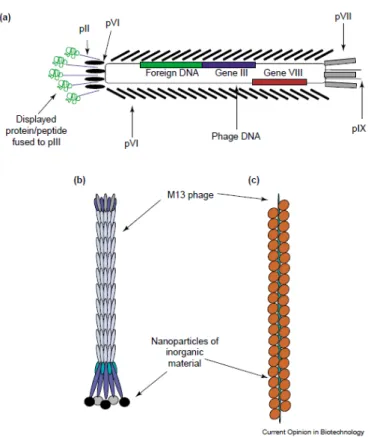
Phage display is the presentation of (poly) peptides on the surface of bacteriophage (bacterial virus). The gene encoding the protein product will be found inside the virus particle while the function encoded in the gene (such as the binding properties of an antibody) will be displayed on the surface of the
virus. Year 2012 marks the 27th anniversary of the invention of this powerful technique. It has allowed rapid, specific, controlled in vitro selection of
proteins/peptides as binders for various targets.
1.2 An introduction to display techniques
Display technique is a family of molecular biology techniques that allow for the in vitro selection of protein/peptide binders from random protein/peptide libraries for many kinds of targets. The concept of display technology is based on physical linkage between the genotype and the phenotype. A protein with the desired binding ability is selected;
simultaneously the genetic sequence corresponding to this protein is co-selected. This concept was successfully applied to small peptides by G P Smith by using phage as display system (Smith, 1985). Display of functional folded protein required several methodological improvements, which were initially done by groups at the MRC laboratory of molecular biology with Winter and McCafferty and the Scripps Research Institute with Lerner and Barbas for display of functional proteins such as antibodies. Later display method also extended to various forms such as ribosome display, mRNA display, cell surface display on bacteria and yeast. In this thesis we have used phage display of antibodies for screening against inorganic targets.
1.2.1 Antibody phage display technique
Antibody phage display technique empowers display of many antibody molecules on surface of small bacterial virus (bacteriophage). Collection of these displayed molecules is known as library. Initially phage display of antibodies was demonstrated by using small fragment of antibody molecules (scFv). ScFv fragment was displayed as a fusion protein on the surface of bacteriophage (see figure 1.2a) (McCafferty et al., 1990). Now this technology has been adapted for other antibody molecules including Fab fragment (Wieland, Orzáez, Lammers, Parmentier, & Schots, 2006), diabody (Takemura et al., 2000).
Library of phages displaying different antibodies can be used to rapidly identify antibodies that bind with high specificity and affinity to any desirable target molecule. This is possible because of presence of random antigen binding region.
Figure 1.2a: Diagram of filamentous phage displaying scFv molecules. The phage consist of circular ss DNA surrounded by coat protein. p8 is major coat protein and p3 (on tip of the phage) is one of the minor coat proteins. The genes encoding variable domains of scFv are fused to gene3 (g3) in the genome of the filamentous phage, as a result scFv is displayed as a fusion to p3 protein at the tip of the phage. Source - (Azzazy & Highsmith, 2002)
1.2.2 The link between genotype and phenotype: bacteriophage
The display concept is based on the link between the phenotype and the genotype and this link is provided by the bacteriophages or simply phages. The link is created by a foreign protein in frame with the coat protein gene of the phages. The filamentous phages are most widely used for phage display systems, among them M13 is most commonly used. The M13 phages can infect a variety of gram-negative bacteria using their pilus as receptors (Carmen
& Jermutus, 2002). The filamentous phages do not produce lytic infection in E. coli. After infection, they replicate inside E. coli and make them phage production factory. The bacteria, which are infected, do not lyse, but undergo reduced growth due to stress of phage production.
The phages are cylindrical shaped particles, which are approximately 7 nm wide and 900 to 2000 nm in length. The M13 phage has 6.4-kb genome, which contains circular, single- stranded DNA. The phage genome codes for 11 phage proteins. Five of these proteins are coat proteins. The major coat protein (p8) is present in approximately 2,700 copies and protects the genome in a cylindrical manner. The minor coat proteins p7 and p9 are necessary for efficient particle assembly, while other minor coat proteins p3 and p6 are necessary for particle stability and infectivity. The p3 protein mediates the binding of the phage to the F pilus and is necessary for viral uncoating and phage DNA transfer to the
cytoplasm of the bacterium. Host enzymes then convert the ssDNA into supercoiled dsDNA, known as the replicative form (RF) (source- chapter 1, phage display of peptide and protein, Winter, McCafferty et al.). The RF is essential to the phage display system because it can be purified and manipulated just like a plasmid. Through the manipulation of the RF of M13, some of the earliest cloning vectors were created. (Messing, 1991) (See next paragraph for phage display cloning vector).
The minor coat protein 3 (p3) and the major coat protein 8 (p8) are the most common proteins used for fusions. The Phage display system that uses the p3 and the p8 proteins has different advantages. Use of p8 protein as the fusion protein enables high copy display of the recombinant protein because there are over 2,700 copies of the p8 protein on the surface of the phages. However, the p8 protein can only display small size peptides. When p8 protein displays large size particles, the function of the coat protein becomes compromised and the number of infectious particles gets decreased (larger size peptide fusion become possible by use of phagemid vector, described in next paragraph). The p3 protein is present in 5 copies at the tip of bacteriophage. A recombinant fusion of larger peptide is possible using the p3 protein. Potential disadvantage is possibility of less infection due to sterical hindrance by displayed peptides. This can be better described as polyvalent display and this problem was solved by use of the phagemid vectors. (Source- book chapter 1, Introduction to phage biology, and phage display, Marjorie Russel et al.) 1.2.3 Phage display cloning vector: phagemids
Phagemids are cloning vector created by combining best features of plasmids and phages.
They are designed to have origin of replication for both M13 phages and E. coli. The important difference between phage based vector and phagemid vector is phagemids code for only fusion gene protein and not for other phage proteins. This feature was gained in the phagemid vectors by fusion of displayed protein gene under control of weak promoter which led to monovalent display of fusion proteins. However, monovalency is possible only for p3 fusion, p8 fusion still remains polyvalent. The valency of display can be an important factor as monovalency lead to selection of strong binders.
The phagemids vector alone can not generate complete phage particles because they do not code for other structural proteins. The other structural and functional proteins necessary to accomplish life cycle of phagemid are provided by the helper phages (e.g. M13KO7 or VCSM13) as co infection to E. coli cells harbouring phagemids. The helper phages have slightly defected origin of replication that causes less effective replication than phagemids.
This process is termed as “phage rescue”. In addition, other elements such as molecular tags and selective markers are introduced into the phagemids to facilitate the subsequent
operations, such as gene manipulation and protein purification. (Source- book chapter 1, Introduction to phage biology, and phage display, Marjorie Russel et al.)
Figure 1.2b: General scheme for phage display using phage and phagemid vectors. The difference in both vectors is illustrated by using p3 fusion protein as an example. The sequences of display proteins are introduced in the vector between the signal peptide and the gene3. Both vector carry Ff origin of replication, which allow them to replicate as M13 phages.
The phagemid vectors also carry origin of replication in E. coli (pBR322 in this figure) and an antibiotic selection marker, which allows them to replicate as plasmids in E. coli. In the phagemid vectors an amber stop codon (TAG) is introduced between the display protein gene and the gene3, which facilitates soluble protein expression by transforming phagemids in a non-supE suppressor strains. Source- book chapter 1, Introduction to phage biology, and phage display, Marjorie Russel et al.
The phages and the phagemids are the most common vectors used in phage display systems, while the phagemids are more widely used than the phages due to the following reasons. The smaller genome of the phagemids in comparison to the phages might facilitate efficient transformation. The phagemid vectors have wider choices of restriction sites. The phagemids are generally genetically more stable than the recombinant phages (Qi, Lu, Qiu, Petrenko, & Liu, 2012). The phagemids are more desirable if the monovalent display is preferred (in case of p3). The polyvalent display of larger peptides can be achieved by using
the phagemid p8 display system. The expression level of fusion proteins can be controlled easily in the phagemid vectors.
1.2.4 Antibody display libraries
It is possible to create a large library in which the proteins displayed on each phage are slightly different from each other; the process is called as library construction. Total number of different phage/phagemid particles displaying unique antibody fragments in the library defines the size of the library, which is a critical parameter for the success of the antibody phage technology. The larger size of a library increases chances of finding antibodies that bind to any given target with higher affinities. The second parameter that defines library performance is the number and location of variable positions. As in the immune system, the antibodies have a common framework, while diversity is present in the CDR loops that determine the specificity of binding. CDR3 loops represents the antibody region in which diversity is mainly concentrated in naturally occurring antibodies. Therefore, also for synthetic libraries, the amino-acid diversity is generally localized in the CDR3 residues (Griffiths et al., 1994). The other important parameters for library construction are: antibody gene should be well expressed, design of library should allow easy engineering of antibodies and finally overall handling of library should be easy (Kretzschmar & Von Rüden, 2002).
There are different ways to create diversity while building an antibody phage display libraries. On the basis of the strategy followed to obtain diversities, it can be classified in
“Immune repertoires” (antigen-biased), and “Singlepot” libraries (antigen-unbiased). Both kinds of libraries are explained in the following paragraph.
Immune antibody phage display libraries
This type of libraries takes advantage of diversity created in vivo by the immune system.
The source of variable immunoglobulin gene in this case is the B cells from immunised animal or immune patient (for human origin B cell) (Clackson, Hoogenboom, Griffiths, &
Winter, 1991). This library needs custom preparation for each antigen, which slims its scope for universal use. Another limitation with this library is the requirement of an anitigen to be immunogenic to provoke immune response. This feature makes use of such library less convenient for targets like inorganic materials used during this thesis, for which the immune responses are not yet fully known.
Single-pot libraries
The single-pot libraries contain virtually all possible binding specificities and are not biased for a particular antigen (Pini & Bracci, 2000). This type of library is suited for our application to find binders for various inorganic materials.
The single-pot libraries can be classified as naïve or synthetic. In the naïve library, variable genes are isolated from unimmunized animals or human donors and are assembled to create large diversity of antibodies. It is theoretically possible to select antibody against any target (antigen) by using this library. The major disadvantage is the requirement of very large library size to isolate high affinity binders as a consequence library has many unknown and uncontrolled contents. In the next step of artificial affinity maturation, these unknown contents need to be sequenced and that require preparation of customised primers.
In the synthetic library, diversity is entirely created outside the natural host. To construct a synthetic antibody library, germ line variable region is isolated from source and amplified;
subsequently CDR region is randomised, and assembled (Griffiths et al., 1994). The source of germ line gene could be any organism. To mimick natural antibodies, generally VH CDR3 region is chosen to insert maximum diversities. In the synthetic library, contents are well defined: antibody structure, knowledge of the antibody regions that are randomized and those that are kept constant. This library is not biased against self antigens. Main disadvantage of this library is that it needs to be highly diverse to obtain high affinity binders and while increasing the diversity of library, the chances of accumulating nonviable antibodies sequences also increase (Carmen & Jermutus, 2002), (Pini & Bracci, 2000).We have used Tomlinson (I+J) library for screening of inorganic materials. The Tomlinson library is single pot, synthetic library comprised of approximately 1 x 10⁸ random phagemids derived from non-immunized human donors (see appendix A for details).
1.2.5 In vitro selection of binders in controlled conditions
The phage display of antibodies provides selection of the binders against desired target under in vitro controlled conditions. Selection process can be divided into five main steps: 1) preparation of antigen/target 2) blocking 3) incubation of phage with antigen 4) washing to remove nonspecific phage and 5) elution. We can have great control during each step of selection, which are described in the following paragraph.
During the in vitro incubation step of the library with the target, physical (e.g. temperature), chemical (e.g. buffer composition, salt concentration, pH) and other parameters such as the
time of incubation (typically 1h) and the amount of target can be controlled to select antibodies which are able to bind the antigen under these defined conditions. Any molecule that remains stable in aqueous buffer for 1h can be used as a target. All these features make this technique applicable beyond biological targets. By playing with these selection conditions, it is possible to isolate binders (antibodies) with controlled affinity (nanomolar concentrations of the target will select for subnanomolar affinity), pH-dependence (selection at low pH and elution at high pH will select for pH-sensitive binders) etc.
1) There are many possibilities to prepare antigen/ target for screening: antigen/ target can be directly mixed (like in figure 1.2c (box2), for e.g. polycrystalline inorganic material is directly mixed with library of phages), or can be immobilized by direct adsorption to a plastic surface or can be used in solution (by using biotinylated antigen).
2) Blocking is done to avoid nonspecific binding which are mostly hydrophobic interactions with target entity. Generally, the target and the incubation device are immerged in buffer containing milk, BSA (Bovine Serum Albumin) or casein.
3) The mixture of library and target can be shaken or agitated for improving chances for phages to bind to the target. Phages which show affinity for target through displayed scFvs remain bound and those which do not show any affinity, either remain in solution or may bind non specifically (as shown in figure 1.2c (box 2).
4) Washing step can remove non-specific and unbound phages (see figure 1.2c (box 3).
Washing is done by quick rinsing using appropriate buffer containing mild detergent.
Washing step can also be performed under controlled condition by increasing or decreasing stringency of washes (usually by using different concentration of mild detergent or by increasing or decreasing number of washes or combination of both).
5) Elution is done to isolate target specific clones (see figure 1.2c (box 4).There is enough choices for elution conditions - one can chose pH dependent reagents (triethylamine, glycine/HCl), or enzymatic reagents (trypsin, chymotrypsin) or competitive elution with soluble antigen. After elution phages are infected to E. coli to amplify population of target specific clone, (see figure 1.2c (box 5). Subsequently, the phagemid bearing E. coli are infected with a helper-phage to produce new phage particles that can be used for further selection rounds until a significant enrichment of the target specific clones is achieved.
At the end of screen (typically 10th day), bacterial colonies can be randomly picked (these colonies appear as result of infection of phages from last round of selection). Each bacterium is capable of secreting scFv, which should be ideally specifically bind to target.
ScFv can be purified using bacterial strain (see appendix B). Genetic sequence of scFv can be obtained (by plasmid DNA purification) and analysed using various bioinformatics tools.
Additionally target specific clones can be modified by using various methods of affinity maturation.
Figure 1.2c: Different steps of phage display technique and selection of scFv specific for target.
screening box1-phage library, box2- incubation with target, box3 – washing step, box4- elution step, box5- infection and amplification in bacteria, box 6,7,8 – further choices after obtaining target specific scFv.
With advanced genetic engineering it is possible to remove or add any key protein domain or any other entity in order to create engineered antibodies.
1.3 Antibody engineering
The modular structure of antibodies allows a wide array of domain rearrangements by performing antibody engineering. The antibody engineering can provide more value to applications in nanoscience based on antibody fragments as inorganics binders.
Engineering of this molecule allows us to modify an original antibody fragment (scFv) selected by phage display for controlled valency, geometry and many other functionalities (refer section 1.1.5 for structural details of scFv molecule).
Engineered antibody constructs are not only useful for applications in nanoscience but also in providing effective pharmacokinetic properties to therapeutic antibodies. Some of the engineered constructs have already been taken to the clinic to serve the purpose as better therapeutics (Jain, Kamal, & Batra, 2007).
* There are many ways to engineer antibody molecules, the examples given here are more based on the work done during this thesis.
1.3.1 Engineering IgGs into small fragments
The large size of IgG can slim its scope for use as a small size binder for nanomaterials and as a therapeutic tool in few cases. Few limitations of using IgG are inefficiency to produce full size antibody, inefficiency to use IgG for display technique, increased circulation time in blood that affects its therapeutic applications and high background in imaging (Jain, Kamal,
& Batra, 2007).These limitations have encouraged development of small antibody fragments. Initial attempts to make smaller fragments were based on digestion of intact IgGs by proteolytic enzymes (pepsin or papain).
With the advancement in various displays system and recombinant DNA technology, it is very easy to generate small antibody fragments. One of the smallest fragments of antibody that can recognise antigen was identified as “Fv” fragment. Due to instability of this unit it was engineered to more stable structure called scFv (explained earlier in section 1.1.5).
Small size of scFv is a favourable characteristic for our aim to use scFv as small sized ligand for surface modification of nanoparticles. Due to small size, most scFvs have short serum half life (<10 min) compared to the intact IgG, which makes it useful for some diagnostic application such as imaging (Colcher, Batra et al.,2002). ScFv is a very interesting molecule for various research purposes.
Another popular fragment is Fab, where VH and VL regions are held together by CH1 and CL
domains as well as by inter chain disulphide bond. These four regions including the inter-
chain disulphide bond comprise of a Fab fragment. A Fab fragment is more stable than Fv or scFv fragment but this stability comes with relatively high molecular weight.
Figure 1.3a: Intact immunoglobulin molecules and its antigen binding fragments. Dark blue color fragment is VH domain, orange color is VL domain. In the structure of a Fab, VH and VL regions are held together by CH1 and CL domains as well as by inter chain disulphide bond. In the structure of an scFv, peptide linker of 15-20 amino acids is used to link VH and VL.. Source (Tikunova & Morozova, 2009)
1.3.2 Engineering the valency of antibody molecules
The Fab and scFv fragments derived from phage display antibody library are monovalent, whereas in many in vitro and in vivo conditions, multivalency of Antibody molecule is preferred.
ScFv expression vector is designed in such a way that the cDNA, which encode for VH and VL chain of an antibody are assembled by a sequence that codes for a peptide linker. The most common linker has 15-20 amino acid residues. Holliger et al have demonstrated that if the peptide linker length is reduced to 5 amino acid, VH and VL of scFv are not able to bind to each other (Holliger, Prospero, & Winter, 1993). The small size of linker causes a displacement in the equilibrium and as a result, scFv dimer is formed (this structure is called diabody, shown in figure 1.3b). The dissociation rate of diabody is lower than its parental scFv. Diabodies are rigid structures and can be expressed in bacteria. Further reduction in linker length to just one residue results in tetrabody formation. scFv tends to form a tribody in absence of a linker (Le Gall, Kipriyanov, Moldenhauer, & Little, 1999). Another approach to create multivalency in antibody fragment is the fusion of CH3 domain to a scFv or Fab fragment and the resulting molecule is called minibody.
Figure 1.3b: Presentation of diabody, tribody and tetrabody with their molecular masses. Source- (Olafsen et al., 2010)
To develop artificial antibody with multiple valencies, in one report, scFv sequence is fused to core streptavidine (Kipriyanov et al., 1996). Resulting fusion protein was termed as scFv::streptavidine. Streptavidine is a stable tetrameric protein and it has high affinity for biotin. This fusion protein was expressed in periplasmic inclusion body of E. coli. After purification, tetrameric complex scFv::streptavidine demonstrated binding with both antigen and biotin. This complex was found stable over wide range of pH and at high temperature.
However, expression level of streptavidine fused protein was observed to be low.
In designing strategies for multimerisation of antibody fragments, several issues need to be addressed like stability, resistance to proteolytic cleavage during in vivo assembly, efficient production of preferably soluble protein, easy gene engineering steps, efficient expression level, sufficient activity and specificity maintenance, thermal stability etc. Among these issues related to multimerisation of antibody fragments, we have attempted to address the issues of easy gene engineering steps by our antibody engineering platform and efficient expression level by using a new expression system (explained in section 1.4).
1.3.3 Bispecific antibodies
Naturally occurring antibodies are directed against a single antigen: they are monospecific.
Advances in antibody engineering have made it possible to combine the specificities of two antibodies into a single molecule - this combined molecule is known as a bispecific antibody. Bispecific antibodies do not occur in nature, it is a man-made construct. Initially, bispecific antibody was designed for therapeutic purposes. The initial use of bispecific antibody was based on the design in which one arm is specific for the tumor cell surface antigen and the other arm recognizes and activates the signalling receptor on the effector cells (Dafne M,RE Kontermann,2010). The combination results in using patient’s own
immune effector cells to target tumor cells which ultimately resulted in the killing of the tumor cells.
Figure 1.3c: Concept of BiTE constructs, Two scFvs having different specificity (one for tumor cell, other for T cell activation) joined together by a linker. This construct can use patient’s own killer cell to eliminate tumor cell. Source - Wikipedia
This particular subset of bispecific antibody, which engages T cell receptor is also known as BiTE construct (Bi-specific T-cell engagers). Concept of BiTE is shown in figure 1.3d. These engineered bispecific molecules also caught the attention of material scientists for building bio-inspired or biomimetic material (explained in details in section 1.5).
There are some technical hindrances related to the construction and production of bispecific antibodies in terms of quantity and quality. Earliest bispecific antibodies were made by fusion of two hybridomas producing antibodies of two desired specificities. This construct had issues related to yield and purity because pairing between heavy and light chains was a random event with this technique. Over the years, many advanced construction and production techniques of bispecific antibody have come up. One of the recent techniques consist of using scFv from phage display selection and engineering them to make bispecific antibody. This engineering can be done by joining VH or VL of one scFv with VH or VL of another antibody by flexible linker. In this thesis we have attempted to make such bispecific antibody construct, using flexible linker. One bispecific antibody having therapeutical importance has already entered into the market and many others are in the clinical phase (www.f-star.com). Recently, Macrogenics and Boehringer Ingelheim have inked a deal of