Endothelial cells are the main stromal cells that play a crucial role in cancer development and seem very good candidates for eliminating cancer. The literature analysis indicates that the immune system is a very promising field of study in terms of prognosis, mostly because the stromal microenvironment in the tumor can provide some information about what may succeed in the future regarding treatment choices and perspectives. So our main scientific question was whether intratumoral endothelial cells present MHCII-cancer antigens complexes and what is the biological significance of this phenomenon in cancer.
Some preliminary data showed that TECs constitute the vast majority of nonhemopoietic MHCII-expressing cells in the lung TME. Lung TECs express high MHCII and co-inhibitory (PDL1) but not co-stimulatory molecules (CD80, CD40, CD86), which is our first indicator that they play an important role as atypical APCs. As a result, their offspring had a deletion of MHCII in lung cancer endothelial cells.
4 Keywords: Endothelial cells (ECs), Tumor-associated endothelial cells (TECs), Tumor microenvironment (TME), tumor, lung cancer (LC), antigen presentation, Antigen-presenting cells (APCs), APC- professional, atypical APCs, MHCII, FAC, mouse model. Finally, I want to thank my family for supporting me all these years.
Introduction
- Immune system: Innate and adaptive immunity
- Innate immunity
- Adaptive immunity
- Antigen presentation
- Professional Antigen presentation
- Non-professional (Atypical) APCs
- Tumor associated endothelial cells
- Endothelial cells’ role in neoangiogenesis
- Endothelial cells as atypical antigen presenting cells
- Tumor associated endothelial cells as atypical antigen presenting cells in TME
- Mouse strains
- Cdh5(PAC)-CreERT2: Β6-Tg(Cdh5-cre/ERT2)1Rha mice
- I-AB-flox : B6.129X1-H2-Ab1tm1Koni/J mice
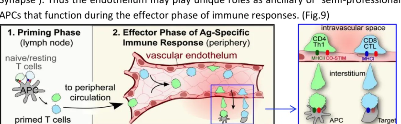
Cytotoxic T cells (CD8) are mainly involved in the destruction of foreign-infected cells. A third type of T cell, known as regulatory T cell (Treg), also plays a role in the immune response. In the first step (Figure 3), oncogenesis generates neoantigens that are released and captured by dendritic cells (DCs) for processing (Step 1).
These activated effector T cells are then recruited into the TME, resulting in targeted killing of tumor cells. We were particularly interested in investigating the role of endothelial cells as potential antigen-presenting cells in the lung tumor microenvironment. Endothelium refers to cells that line the inner surface of blood vessels and lymphatic vessels and form an interface between circulating blood or lymph in the lumen and the rest of the vessel wall.
Hypoxia often results in excessive production of angiogenic factors by several stromal cells (Figure 5). Endothelial cells are even more important cells because there is evidence that they can influence the immune system. Compared to professional antigen-presenting cells, which can provide 3 signals, endothelial cells are equipped with all the necessary capabilities, with one key exception, to provide APC signals 1-3 already mentioned in professional antigen-presenting cells.
All these findings on the multifaceted role of tumor-associated endothelial cells in cancer growth and progression highlight the need to further characterize the interaction of TECs in the tumor stroma in the context of cancer.
Materials and Methods
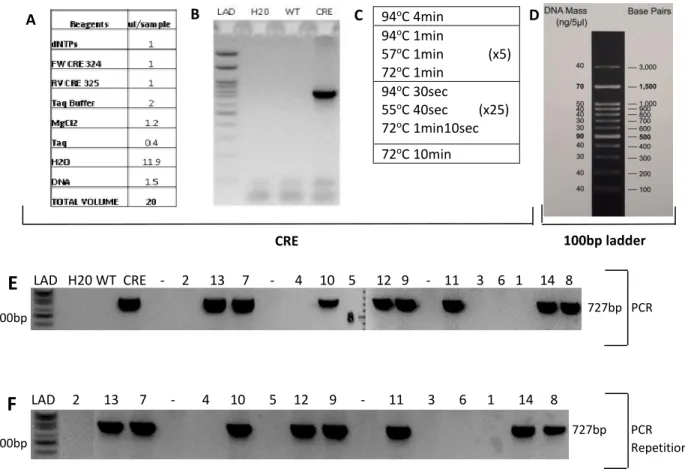
Finally, the tubes were stored in the cold room until used for PCR. A master mix was made containing dNTPs, primers, Taq buffer, MgCl2, Taq, and water, and then each DNA was added in the appropriate amount based on Fig16A. This mixture was heated in the microwave until the powder needed to be diluted and the mixture was homogeneous.
4 μl of orange G (loading buffer) was added to the total volume of 20 μl of each PCR product and 12 μl was added to each well. A 100 bp ladder was added to the first well that was an indicator of the bp of each different band. 200 mg of tamoxifen powder and 2 ml of 100% ethanol (mixture) were also placed in the water bath.
The LLC (Lewis lung carcinoma) cell line was used to induce a cancer tumor in the lung. Before operations, LLC cells had to be manipulated in a cell culture hood in sterility. 2 weeks after the last tamoxifen injection, Lewis lung cancer (LLC) cells were injected into the left lung lobe of each mouse to induce cancer after 2 weeks.
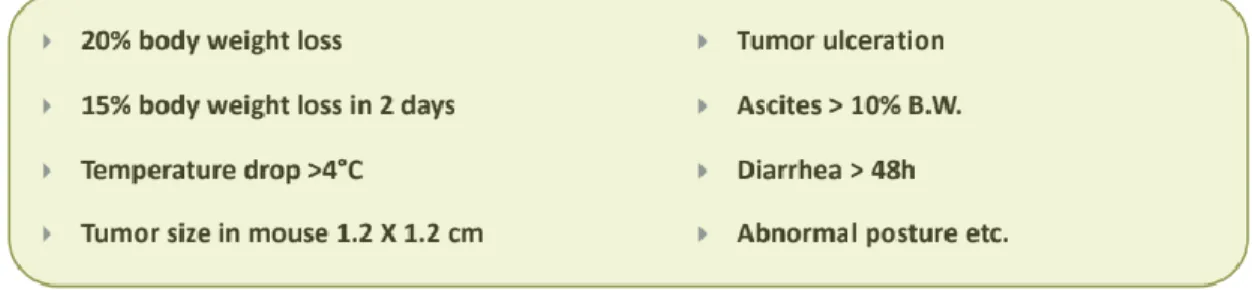
More specifically, the mice were anesthetized and a necropsy was performed to allow injection into the left lung lobe with the appropriate amount of LLC cells. As already mentioned, we had to wait two weeks before the lung cancer could be induced and possibly isolated. The cells proliferated and the mice were sacrificed 15 days after the injection of LLC-OVA cells into the lung.
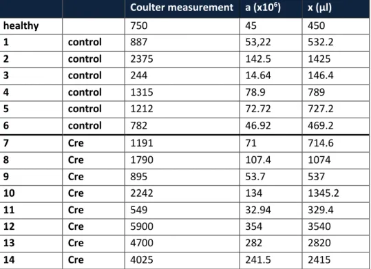
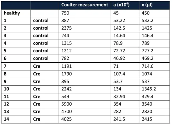
So, the volume of each sample had to be x μl after addition of PBS+ according to Table 4. The correct concentration of cells was finally used for staining for FAC analysis. It was necessary to choose the best antibodies and their correct amount in order to have the best experimental setup.
Results
Mice were sacrificed 15 days after LLC-OVA injections and the tumor was analyzed by FACS. In vivo, it is shown that LLC cells generally grow faster in MHCII F/F Cdh5aCre mice (Figures 18A and B) and that the mice have statistically significant larger tumors compared to control (MHCII F/F). Tumor burden was measured with Zombie FITC, a dead cell discriminator, and mcherry APC (eBioscience, M11241), an intracellular stain.
Expression of MHCII in CD31 PDPL NEG TM is statistically significantly higher in homozygous loxP-flanked MHCII (MHCII F/F) than in the MHCIIF/F CDdh5aCre, so the Cdh5aCre resulted in a deletion of MHCII allele in endothelial cells (19A). MFI (Mean Fluorescence Intensity) refers to the fluorescence intensity of each event on average, represents the expression amount of the parameter you selected on each event. At 2nd and 4th image on the lower part of the control (MHCII F/F) lung are the infiltrating lymph nodes of these mice.
Podoplanin (PDPN) is expressed on many tumors and participates in tumor metastasis. CD16/CD32 is expressed on B cells, monocytes/macrophages, NK cells, granulocytes, mast cells and dendritic cells. Absolute CD4 and CD8 (ABS.NO) counts are comparable in two different groups of mice. Specifically, LLC-OVA cells will trigger anti-tumor T-cell responses (detectable by dextramers).
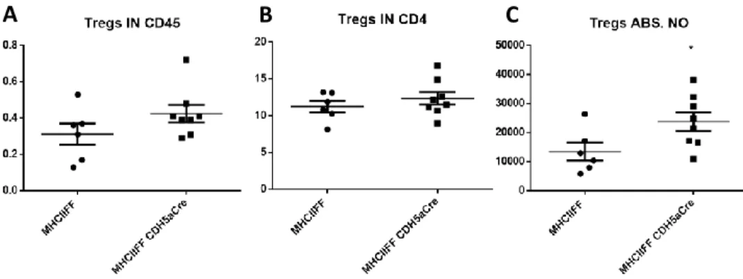
As for TIGIT and PD1, they were slightly lower on the surface of the vascular endothelium. The only difference was in TIM3 being higher in CRE mice on the surface of CD4, but the difference was not statistically significant. The scatter plots show that Tregs were higher in hemopoietic cells (CD45) as well as CD4 T cells in the experimental group versus the control group. Fig.23) The absolute number of Tregs in the tumor of the experimental mice was significantly higher indicating the activation of the immune system to eliminate the larger tumor.
As for intracellular staining for FOXP3, FOXP3 APC (Biolegend, 126407) was used as well as counting beads. As for B cells, they were higher in the experimental group compared to controls, but the difference was not statistically significant. B220 also known as CD45R is expressed on B cells (at all stages of development from pro-B cells through mature B cells), activated B cells and subsets of T and NK cells.
Discussion
The surface of endothelial cells in the lung tumor had larger tumors, providing evidence that MHCII in endothelial cells actually plays a role in tumor control (Fig. 17A). In vivo LLC cells grow faster in MHCII F/F Cdh5aCre mice (Fig. 17B) that lack MHCII in tumor endothelial cells. Expression of MHCII in endothelial cells is statistically significantly higher in homozygous, loxP-flanked MHCII (MHCII F/F) than in the MHCIIF/F CDdh5aCre.
This is an indication that tamoxifen worked correctly, inducing Cdh5aCre and leading to a deletion of MHCII allele in the surface of endothelial cells. Fig.20 D-F) So removing MHCII in the surface of endothelial cells did not affect CD4 or CD8 T cell number. Ideally, we would expect CD4 and CD8 to be less in the mice with no MHCII in endothelial cells that had larger tumors.
There is a trend on the surface of CD4 and CD8 cells when MHCII is expressed on endothelial cells, but still not statistically significant. MHCII was significantly higher in intratumoral endothelial cells in control mice with a significantly smaller tumor (Fig. 18B). More specifically, we propose that endothelial cells extract MHCII from the surface of CD4 T cells.
In preliminary data it was shown that endothelial cells express only co-inhibitory molecules (Fig. 17). 42 inhibitory molecules and their expression is different on the surface of CD8 T cells showing a possible explanation of the whole idea of atypical presentation via MHCII by endothelial cells in the context of lung cancer. More specifically, we can use the innovative mNFLs carrying antigens and adjuvants in order to specifically target the endothelial cells in the tumor region and fight against cancer.
The entire procedure used to target endothelial cells should ideally be used for therapeutic purposes in the near future. Human effector memory CD4+ T cells directly recognize allogeneic endothelial cells in vitro and in vivo. Steady-State Antigen Scavenging, Cross-Presentation, and CD8+ T Cell Priming: A New Role for Lymphatic Endothelial Cells.
Efficient presentation of exogenous antigen by free endothelial cells to CD8+ T cells results in antigen-specific T cell tolerance. Antigen presentation by endothelial cells and chemoattraction are required for homing of insulin-specific CD8+ T cells.