INTRODUCTION
- A N OVERVIEW
- W HY IT IS IMPORTANT TO STUDY THE STRUCTURE AND FUNCTION OF THE CFTR CHANNEL ?
- R EGULATION OF CFTR CHANNEL GATING BY INTRACELLULAR ATP
- C ONDUCTION PROPERTY OF CFTR
- CFTR CHANNEL BLOCKERS PROVIDE THE INFORMATION OF PORE STRUCTURE
- A NEWLY DISCOVERED SINGLE PEPTIDE TOXIN , G A T X 1, INHIBITS CFTR
- A THREE - DIMENSIONAL (3-D) MODEL OF THE CFTR CHLORIDE CHANNEL P ORE
- C OVALENT LABELING PROBES THE STRUCTURE AND FUNCTION OF CFTR
- W HAT FORMS FUNCTIONAL UNIT OF CFTR?
- CFTR PORE IS NOT STATIC
Studies by Akabas and colleagues provided limited information about CFTR channel structure and function. Recently, Beck and colleagues reported that there is a conformational change on the extracellular side of the CFTR pore [132].

OBJECTIVES
T O DETERMINE WHAT FORMS THE PORE OF CFTR AND HOW MANY PORES CFTR HAS ?
T O DETERMINE HOW BINDING AND HYDROLYSIS OF ATP AT THE NBD S CONTROLS THE
METHODS AND MATERIALS
- P REPARATION OF OOCYTES AND C RNA
- E LECTROPHYSIOLOGY
- A NALYSIS OF SINGLE CHANNEL EXPERIMENTS
- A NALYSIS OF MACROPATCH EXPERIMENTS
- S OURCE OF REAGENTS
- S TATISTICS
Over time, MgATP and PKA diffused to the intracellular face of the outer patch; CFTR channels were fully activated for ∼75 min ( Fig. 6B ). For the outside-out macropatch experiments in which CFTR channels were exposed to MTSET+ twice (see Fig. 12), we determined the relationship between the magnitude of the fractional increase in current upon first exposure (fractional ΔI) and the rate coefficient of MTSET+ modification during the second exposure (k2, is equal to converting the time constant (τ2) of the second exposure to a rate coefficient with units of sec-1M-1 by dividing by [MTSET+])).

RESULTS
T O DETERMINE WHAT FORMS THE PORE OF CFTR AND HOW MANY PORES CFTR HAS
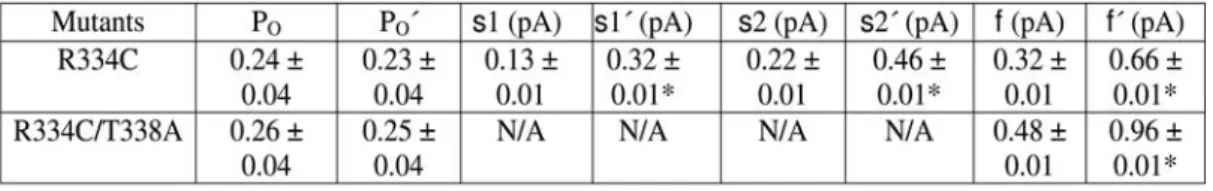
- Wildtype and mutant CFTRs exhibit comparable subconductance states with differing
- Deposition of a positive charge at 334 amplifies all conductance states proportionately
- Covalent modification of R334C-CFTR did not alter gating
- How many cysteines in one pore?
- Does modification of one cysteine absolutely prohibit modification of a second cysteine in the
We previously showed that covalent modification of R334C-CFTR channels with MTSET+ increased the amplitude of the most prominent single-channel conductance (here called s2) without altering the transition (Figure 5) [79]. The extreme shortness of both subconductance states in this mutant results in these images. The amplitudes of short underconductance states and full conductance states increased ~2-fold after the change induced by MTSET+ (Fig. 10C,E and Table 1).
9G, the fractional abundance of the s1, s2, and f conductance states did not change with MTSET+-induced modification in R334C-CFTR channels. To investigate the number of cysteines per pore, we examined the time course of the modification of CFTR channels by MTSET+ in inside-out macrospots using a rapid perfusion system. Consequently, the short exposure to MTSET+ resulted in alteration of a subset of the available cysteines.
12B contains a plot of the modification rate coefficient (k2) during the second MTSET+ exposure as a function of the fractional change in current resulting from the first MTSET+ exposure (see Methods). To determine whether any engineered cysteine remains unchanged in R334C-CFTR channels after long-term exposure to MTSET+, we exploited the sensitivity. We can see that the single-channel amplitude of the last modified aperture is almost the same as the amplitude of the first modified aperture.

T O DETERMINE HOW BINDING AND HYDROLYSIS OF ATP AT THE NBD S CONTROLS THE
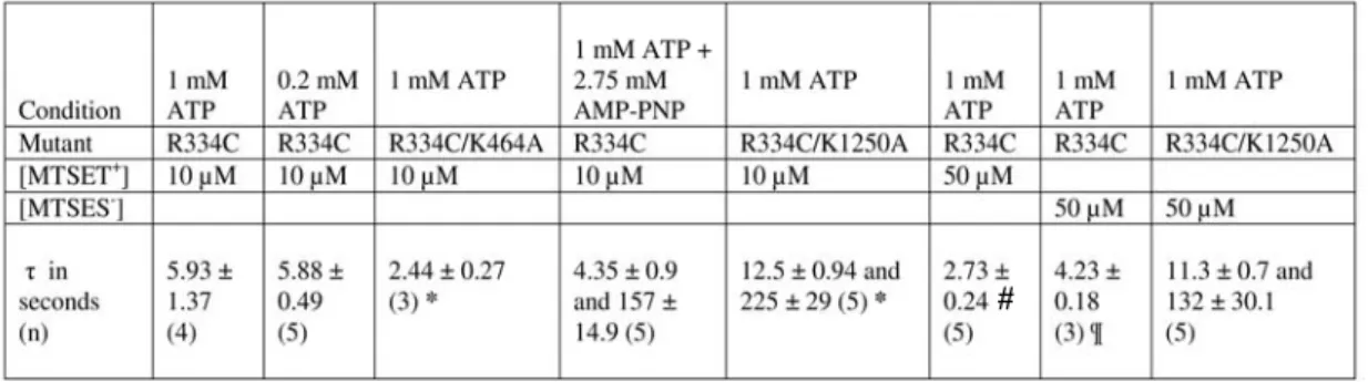
- R334C channels are modified by MTSET + only in the closed state
- Kinetics of macroscopic modification were altered in NBD mutants
- Kinetics of modification by MTSES -
These observations strongly suggest that modification of R334C-CFTR by MTSET+ is favored by the closed state. Accordingly, modification of R334C-CFTR in the presence of mixtures of ATP and AMP-PNP occurred in two phases. The value of the time constant describing the faster phase of modification in the presence of AMP-PNP (τ1) was not statistically different from the single time constant (τ) in the presence of ATP alone (p = 0.362), and was also not the modification rate coefficients differ (p = 0.286).
The slower phase of macroscopic modification in the presence of AMP-PNP may reflect the modification process of those channels locked in a long open burst. The faster modification phase in the presence of AMP-PNP may reflect the modification process of channels showing open bursts of normal duration. This individual patch contained at least three active R334C-CFTR channels included in the presence of ATP +AMP-PNP.
In a manner similar to the experiments using R334C-CFTR in the presence of ATP + AMP-PNP, the shorter open bursts were always modified earlier than the longer bursts (data not shown). To test this hypothesis, we first studied outside-out macrospots of R334C-CFTR channels in the presence of 0.2 mM ATP and measured the kinetics of modification. The kinetics of modification of R334C-CFTR by MTSES- was best fitted with a first-order exponential function (Table 2).
DISCUSSION
A NION CONDUCTION BY CFTR: O NE PORE PER POLYPEPTIDE
Three proposed schemes for the structure of the minimal functional unit for the CFTR channel. First, it unequivocally demonstrates that the subconductance states reported here are indeed a property of the CFTR channel. We have shown that even the effects of a neutral compound such as NEM can be understood in terms of the charge that is neutralized upon formation of the thioether bond.
Others have also suggested that R334 provides a fixed positive charge in the outer mouth of the pore, which plays a role in anion permeation [167]. While these findings do not allow us to unequivocally place R334 in the outer vestibule of the CFTR pore, it seems entirely appropriate to conclude that the available evidence is consistent with a model placing R334 in the pore. The simplest interpretation of the results presented here is that a single CFTR polypeptide folds in such a way as to form a single, anion-conducting pore (Fig. 20A).
However, it is unclear whether musculoskeletal disorders from both interacting CFTR peptides contributed to chloride permeability. Furthermore, since the channels were evident in the recording prior to the addition of PDZ peptides, it is apparent that the formation of a minimal functional unit does not require PDZ-mediated interactions between multiple CFTR peptides. CFTR is a member of the large ABC Transporter protein superfamily that has been extensively studied.

S UBCONDUCTANCE STATES : I MPLICATIONS FOR CHANNEL STRUCTURE AND FUNCTION
Two different models for the composition of the permeation pathway in CFTR, based on the results of this study. In (A), two pathways diverge at the cytoplasmic end of the channel but share a common conduction pathway at the extracellular end. We speculate that conformational changes in the pore, resulting in the four stable configurations shown, result in different permeation rates.
Interestingly, the openings of the R334C-CFTR channel also appear to be non-random in the order of positions in the three conductance states, as they switch to the full conductance state at the end of most bursts [Fig. Other researchers have reported that subconductance states are more frequently visited in channels that have deletions in one of the intracellular loops connecting the TM domains, suggesting that these loops may interact with NBDs to stabilize the open state [142; 143]. These results suggest that the pore can exist in multiple conformations, with different selectivity characteristics, determined by the activity of the nucleotide binding folds.
In the case of S1118F-CFTR, channel pore selectivity changes during these relaxations, and the rate of relaxation depends on the nature of the permeating anion [144]. In this regard, it is important to note that amino acid substitutions in the TM can affect the transition by changing the distribution of a particular substance. Changes in the conformation of the outer vestibule of the CFTR channel pore between the open and closed states.
C HANGES IN THE CONFORMATION OF THE OUTER VESTIBULE OF THE CFTR CHANNEL PORE
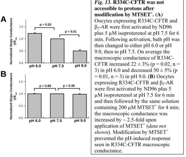
However, we previously found that the macroscopic conductance of whole oocytes expressing R334C-CFTR channels was sensitive to bath pH, due to titration of the partial negative charge on the unmodified cysteine (Fig. 13) [79 ]. Nevertheless, these data suggest that changes in the rate coefficients for SH-modifying reagents at R334C under different experimental conditions reflect conformational changes in the outer vestibule of the CFTR pore, which are associated with ATP-dependent gating action. In addition, in this study we used maneuvers to enhance PO that lock open R334C-CFTR channels in the prominent s2 subconductivity state (Figures 15B and 17B).
Results of the present study show that when R334C-CFTR channels are locked open by AMP-PNP or by addition of the K1250A mutation, they are locked in the s2 state. In contrast, previous studies indicate that WT-CFTR channels opened by the same maneuvers are also locked in the f-state. These observations suggest that the pore's most stable conductive state reflects the fully occupied, pre-hydrolytic state of the NBDs.
Consistent with this notion, we recently reported that WT-CFTR channels gated open by either AMP-PNP or vanadate (and K1250A-CFTR channels by ATP alone) have a reduced frequency of pulsatile closure compared with WT-CFTR channels in presence of ATP alone [106]. Therefore, it seems that the stability of the open conduction states is determined by the processes of binding and hydrolysis to the NBD. The mechanism that couples conformational changes at the NBD to conformational changes in the pore is an interesting subject for future studies.
SUMMARY AND CONCLUSIONS
Modification by MTSET+ and MTSES- was much slower when channels were locked open by addition of non-hydrolysable nucleotide AMP-PNP, or when the R334C mutation was coupled to a second mutation, K1250A, which significantly reduces the rate of channel closure. These data indicate that the reactivity of the engineered cysteine in R334C-CFTR is state-dependent, providing evidence of pore conformation changes coupled to ATP binding and hydrolysis at the NBDs. The data also show that maneuvers that lock the open R334C-CFTR do so by locking channels in the prominent s2 subconductivity state, suggesting that the most stable conductive state of the pore is the fully occupied, pre-hydrolytic state of the NBDs. reflects.
Taken together, our data showed that the minimal functional unit is formed by a single CFTR polypeptide that constitutes a single pore and provided direct evidence that there is a conformational change in the outer vestibule of the pore associated with ATP-dependent NBD switching events.
ACKNOWLEDGEMENT
BIBLIOGRAPHY
Cut channels probe regulation of gating of the cystic fibrosis transmembrane conductance regulator by its cytoplasmic domains. The two nucleotide-binding domains of cystic fibrosis transmembrane conductance regulator (CFTR) have distinct functions in controlling channel activity. 1998 ) Adenosine triphosphate-dependent asymmetry of anion permeation in the cystic fibrosis transmembrane conductance regulator chloride channel.
A single chloride ion conduction pore formed by two cystic fibrosis transmembrane conductance regulator molecules. 1997) Locating the anion selectivity filter of the cystic fibrosis transmembrane conductance regulator (CFTR) chloride channel. Molecular determinants of anion selectivity in the cystic fibrosis transmembrane conductance regulator chloride channel pore.
Arg352 is a major determinant of charge selectivity in the cystic fibrosis transmembrane conductance regulatory chloride channel. 2001) Disruption of the cystic fibrosis transmembrane conductance regulator (CFTR) pore inhibits its atpase activity. 1994) Regulation of cystic fibrosis transmembrane conductance regulator C1 channels by ATP phosphorylation and hydrolysis.
Truncated molecules functionally define the boundaries of the cystic fibrosis transmembrane conductance regulator's NH2-terminal nucleotide binding domain. A monomer is the minimum functional unit required for channel and ATPase activity of the cystic fibrosis transmembrane conductance regulator.
PUBLICATIONS OF PH.D. CANDIDATE
O THER PUBLICATIONS
Eaton and H-P Ma (2007) Ceramide mediates renal epithelial sodium channel inhibition by tumor necrosis factor α via protein kinase C.
ABSTRACT
ABSTRACT IN HUNGARIAN (ÖSSZEFOGLALÁS)
THE THREE MOST IMPORTANT ARTICLES OF APPLICANT


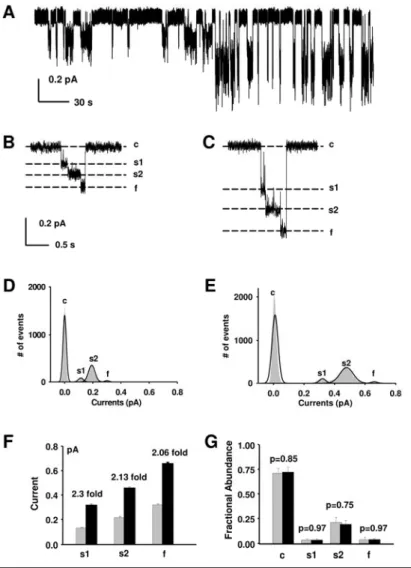
![Fig. 8. Representative trace from an oocyte expressing R334C-CFTR in the detached, inside-out patch configuration; V M = -100 mV, with asymmetrical [Cl - ]](https://thumb-eu.123doks.com/thumbv2/9dokorg/19337260.0/35.892.132.671.187.683/representative-oocyte-expressing-r334c-detached-inside-configuration-asymmetrical.webp)