3d Evaluation Of Soft Tissue Changes Following Class Ii Orthognathic Surgery– A Systematic Review
Abinaya Somaskandhan, Janani Jayapal, Devaki Vijayalakshmi, Ratna Parameswaran
Dept of orthodontics and dentofacial orthopaedics, Meenakshi academy of Higher education and research
Meenakshi Ammal Dental College, Tamilnadu, Chennai, India
ABSTRACT
AIM: To evaluate soft tissue changes following class II orthognathic surgery using three-Dimensional imaging.
MATERIALS AND METHODS: This review was conducted according to preferred reporting Items for systematic reviews and Meta-analysis guidelines by systematically searching the six databases including PubMed, Cochrane,Google scholar,LILACS,Directory of Open Access Journals and Opengrey.
RESULTS vertical: This systematic review comprises of most up-to-date evidence from twelve articles answering the review questions.
CONCLUSION: Maxillary setback shows significant decreased in nasolabial and alar width and posterior movement of point A. Mandibular advancement shows significant reduction in mento-labial angle with increase in the volume of lips and cheeks. The largest changes occurred on the anterior and inferior surfaces of the chin. Labii Inferioris showed a statistically significance change at horizontal lines with an increase in the Cutaneous bi-gonial distance. In case of Bi-jaw surgeries, the lip width had decreased significantly.
KEY WORDS: Three-Dimensional, class II skeletal base, Soft tissue, orthognathic surgery Introduction
Adult patients seeking orthodontic treatment is predominantly due to esthetic concerns.
Combined orthodontic-orthognathic treatment is done routinely in non-growing patients in order to obtain a stable occlusion and esthetic profile. The facial soft-tissue drape does not follow the movement of the underlying skeletonaccurately. The final soft tissue appearance post-surgically might not exactly simulate the surgical movements of the jaws[1].
Considering the soft tissue profile changes during orthodontic treatment was reported by Angle at the beginning of the 20th century. Along with establishing a balanced and stable dento-skeleto-facial complex, achieving anesthetically pleasing soft tissue envelope has become a major goal for orthognathic surgeries. During surgical treatment plans, predicting the soft tissue changes is an important component in order to provide aesthetic and psychological benefits and also to avoid unrealistic expectations of the patients. In order to understand and determine the amount of soft tissue changes that will occur following orthognathic surgery, evaluation of soft tissue changes in already surgically treated cases is a necessity.
Initially, the assessment of surgical outcomes was done using two-dimensional radiographs[2]. Prediction and evaluation of surgical outcomes has evolved from manual tracing of skeletal segments and soft tissue parameters to digitized imaging and computerized line drawings.
The usage ofThree-dimensional imaging techniques has been increasingly in the recent years.
They help us to determine the hard and soft tissue relationships in complex facial structures.
CBCT notably has been gaining popularity specially to assess the orthognathic surgical outcomes. It also aids in determining the correlations and proportions of the soft-to-hard tissue movements. Recent advances in 3D evaluation of soft tissue include 3D stereophotogrammetry[3,4],3D facial image scans using LED white light scanning system[5], Computed tomography (CT)[6], Cone Beam Computed Tomography (CBCT)[7,8,9] etc.
The primary aim of this current systematic review is to evaluate three dimensionally, the soft tissue changes that will take place following class II orthognathic surgery such as Bilateral Sagittal Split Osteotomy (BSSO) advancement, maxillary setback or a combination of both.
Materials and methods Protocol and registration
This review was based on a specific protocol developed and piloted following the guidelines outlined in the Preferred Reporting Items for Systematic Reviews and Meta-Analyses (PRISMA)statement[10,11]. Prospero registration number: CRD42020152338
Eligibility criteria:
Eligibility criteria was based on the research question defined in the PECO format. Do patients who have undergone mandibular advancement or maxillary setback or a combination of both (P) and evaluated using 3D imaging techniques (I) exhibit before and after (C) changes in the facial soft tissue (O).
Inclusion and exclusion criteria were formulated based on participant, intervention, comparison, outcome and study design. The inclusion criteria comprised of human adult participants of either gender who were over 18 years of age. They should have undergone single jaw surgery (BSSO advancement or Le Forte I setback) or bi-jaw surgery for correction of skeletal class II malocclusion.All participants should have 3D soft tissue assessment records before and after surgical procedure. Data published in English during 2009-2019 were included.
The exclusion criteria comprised of animal or in vitro studies, case reports or case series, patients who had cleft lip or palate, craniofacial disorders, degenerative conditions, trauma, Temporo-Mandibular Joint disorders, any other type of orthognathic surgery undertaken, inflammatory conditions and facial asymmetries. Studies using 2D images were also excluded from the review.
Search strategy for identification of studies
A detailed search was conducted in two parts. Firstly, an electronic search was done based on a search strategy developed on PICO format and was checked using PRESS checklist for
systematic reviews. The search terms included controlled vocabulary, author keywords, Boolean operators, and truncations which were appropriately used and revised for each database. The search was carried out in the following electronic databases: PubMed, Google Scholar, LILIACS, Cochrane registry of clinical trials, and Directory of Open Access Journals, and unpublished literature was searched on opengrey.eu.
The second part of the search was hand search of the relevant orthodontic journals. The following journals were searched:
• American Journal of Orthodontics and Orthopedics.
• British Journal of Orthodontics (Journal of Orthodontics).
• European Journal of Orthodontics.
• Journal of Indian Orthodontic Society.
• Korean Journal of Orthodontics.
• The Angle Orthodontist.
• World Journal of Orthodontics.
• Journal of Aligner Orthodontics.
The data collection was performed by two researchers (AS and JJ)who were not blinded to the identity of the authors of the studies, their institutions, or the results of the research. The researchers assessed the participants, intervention, evaluation, statistical analysis and outcome of the studies. Authors were contacted whenever it was necessary in order to obtain more elaborate details pertaining to the study design and also for clarification of the data.
The reference lists of all eligible studies were hand searched for additional studies.
Potentially eligible studies were uploaded to MENDELEY 1.19.2, Elsevier, 2018, New York, USA software in order to remove duplicate articles. The non-eligible studies were excluded and the eligible studies were assessed independently by both the authors. In case of disagreements between the authors (A.S. and J.J), consultations with a third reviewer (R.P) was made to resolve.
Assessment of Risk of Bias:
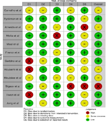
Two reviewer authors (A.S. and J.J) independently assessed the risk of bias of the eligible trials according to the Cochrane Collaboration’s risk of bias tool (Figure 2). In case of discrepancy, consensus was obtained by consulting a third reviewer (R.P). The domains assessed were (1) random sequence generation; (2) allocation concealment; (3) blinding of the participants; (4) blinding of the personnel; (5) blinding of the outcome assessment; (6) incomplete outcome data; (7) selective reporting; (8) other biases (baseline imbalance, similarity in using cointerventions between groups, and inadequate statistical analysis). The potential risk of bias for each study was classified as high, unclear or low.
Figure 1: Risk of bias
Data extraction
The data collection was performed by two researchers (AS and JJ) who were not blinded to the identity of the authors of the studies, their institutions, or the results of the research. The researchers assessed the participants, intervention, evaluation, statistical analysis and outcome of the studies. Authors were contacted whenever it was necessary in order to obtain more elaborate details pertaining to the study design and also for clarification of the data.
Results
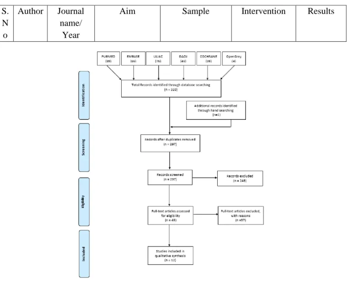
A PRISMA flow diagram of the article selection process has been illustrated in Figure.2.
Figure 2: Prisma flowchart
The results of this systematic review are detailed and tabulated in Table 1 with twelve included articles which answer the review question.
Discussion
Among the Indian population, severe skeletal malocclusions especially class II malocclusion is seen commonly. Treatment of impaired facial esthetics in adults requires careful assessment of the skeletal and soft tissue problems. Cautious treatment planning predicting the amount of soft tissue changes that will take place post surgically is obligatory to achieve a pleasing facial appearance. The current systematic review delineates the three-dimensional soft tissue changes that will occur during surgical correction of class II malocclusion by Le Fort I maxillary setback, Bilateral sagittal split osteotomy with mandibular advancement or a combination of these. In the 12 studies included in the current systematic review, only one study by Rahul Tiwari et al (2018) [6] had discussed about all three approaches for correction of class II malocclusion namely maxillary setback, mandibular advancement and Bi-jaw surgeries using Three Dimensional Computed Tomography scan (3DCT)
S.
N o
Author Journal name/
Year
Aim Sample Intervention Results
1) Felipe de Assis Ribeir o Carval ho et al
American Journal of Orthodont ics and Dentofaci al
Orthopedi cs April (2010)
to evaluate the 3D changes in the position and remodeling of the
mandibular rami, condyles, and chin at splint removal and 1 year after mandibular advancement surgery.
Twenty-seven patients (9 men, 18 women; mean age, 30.04 ± 13.08 years;
range, 17.2-48.1 years)
All patients underwent orthodontic treatment and had
mandibular advancement surgery with bilateral sagittal split osteotomy.
nine
participants also had genioplasty as an adjunctive procedure.
e excluded.
CBCT scans were taken before surgery, at splint
removal (4-6 weeks postsurgery), and 1 year postsurgery (after orthodontic treatment)
-nearly half of the patients had >2 mm change in chin position from splint removal to the 1-year follow-up, with approximat ely equal chances of anterior and posterior movement.
-Torque of the rami usually occurs with mandibular advanceme nt surgery.
2) Micha el S.
Ryck man et al
Am J Orthod Dentofaci al Orthop (2010)
to quantify both
anteroposterior and transverse facial soft- tissue changes with respect to underlying skeletal movements after
maxillomandib ular
30 white patients- the average patient age was 27.9 years (range,16- 63 years); the sample included 10 male and 20 female subjects
all patients received alar base cinches, and V-Y advancement mucosal closures were performed as necessary for patients
requiring more upper lip fullness.
-For patients who received mandibular advanceme nts less than 10.0 mm, the mean ratio for
transverse softto-hard
advancements by using cBct.
- all patients had 3 cBct scans: within 1 week
presurgery (t0), within 1 week
postsurgery (t1), and at least 8 weeks postsurgery (t2).
tissue movement in the subcommis sural region was 95.2% ± 66.4%.
- Patients who received advanceme nts greater than or equal to 10.0 mm, on the other hand, had a mean ratio of 57.0% ± 4.6%. a statistically significant difference was found for the transverse softto-hard tissue movement in the subcommis sural region between these 2 groups - there were no significant differences for the
ratios of soft-tohard tissue movement in the anteroposte rior chin region, anteroposte rior
subcommis sural region, or transverse gonial region between patients who received mandibular advanceme nts less than 10.0 mm and those who received advanceme nts greater than or equal to 10.0 mm 3) Almei
da et al
Internatio nal journal of oral and maxillofac ial surgery (2011)
To assess the stability of 3D soft tissue changes following mandibular advancement, and the association between soft and hard tissue
25 patients (7 men, 18 women;mean age 30.8 +/- 13.08 years) scheduled for
mandibularadvan cement surgery were recruited.
In 5 cases, the
CBCT scans were taken before surgery, at splint
removal (4–6 weeks
postsurgery), and 1 year postsurgery (after orthodontic
-anterior- inferior displaceme nt of the hard chin at splint removal as an outcome of surgery.
-The correlation
changes CBCT imaging field of view did not include all soft tissue structures,resulti ng in data for 21 patients for the lower lip and 20 patients for the soft tissue chin
(mandibularadva ncement alone n = 11; and mandibular advancement and
genioplasty n = 9).
treatment) with the NewTom 3G (AFP Imaging, Elmsford, NY, USA).
between the soft and hard tissue chin displaceme nts were statistically significant (P <
0.0001) for presurgery to splint removal (r
= 0.92), splint removal to 1 year postsurgery (r = 0.77) and
presurgery to 1 year postsurgery (r = 0.86).
-The average displaceme nt of the soft tissue chin was greater than that of the hard tissue chin for all three time
intervals, but the average difference between the hard and soft
tissue displaceme nts from splint removal to 1 year after surgery was not statistically significant.
-For 10%
of the subjects, the soft tissue chin changes between presurgery and splint removal in an anterior inferior direction were more than 2 mm larger than the hard tissue chin changes.
4) Alexa ndre T.
Motta et al
J Oral Maxillofa cSurg (2011)
to evaluate the association of 3D changes in the position of the condyles, rami, and chin at splint removal and 1 year after mandibular advancement surgery
A total of 27 patients (9 men and 18 women, mean age 30.04
± 13.08 years).
The patients underwent orthodontic treatment and mandibular advancement surgery with bilateral sagittal split ramus
osteotomy, and 9 also
underwent
-The mean chin advanceme nt at splint removal (chin T1 to T2 changes 6.8 ± 3.2 mm) was maintained at 1 year after surgery
genioplasty as an adjunctive procedure.
CBCT scans were taken before surgery (time 1 [T1]), at splint removal 6 weeks after surgery (T2), and 1 year after surgery (T3) using the NewTom 3G scanner
(mean chin T1 to T3 changes 6.4 ± 3.4 mm).
-For all other anatomic regions evaluated, only the inferior rami (left 3.0 ± 2.7 mm and right 2.3 ± 2.4 mm) had a mean displaceme nt of 2 mm or more with surgery.
Between splint removal and 1 year, a slightly higher percentage (15%) of the subjects had a soft tissue displaceme nt that exceeded the hard tissue displaceme nt by 2 mm or more,
while 15%
had soft tissue changes that were more than 2 mm smaller than the hard tissue change.
-The chin displaceme nt from presurgery to 1 year postsurgery explained 73% of the variability in the soft tissue chin changes, which is less than the 85%
that was observed for
presurgery to splint removal.
-Regarding changes in the chin area 1 to 3 years after surgery, 17% of cases presented inferior displaceme
nt and 17%
of cases presented posterior displaceme nt from 2 to 4 mm.
5) T.J.J.
Maal et al
Int. J. Oral Maxillofa c. Surg.
2012
Using cone beam computed tomography (CBCT) imaging and 3D stereophotogra mmetry, to accurately compare the 3D soft tissue changes caused by skeletal
transformations after a bilateral sagittal split osteotomy (BSSO) 1 year after surgery
Eighteen Caucasian patients with a symmetrical mandibular hypoplasia without a maxillary
hypo/hyperplasia or an anterior open bite (6 males and 12 females) were prospectively enrolled in this study.
-All patients were treated with a mandibular advancement using a BSSO according to Hunsuck modification -
Preoperatively and 1 year postoperatively , an extended height CBCT scan was acquired (i- CATTM, Imaging Sciences International, Inc., Hatfield, USA). Apart from the CBCT scans, 3D
photographs were acquired preoperatively and 1 year postoperatively using a 3D
- For the soft tissue, a mean volume increase of 10029 mm3 (95%
CI _2.2 to 137.2 mm3)was found.
- 3D curvature changes of the labio- mental Fold: A mean preoperativ e curvature of 3.57 (radius in cm), with a 95%
confidence interval of _0.08 cm to 0.13 cm, was found in contrast to a postoperati
camera (3dMDCranial TM System, 3dMD LLC, Atlanta, USA).
ve mean value of 5.24 (radius in cm) - A mean volume increase of 4660 mm3 was found in the region of the chin.
- The lip region increased with a mean volume of 1540 mm3.
-The remaining soft tissue volume increase was visible on the left (4443 mm3) and right (4533 mm3) sides of the mandible.
6) Alexa ndre A.
Franco et al
J Oral Maxillofa cSurg (2013)
to analyze long-term 3D alterations in the rami, condyles, and chin 1 to 3 years after surgery in patients treated with mandibular advancement.
27 patients (18 female and 9 male)
with an average age of 26.7 _ 13.2 years
All mandibular advancement surgerieswere performed using bilateral sagittal split osteotomyand rigid fixation with plates and screws.
Forty percent of subjects had a genioplasty.
CBCT scans were obtained before surgery, 1 year
after surgery, and 3 years after surgery with the NewTom 3G scanner
-average displaceme nt was largest for the chin -The largest average changes occurred on the anterior and inferior surfaces of the chin even after adjusting for the presence of a
genioplasty , age at time of surgery, and gender.
-The inferior border of the mandible was the only area that had a statistically significant average change.
-The 1.11- mm average change indicated
inferior displaceme nt
of the chin.
-Virtually all patients had greater than 2 mm of anterior movement of the chin at 1 year after surgery.
Approxima tely 40%
had greater than 4 mm anterior displaceme nt of the anterior surface of the chin 7) Giova
nni Gerbin o et al
Journal of cranio- maxillo- facial surgery (2014)
0three- dimensional (3D) analysis of the soft tissue changes in typical OSAS patients before and after MMA, in order to improve treatment planning and increase predictability of the esthetic outcome.
27 patients with severe OSAS underwent MMA surgery.
Patients with dento-
skeletaldiscrepan cies leading to facial deformity (mainly severe class
IIdeformities), in which occlusion correction and pre-operative orthodontic treatment were incorporated in the treatment
standardized surgical treatment consisting of a LeFort I osteotomy and bilateral sagittal split- ramus
osteotomies), with skeletal advancement planned between 10 and 12 mm.
Soft-tissue 3D data were also obtained
-The comparison of
measureme nts of the cutaneous landmark distances on T0 and T1
revealed an increase of inter- cheilion width.
-Increased bulking of the upper lip was
plan, were not included in the study. Thus, 10 patients were enrolled in the present study.
before and 1 year after surgery using a Head and Face Color 3D Scanner Patients’
satisfaction with facial appearance after surgery was
subjectively evaluated by a questionnaire.
also observed -The comparison between T0 and T1 showed a post-op overall increase of the sagittal projection of soft tissue A point, B points, lips and of the chin -At the questionnai re, six out of the ten patients gave favorable responses to their facial changes (i.e., that they appeared either more attractive or younger;
four patients felt neutral regarding their facial esthetic results.
None of
the patients responded unfavorabl y).
8) Salah Al-Din Al- Housa mi et al
The Journal of Craniofaci al Surgery (2015)
to quantify anteroposterior and transverse facial soft tissue changes with respect to underlying skeletal movements after bilateral sagittal split osteotomy by using cone beam computed tomography (CBCT)
6 patients (4F and2M) who required bilateral sagittal split osteotomy for correction of mandibular retrognathism
The patients were scanned using CBCT 1 week
preoperatively, and 6 months postoperatively
The facial profile was improved due to advanceme nt of the mandible, the mentolabia l fold MLF become shallower in depth and the mentolabia l angle MLA approached the
standard norms.
- the tip of the nose and the chin assumed a better relationshi p
concerning the facial balance and the E- Line.
- The ratio of the mean hard to soft tissue movement was 1:0.97, respectivel y after
mandibular advanceme nt
- LI showed a statistically significanc e change at both the vertical and horizontal lines, the ratio of the mean movement in the horizontal direction was 1:0.80 - The soft tissue thickness at B-MLF and POG- POG’
showed a non-
statistically significant increase in the
measureme nts
postoperati vely.
- As for the MLF depth, there was a statistically significanc e decrease
in the measureme nts
postoperati vely - For angular measureme nts, there was
statistically significant increase in the mean measureme nts
postoperati vely for MLA and facial convexity angles; the mean increase in the facial convexity was (2.1o) and the mean increase in the MLA was (27.7)
9) Jene Meulst ee et al
Journal of Cranio- Maxillo- Facial Surgery (2015)
to evaluate changes in the soft tissue facial profile in patients who underwent bilateral sagittal split osteotomy (BSSO) using 3D
stereophotogra mmetry and principal component analysis (PCA).
Female patients with dentofacial deformities who underwent a bilateral sagittal split osteotomy (BSSO).
-total of 95 women were enrolled for the study; 25 were patients (mean age, 24 years;
range: 18e26 years) and 70 were controls (mean age, 24 years; range:
18e26 years).
Three- dimensional photographs of all patients and controls
were acquired using the 3DMD
stereophotogra mmetry facial system
(3dMDFace, 3dMD, Atlanta, GA, USA).
The acquired 3D
photographs were imported into the 3DMDPatient software (3dMDPatient, 3dMD).
- A clockwise rotation of the
mandible and a shortening of the lower part of the face were the most prominent differences between the two groups.
-
protrusion of the upper lip and
retrusion of the
mandible were observed among the preoperativ e BSSO patients compared with the control group.
- an overaccent uation of the labial- mental fold was
present in the
preoperativ e BSSO patient group compared with the control group.
- the postoperati ve group did not overlap the control group completely , indicating that many patients who had undergone BSSO maintained some characterist ics of a Class II facial profile despite the surgery.
- despite BSSO advanceme nt surgery, some patients still possess some dysgnathic facial characterist
ics
1 0)
Nicola s Sigaux et al
Journal of Oral and Maxillofa cial Surgery(2 018)
to assess transversal changes in mandibular advancement by comparing 3D (three- dimensional) photogrammetr ic
modifications and 2D (two- dimensional) radiographic enlargement.
Fourteen patients (5M 9F) were included. Mean mandibular advancement was
6 mm. Both BGD (+6.1 mm;
p<10-3) and CBGD (+4.2 mm; p=0.0017) were
significantly increased.
All patients had
standardized 3D
photogrammetr ic and 2D radiographic evaluations on a 100% scale (frontal cephalogram radiograph, lateral cephalogram radiograph and panoramic radiograph) before and after surgery.
cutaneous bi-gonial distance CBGD was increased postoperati vely in thirteen patients (unchanged in one patient), with a mean increase of 4.2 + 2.9 mm.
- In most cases, morphologi cal changes were observed in the full lower face, including the lateral regions.
-The mean ratio of soft tissue response to
transversal skeletal changes was 0.81.
1 1)
Rahul Tiwari et al
The Open Dentistry Journal, 2018
to assess and compare pre and post- operative perioral soft tissue changes of lip width, nasolabial and mentolabial angle using Three Dimensional Computed Tomography scan (3DCT).
- Total of 10 (4 males and 6 females) patients with age range of 18 to 26 years -
Pre and post- operative 3DCT scan were taken after 12 months using iCT 256 slice whole body CT scanner and evaluated for changes using Dicom PMS D view
-Changes in
Nasolabial Angle After Maxillary Setback: A total of five patients have undergone maxillary setback of 2 mm to 3 mm in which the nasolabial angle has decreased by 4.1 to 11.5°, respectivel y. So, the mean setback in the maxilla was 2.6 mm and the mean difference was 7.12°.
Hence,
1mm movement of maxilla setback is a decrease in the
nasolabial angle by 2.73°
- Changes in
Mentolabia l Angle after
Mandibular Advancem ent:
Among six patients, three patients underwent mandibular advanceme nt of 2 mm to 8 mm. In three patients, the
mentolabia l angle was decreased by 7.6° and in
remaining three patients, mentolabia l angle increased by 3.6°
- . Changes in the Lip
Width in Bi Jaw Surgeries:
Four patients underwent maxillary setback and
mandibular advanceme nt in which the mean maxillary setback was 2.75 mm. and the mean mandibular advanceme nt was 3.0 mm.
- The mean decrease in the lip width was 2.15 mm 1
2)
Junho Jung et al
Head &
Face Medicine (2018)
to assess and describe the nasolabial soft tissue changes three-
dimensionally, after bilateral sagittal split osteotomy (BSSRO) or Le Fort I
osteotomy with BSSRO, using structured light systemone of the LED white
- 32
malocclusion cases (17 men, 15 women; mean age, 23.8±3.60 years; range, 17–
33 years) who had undergone BSSRO or/and Le Fort I
advancement or setback
osteotomy - The patients were divided into 3 groups:
- 3D facial image scans using a LED white light scanning system
(Morpheus 3D, Morpheus Co., Ltd., Seoul, Korea) were acquired preoperatively and at 3 months postoperative (scan time: 0.8
-After the Le Fort I setback osteotomy, on an average, point A moved posteriorly by about 2.1 mm (±1.0).
-
postoperati vely the
The nasolabial angle had decreased by 4.1 to 11.5O for 2mm to 3mm of maxillary setback.
The mentolabial Angle after Mandibular Advancement was decreased by 7.6° in 50% of the patients and increased by 3.6° in the other 50% of the patients. The lip width in Bi Jaw Surgeries was decreased with a mean of 2.15mm.
Two studies have quantified both anteroposterior and transverse facial soft-tissue changes with respect to underlying skeletal movements after Bilateral Sagittal Split Osteotomy using CBCT scans. A study by Michael S. Ryckman et al (2010)[1] and Salah Al-Din Al-Housami et al (2015)[12] concluded that the mean change in the anteroposterior soft tissue movement was 3.9mm of mandibular advancement. The ratio of the mean hard to soft tissue movement was 1:0.97 respectively after mandibular advancement. Labii inferioris showed a statistically significance change at both the vertical and horizontal lines with the ratio of the mean movement in the horizontal direction being 1:0.80. The mentolabial fold depth decreased by 1.4mm and the facial convexity had increased by 2.1o with 27.7O increase in the mentolabial angle.
Junho Jung et al (2018)[5] conducted a study to assess and describe the nasolabial soft tissue changes by three-dimensional facial image scans using a LED white light scanning system and CBCT scans in patients undergoing Le Fort I osteotomy. He concluded by saying that on an average, point A moved posteriorly by about 2.1 mm (±1.0). postoperatively the alar width decreased about 4.7 mm. In the upper lip area, the soft tissue movement was 3–52%
compared to the bony movement, and it was 15% at the nasal tip.
Three studies have used 3D stereophotogrammetry for evaluation of soft tissue changes following Bilateral Sagittal Split Osteotomy (BSSO) advancement. T.J.J. Maal et al
light scanning system
BSSRO only (9 patients; mean age, 23.2±3.5;
range, 19–31), Le Fort I
advancement (13 patients; mean age, 24.0±3.4;
range, 17–31), and Le Fort I setback (10 patients; mean age, 24.1±4.1;
range, 19–33
s, 33 frame rate: 15 frames/ s, data accuracy: ±0.2 mm
-CBCT scans were acquired preoperatively and at 3 months postoperative, using the Alphard 3030 Dental CT system
alar width decreased about 4.7 mm -In the upper lip area, the soft tissue movement was 3–52%
compared to the bony movement, and it was 15% at the nasal tip -
(2012)[13] suggested that there was a mean increase of 10029 mm3 in the volume of soft tissue. The lip region increased with a mean volume of 1540 mm3 and the chin region increased by 4669 mm3. Jene Meulstee et al (2015)[14] observed a clockwise rotation of the mandible and shortening of the lower part of the face. However, the study concluded that despite BSSO advancement surgery, some patients still possessed some dysgnathic facial characteristics. Nicolas Sigaux et al (2018) [15]assessed transversal changes by comparing 3D photogrammetric modifications and 2D radiographs and concluded that the cutaneous bigonial distance was increased by 4.2 +_ 2.9 mm. The mean ratio of soft tissue response to transversal skeletal changes was 0.81.
The remaining 5 out of the 12 studies evaluated 3D soft tissue changes following BSSO advancement using CBCT scans. Felipe deAssis Ribeiro Carvalho et al (2010)[7] concluded that nearly half of the 27 patients had greater than 2mm change in chin position and there was a torque of the rami post surgically. Almeida et al (2011) [8]concluded that the changes in the soft tissue chin was more than 2mm in 15% of the patients while 15% had less than 2mm changes. A statistically significant displacement of the lower lip was found which was suggested to be due to the change in the lower incisor position. Alexandre T. Motta et al (2011)[16] proposed that the mean chin displacement was 1.57mm. Alexandre A. Franco et al (2013)[9] concluded that the largest soft tissue changes occurred on the anterior and inferior surfaces of the chin with an average of 1.11mm. 1 year post surgically, 2mm anterior movement of the chin was noted with approximately 40% demonstrating greater than 4mm anterior displacement of the chin. At 1 to 3 years post surgically, 17% displayed inferior displacement and 17% displayed posterior displacement of the chin from 2 to 4mm.
Giovanni Gerbino et al (2014)[17] analysed soft tissue changes in Obstructive Sleep Apnea Syndrome (OSAS) patients using 3D laser scanning and deduced that there was an overall increase in the projection of the cheeks, lips and chin in the sagittal dimension. There was also an increase in the cheeks at the cross section through chelion.
Conclusion
The following conclusions can be made from this review:
During maxillary setback:
- The nasolabial angle had decreased by 4.1 to 11.5O for 2mm to 3mm of setback - On an average, point A moved posteriorly by about 2.1 mm (±1.0).
- postoperatively the alar width decreased about 4.7 mm.
- In the upper lip area, the soft tissue movement was 3–52% compared to the bony movement, and it was 15% at the nasal tip
For mandibular advancement:
- The Mentolabial Angle was decreased by 7.6° in 50% of the patients and increased by 3.6° in the other 50% of the patients.
- The soft tissue changes related to mandibular advancement would appear to be fairly predictable and follow their underlying skeletal structures in 1:0.97 ratio in the chin area.
- The lip region increased with a mean volume of 1540 mm3 and the chin region increased by 4669 mm3.
- clockwise rotation of the mandible and shortening of the lower part of the face - the cutaneous bigonial distance was increased by 4.2 +_ 2.9 mm. The mean ratio of
soft tissue response to transversal skeletal changes was 0.81.
- the largest soft tissue changes occurred on the anterior and inferior surfaces of the chin with an average of 1.11mm
- There was an increase in the cheeks at the cross section through chelion.
For Bi Jaw Surgeries:
- The lip width decreased with a mean of 2.15mm.
A change in the facial appearance relies on the underlying skeletal movement.
Comprehensive understanding of the relationship between the bone movement and soft tissue response is crucial for predicting postoperative facial change and useful for treatment
planning and patient consultation.
Funding:
There was no sources of funding for this research Conflict of interest:
There are no conflicts of interest pertaining to this study.
Ethical approval:
Ethical approval was not necessary for this systematic review.
References
1) Michael S. Ryckman,a Steven Harrison,b Don Oliver,b Christian Sander,c Andrew A.
Boryor,d Ansgar A. Hohmann,d Fatih Kilic,d and Ki Beom Kime; Soft-tissue changes after maxillomandibular advancement surgery assessed with cone-beam computed tomography; (Am J Orthod Dentofacial Orthop 2010;137:S86-93
2) Kim NK, Lee C, Kang SH, Park JW, Kim MJ, Chang YI. A three-dimensional analysis of soft and hard tissue changes after a mandibu- lar setback surgery. Comput Methods Pro-grams Biomed 2006;83:178–87.
3) Moss JP, Ismail SF, Hennessy RJ. Three-dimensional assessment of treatment out- comes on the face. OrthodCraniofac Res 2003;6(Suppl 1):126–31. discussion 179–
182.
4) Ayoub AF, Xiao Y, Khambay B, Siebert JP, Hadley D. Towards building a photo- realistic virtual human face for craniomaxillofacial diagnosis and treatment planning.
Int J Oral MaxillofacSurg2007;36:423–8.
5) Junho Jung, Chi-Heun Lee, Jung-Woo Lee and Byung-Joon Choi; Three dimensional evaluation of soft tissue after orthognathic surgery; Head & Face Medicine (2018) 14:21
6) Rahul Tiwari, P. Srinivas Chakravarthi, Vivekanand S. Kattimani* and Krishna Prasad Lingamaneni; A Perioral Soft Tissue evaluation after Orthognathic Surgery Using Three-Dimensional Computed Tomography Scan; The Open Dentistry Journal, 2018, 12, 366-376.
7) Felipe de Assis Ribeiro Carvalho,a Lucia Helena Soares Cevidanes,b Alexandre Trindade Simões da Motta,c Marco Antonio de Oliveira Almeida,d and CeibPhillipse;
Three-dimensional assessment of mandibular advancement 1 year after surgery; (Am J Orthod Dentofacial Orthop 2010;137:S53.e1-S53.e12
8) R. C. Almeida, L. H. S. Cevidanes, F. A. R. Carvalho, A. T. Motta, M. A. O.
Almeida, M. Styner, T. Turvey, W. R. Proffit, C. Phillips: Soft tissue response to mandibular advancement using 3D CBCT scanning. Int. J. Oral Maxillofac. Surg.
2011; 40: 353–359.
9) Alexandre A. Franco, Lucia Helena S. Cevidanes, Ceib Phillips, Paul Emile Rossouw, Timothy A. Turvey, Felipe de Assis R. Carvalho, Leonardo K. de Paula, C_atia Cardoso and Marco Antonio O. Almeida; Long-Term 3-Dimensional Stability of Mandibular Advancement Surgery; J Oral MaxillofacSurg 71:1588-1597, 2013
10) Higgins JP, Green S. Cochrane Handbook for Systematic Reviews of Interventions Version 5.1.0. Cochrane Collaboration; 2011. 8.
11) Liberati A, Altman DG, Tetzlaff J, Mulrow C, Gøtzsche PC, Ioannidis JP, et al. The PRISMA statement for reporting systematic reviews and meta-analyses of studies that evaluate health care interventions: explanation and elaboration. J Clin Epidemiol 2009;62:e1-34.
12) Salah Al-Din Al-Housami, Maha Shawky, MD,y Khairy El-Morsy, and Hassan Abdel-Ghany; Three-Dimensional Soft Tissue Assessment Following Mandibular Bilateral Sagittal Split Osteotomy; The Journal of Craniofacial Surgery , Volume 26, Number 8, November 2015
13) T. J. J. Maal1, M. J. J. de Koning1, J. M. Plooij1,3, L. M. Verhamme, F. A.
Rangel2,3, S. J. Berge, W. A. Borstlap; One year postoperative hardand soft tissue volumetricchanges after a BSSOmandibular advancement; Int. J. Oral Maxillofac.
Surg. 2012; 41: 1137–1145
14) Jene Meulstee , Jeroen Liebregts , Tong Xi*, Frank Vos, Martien de Koning, Stefaan Berge, Thomas Maal; A new 3D approach to evaluate facial profile changes following BSSO; Journal of Cranio-Maxillo-Facial Surgery 43 (2015) 1994-1999
15) Sigaux N. MD, MSc Mojallal A. MD, PhD Breton P. MD, PhD Giai J. MD, MSc Louvrier A, MD, MSc Bouletreau P. MD, PhD; Mandibular advancement means lower facial enlargement: a two-dimensional and three dimensional analysis; Journal of Oral and Maxillofacial Surgery (2018)
16) Alexandre T. Motta, DDS, Lucia H. S. Cevidanes, Felipe A. R. Carvalho, Marco A.
O. Almeida, and Ceib Phillips; Three-Dimensional Regional Displacements After
Mandibular Advancement Surgery: One Year of Follow-Up; J Oral Maxillofac Surg.
2011 May; 69(5): 1447–1457
17) Giovanni Gerbino, Francesca Antonella Bianchi, Laura Verze ́, and Guglielmo Ramieri; SOFT TISSUE CHANGES AFTER MAXILLO-MANDIBULAR ADVANCEMENT IN OSAS PATIENTS: A THREE-DIMENSIONAL STUDY;
Journal of cranio-maxillo-facial surgery, Volume 42, Issue 1, January 2014