© Senckenberg Gesellschaft für Naturforschung, 2019.
Revision of Agraptocoris Reuter (Heteroptera: Miridae:
Phylinae), with description of five new species and a review of aedeagal terminology
Fedor V. Konstantinov
Department of Entomology, Faculty of Biology, St. Petersburg State University, Universitetskaya nab 7/9, St. Petersburg 199034, Russia [f.konstantinov@spbu.ru] — Zoological Institute, Russian Academy of Sciences, Universitetskaya nab. 1, St. Petersburg 199034, Russia Accepted on February 21, 2019.
Published online at www.senckenberg.de/arthropod-systematics on May 17, 2019.
Published in print on June 03, 2019.
Editors in charge: Christian Schmidt & Klaus-Dieter Klass.
Abstract. The predominantly Central Asian genus Agraptocoris Reuter is revised. Eight valid species are recognized, five of those being described as new to science, namely A. eugeniae, A. nigrisetosus, A. pallescens, A. subconcolor (all Mongolia), and A. pamiricus (Tajik
istan and Kyrgyzstan). A phylogenetic analysis based on 37 morphological characters is presented for all Agraptocoris species and 13 outgroup taxa. This analysis establishes Agraptocoris as monophyletic and rendered the subtribes Phylina and Oncotylina as non-mono
phyletic. The differential diagnosis for the genus and a key to all species are given. Habitus photographs, illustrations of male genitalic structures, scanning micrographs of morphological structures, host and distributional information are provided for all species. Homologies and terminology of the aedeagal structures in the subfamily Phylinae are discussed.
Key words. Taxonomy, phylogeny, hosts, male genitalia, distribution, key.
1. Introduction
The genus Agraptocoris Reuter, 1903 belongs to the sub
family Phylinae of the family Miridae. With more than 11000 described species, this family belongs to the top 20 most diverse families of insects (Cassis & sChuh 2012).
The majority of plant bugs are herbivorous and often closely associated with particular host plants (Wheeler 2001). Phylinae, being the second largest subfamily of plant bugs, remains a taxonomically challenging group with many genera lacking adequate diagnoses. The sub
family is especially species-rich in the Mediterranean ecosystems, steppes, shrublands, and deserts. In the Pa
learctic Region phylines are represented by more than 1300 described species (Kerzhner & Josifov 1999) and many more remain undescribed.
Menard et al. (2014) provided a total-evidence phy
logenetic analysis of the Phylinae based on 164 terminal taxa representing all tentative lineages of the world fauna.
The resulting phylogeny allowed the authors to propose a revised suprageneric classification of the subfamily
(sChuh & Menard 2013). Although the novel classifica
tion has significantly expanded our understanding of phy
lines and undoubtedly will have a huge impact on future studies, available data are insufficient to reveal the place
ment of many genera not included in the initial analysis.
This is the case of Agraptocoris, which remained within the nominative subtribe Phylina “for lack of information that would allow us to comment further on its subtribal placement” (sChuh & Menard 2013).
Agraptocoris was originally erected by reuter (1903) to accommodate the single species Agraptocoris concol
or described in the same paper from Mongolia. The ge
nus remained monotypic for almost a century until vino
Kurov (in vinoKurov & KanyuKova 1995a) described A. oncotyloides from Russian Altai and Mongolia.
Kerzhner (1997) compared the original description and figures of the monotypic genus Tibetocoris Hutchin
son, 1934 described from Tibet with specimens of Agra
ptocoris and synonymized the former genus with the lat
ter. Thus, Agraptocoris currently comprises three species known from Mongolia, Tibet, and single locality in Altai (Russia).
The examination of extensive and previously unsor
ted plant bug specimens from numerous surveys in Mon
golia between the 1960s and 1970s held in the Zoological Institute, Russian Academy of Sciences, yielded five new species. This paper provides a revision of Agraptocoris and summarizes new data on morphology, distribution and host associations for all species. The key is designed to be used for male specimens, although it will work for females of most species. Species treatments are presen
ted in alphabetical order.
In order to test the monophyly of the genus and to evaluate host and biogeographical patterns, a phylogene
tic analysis for Agraptocoris is performed. Thirty-seven morphological characters were coded for eight Agrapto
coris species and 13 outgroup taxa.
2. Material and methods
2.1. Specimens and collections
Slightly more than 2000 specimens were examined for this study. This material is mainly retained at ZISP – Zoological Institute, Russian Academy of Sciences, St.
Petersburg, with some specimens borrowed from the following collections: AMNH – American Museum of Natural History, New York (R.T. Schuh and R. Salas);
BMNH – Natural History Museum, London (M. Webb);
NMPC – National Museum of Natural History, Prague (P. Kment); YPM – Yale Peabody Museum of Natural History, New Haven (R.J. Pupedis); ZMUH – Zoologi cal Museum, University of Hamburg (F. Wieland, M. Huse
mann). Holotypes of all species described in this paper are kept at the Zoological Institute RAS in St. Petersburg (ZISP).
Bar code labels, which uniquely identify each speci
men, were attached to all examined specimens, listed in the “Material examined” sections, and are referred to as unique specimen identifiers (USIs). Generally each USI label corresponds to a single specimen; however, some USI labels correspond to two or three specimens in cases when several specimens are mounted on one pin. Further information such as additional photographs of habitus and genitalic structures, georeferenced coordinates of each locality, specimens dissected, notes, and collecting me
thod can be obtained from the Heteroptera Species Pages (http://research.amnh.org/pbi/heteropteraspeciespage), which assembles available data from the Arthropod Easy Capture Specimen Database (formerly the Plant bug Pla
netary Biodiversity Inventory (http://research.amnh.org/
pbi/databases/locality_database.html). Refer to Supple
ment File 2 for USI numbers of illustrated specimens.
The original locality data is given in square brackets if it differs from currently existing toponyms (see specimens examined).
2.2. Microscopy and illustrations
Observations, measurements, and digital dorsal color images were made with a Nikon SMZ 1500 stereomic
roscope equipped with Nikon D700 digital SLR camera.
Partially focused images of each specimen or structure were stacked using the Helicon Focus 5.3.14 software.
Images of the male genitalic structures were taken with a Keyence VHX – 500F digital microscope (University of Hamburg). Illustrated structures were macerated in potas
sium hydroxide, cleared in distilled water and then trans
ferred to glycerin jelly for proper orientation. Drawings were made with a Leica DM 2500 microscope equipped with a camera Lucida. Scanning electron micrographs of selected structures were taken using a Quanta 250 and Hitachi TM3000 scanning microscopes. The distributio
nal maps were made using SimpleMappr online software (shorthouse 2010).
Unless otherwise stated, all measurements are in mil
limeters. Measurements shown in Table 1 include body length, clypeus to apex of cuneus length, width of head, interocular distance, length of antennomeres I and II, and pronotum median length and posterior width.
2.3. Terminology
The terminology used in this paper for the thoracic pleura and pretarsal structures is illustrated in naMyatova et al.
(2016: figs. 11 – 21). The terminology used for genitalia follows sChWartz (2011) for females and Konstantinov
(2003) for males. Refer to the section 3.5. for additional discussion of the terminology of male aedeagus. The na
mes of host plants are given according to the Internatio
nal Plant Names Index website (http://www.ipni.org) and Czerepanov (1995).
2.4. Phylogenetic methods
2.4.1. Taxa. The main objectives of the phylogenetic analysis were to test the monophyly of the revised ge
nus and to assess its placement within the tribe Phylini.
sChuh & Menard (2013) proposed a novel classification of the entire subfamily based on the comprehensive total- evidence analysis (Menard et al. 2014). Their analysis allowed for recognition of two sister subtribes Oncotyli
na and Phylina, although both clades were weakly sup
ported on the resulting tree. The taxonomic placement of many genera not included in the initial analysis is further hampered by the lack of morphological support for both subtribes. As the genus Agraptocoris possesses many features shared by genera currently assigned to Oncoty
lina, a fairly large sample of both subtribes was included into analysis. Overall, 13 non-Agraptocoris species were added for adequate representation of the morphological diversity and the tree was rooted with Camptotylus reu
teri Jakovlev, 1881 (Exaeretini).
2.4.2. Software and searching strategies. A matrix containing 37 characters (section 2.5.) coded for 21 taxa was prepared using Mesquite, version 3.04 (Maddison &
Maddison 2018). The data were analyzed in PAUP 4.0 (sWofford 2000) and TNT (Goloboff et al. 2000) with all characters treated as unordered and equally weigh
ted. Due to the limited number of terminal taxa, implicit enumeration (equivalent of branch-and-bound of PAUP) search strategy was possible with this study. Successi
ve approximation weighting (farris 1969; Carpenter 1988) was completed in PAUP 4.0 using rescaled con
sistency index and implied weighting (Goloboff 1993) using a wide range of weighting strengths (concavity constants) from K = 3 to 100 was performed in TNT.
All characters were treated as unordered. Character-state optimization and editing of the resulting trees was per
formed by Winclada version 1.00.08 (nixon 2002). The reliability of each branch was assessed using the Bremer support or decay index (breMer 1994). Bremer support values were obtained in TNT from suboptimal trees up to 10 extra steps and shown on the strict consensus tree (Fig. 1).
2.5. Characters
1 Antennomere I, length: (0) shorter than or subequal to width of vertex (Figs. 3, 4, 8); (1) at least twice as long as width of vertex (Konstantinov 2008c: figs.
1 – 4).
2 Post-ocular region of head: (0) not pronounced, eyes encompass lateral sides of head (Fig. 8); (1) promi
nent, eyes remote from anterior margin of pronotum (Konstantinov 2008c: figs. 1 – 4).
3 Woolly silvery setae on pronotum, scutellum and fo
rewing: (0) absent (Konstantinov 2008c: figs. 1 – 4);
(1) present (Fig. 8).
4 Dark simple setae on pronotum: (0) absent (Fig.
8A,G – I); (1) present (Fig. 8C – E).
5 Dark simple setae on hemelytron: (0) absent (Fig.
4D); (1) present everywhere (Figs. 3C – E,G,H, 4A – C); (2) present on apical 2/3 of hemelytron (Figs.
3A,F, 4F,H).
6 Spinelike setae on antennomere I: (0) one ventral and two mesial (Fig. 8A,D,G,I); (1) one ventral and three mesial (Fig. 8E,F); (2) one ventral and four mesial (Fig. 8B,H); (3) numerous (Konstantinov 2008c:
figs. 1 – 4).
7 Subapical spines on hind femur: (0) absent; (1) pre
sent (Konstantinov 2008a: fig. 50).
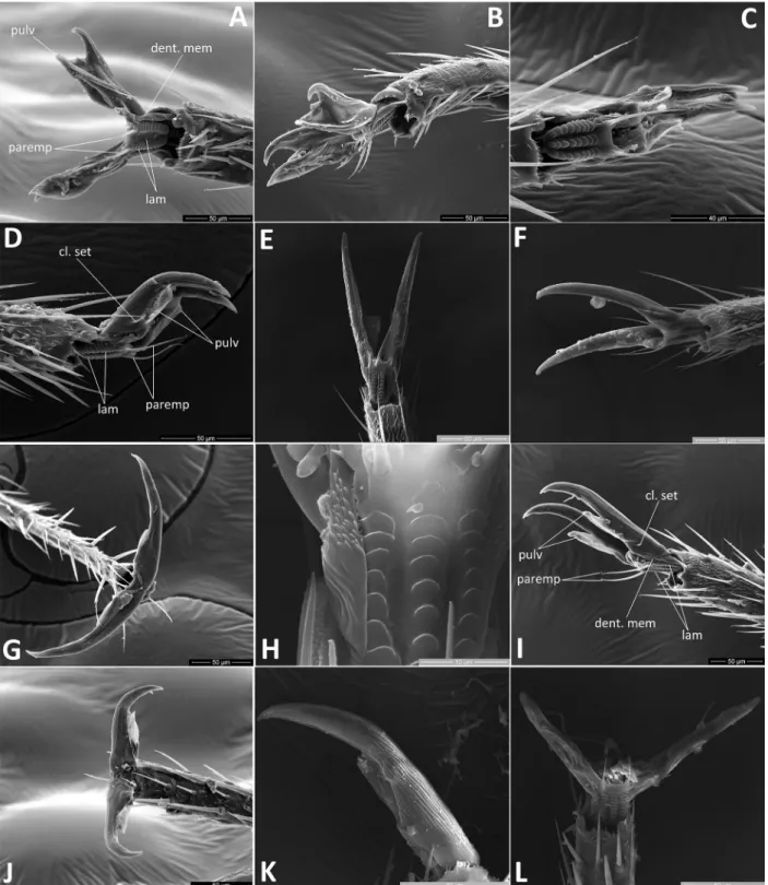
8 Lamellae on unguitractor: (0) broadly rounded, arran
ged in three widely spaced columns (Figs. 5C,H,L, 6H); (1) with almost straight margins, arranged in three adjacent columns (Fig. 6B,C,J); (2) straight in lateral columns and reduced, broadly rounded in cen
tral column (Fig. 5A).
9 Lamellae of central column of ribs on unguitractor:
(0) smooth (Figs. 5C,E,H, 6H,J); (1) finely dentate (Fig. 5A).
10 Membrane between base of claw and unguitractor:
(0) smooth (Figs. 5C,D, 6E,F); (1) finely dentate (Figs. 5A,B,G,I, 6A,G,K,L).
11 Claw setae: (0) absent (Fig. 6A,D,F); (1) present (Fig. 5A,D,F).
12 Claw shape: (0) long, distinctly bent at midpoint (Figs. 5D, 6F,G,I); (1) long and slender, straight, gradually curving close to apex (Figs. 5F,G,I,K, 6A,B,D,K,L); (2) short, with broad base, strongly bent close to apex (Fig. 5B).
13 Pulvilli size: (0) thin, reaching or barely surpassing half-length of claw (Figs. 5D,G,I – L, 6A – F,I – L);
(1) wide, flaplike, distinctly surpassing half-length of claw (Figs. 5A,B, 6G); (2) absent (Fig. 5E,F).
14 Pulvilli apex: (0) attached to claw along entire length (Figs. 5D,G,K, 6G); (1) apically free (Figs. 5A,B,I,J, 6A – F,K,L). — Character not applicable to Cam
ptotylus reuteri Jakovlev, 1881 showing state (2) in character 13.
15 Microsculpture anterior of metathoracic spiracle:
(0) absent (Fig. 7F); (1) well developed along entire length (Fig. 7D,E,H,J,K,L); (2) only dorsally present (Fig7G,I). — In many taxa of Miridae the spiracle is entirely or partly bordered by characteristic evapora
tive or mushroom bodies similar to those on the eva
porative area of metathoracic scent-efferent system (see Konstantinov & Knyshov 2015; naMyatova et al. 2016).
16 Microsculpture posterior of metathoracic spiracle: (0) absent (Fig. 7F,H,L); (1) present (Fig. 7D,E,G,I – K).
17 Peritreme shape: (0) distinctly protruding abo
ve evaporative area and broadly rounded (Fig.
7D,E,H,K,L); (1) flat, not protruding above evapora
tive area, tongue-shaped (Fig. 7F,G,I,J).
18 Head, coloration: (0) uniformly whitish yellow (Fig.
8A,D,G – I); (1) pale yellow with dark markings on frons and vertex (Fig. 8C,E,F); (2) uniformly dark brown.
19 Pronotum, coloration: (0) uniformly pale yellow, wi
thout dark pattern (Fig. 8A – D,G – I); (1) pale yellow with pale brown spots (Figs. 3F, 8E; Konstantinov
& vinoKurov 2011: figs. 1, 2).
20 Hemelytron, coloration, male: (0) pale yellow, with- out dark pattern or with a few pale brown spots (Figs.
3A, 4D,H); (1) pale yellow, with regularly distribu
ted brown minute spots (Figs. 3C – F, 4A – C,F); (2) uniformly dark brown.
21 Claval commissure, coloration, male: (0) uniformly pale (Figs. 3C,D,F, 4D,F,H); (1) darkened (Figs. 3A, 4A,B; Konstantinov & vinoKurov 2011: figs. 1, 2).
22 Rows of minute spinules on hind tibia: (0) present along entire length (as in naMyatova et al. 2016: fig.
18B,D); (1) absent or barely expressed close to ex- treme apex.
23 Tibial spines, color: (0) pale whitish to pale brown (Konstantinov 2008d: figs. 5 – 8); (1) black (Figs. 3, 4).
24 Development of hemelytron, female: (0) macro
pterous (Konstantinov 2008a: fig. 43; Konstantinov
2008d: figs. 7, 8; Konstantinov & vinoKurov 2011:
fig. 2); (1) submacropterous (Konstantinov 2008c:
fig. 6); (2) brachypterous (Figs. 3B,E,H, 4C,E,G,I;
Konstantinov 2008c: figs. 2, 4). — Wing modifica
tions are given according to sChuh & slater (1995).
25 Length of hemelytron in brachypterous females: (0) apex of hemelytron barely reaching abdominal ter
gite III (Konstantinov 2008c: figs. 2, 4); (1) apex of hemelytron reaching abdominal tergite V or at most VI (Figs. 3B,H, 4E,I); (2) apex of hemelytron always reaching, usually surpassing abdominal tergite VII (Figs. 3E, 4C,G). — Character not applicable to spe
cies showing states (0) and (1) in character 24.
26 Hind femur length and thickness, female: (0) dis
tinctly longer but only slightly thicker than middle femur (Konstantinov 2008c: figs. 2, 4, 6; Konstan
tinov 2008d: figs. 6, 8; Konstantinov & vinoKurov 2011: fig. 2); (1) swollen, slightly longer than and at least twice as thick as middle femur (Figs. 3, 4;
Konstantinov 2008a: fig. 43).
27 Ventral keel of genital capsule: (0) absent; (1) pre
sent.
28 Vesica, shape: (0) S-shaped, more or less smooth
ly curved along entire length (Figs. 9A – C, 10A,C;
Konstantinov 2008c: figs. 7 – 10; Konstantinov
2008d: fig. 27; Konstantinov & vinoKurov 2011:
fig. 8); (1) strongly bent distal to secondary gonopo
re, so that apical blade located at almost right angle to body of vesica (Figs. 10F, 11A,B); (2) Cshaped (Konstantinov 2008c: fig. 18).
29 Sclerites around secondary gonopore: (0) absent, se
condary gonopore surrounded by membrane (Figs.
10C – F; Konstantinov 2008d: figs. 27, 28; Konstan
tinov & vinoKurov 2011: fig. 8); (1) present, secon
dary gonopore partly or entirely bordered by sclero
tized straps (Figs. 9, 10A,B, 11A – D; Konstantinov
2008a: fig. 2; Konstantinov 2008c: figs. 7 – 10, 18).
30 Number of apical blades of vesica: (0) 1 (Figs. 9 – 11;
Konstantinov & vinoKurov 2011: fig. 8); (1) 2 (Konstantinov 2008a: fig. 2; Konstantinov 2008c:
figs. 7 – 10; Konstantinov 2008d: figs. 27, 28); (2) absent (Konstantinov 2008c: fig. 18).
31 Secondary gonopore, location: (0) at extreme apex of vesica (Konstantinov 2008c: fig. 18); (1) located subapically (Figs. 10A – D); (2) remote from apex at a distance of at least 1/3 of total vesica length (Figs.
9, 10E,F, 11; Konstantinov 2008a: fig. 2; Konstan
tinov 2008c: figs. 7 – 10; Konstantinov 2008d: figs.
27, 28; Konstantinov & vinoKurov 2011: fig. 8).
32 Outgrowth of vesica distal to secondary gonopore:
(0) absent (Figs. 9, 10; Konstantinov 2008a: fig. 2;
Konstantinov 2008c: figs. 7 – 10, 18; Konstantinov
2008d: figs. 27, 28; Konstantinov & vinoKurov 2011: fig. 8); (1) present (Fig. 11).
33 Anterior margin of dorsal labiate plate: (0) straight, weakly sclerotized (Konstantinov 2008c: figs. 12, 13; Konstantinov 2008d: fig. 33); (1) slightly uptur
ned, folded, sclerotized (Fig. 13J).
34 Sclerotized rings of dorsal labiate plate, shape: (0) broadly oval (Konstantinov 2008c: figs. 11 – 13;
Konstantinov 2008d: fig. 33); (1) oval, distinctly elongate (Fig. 13K); (2) triangular.
35 Vestibulum: (0) short and straight, symmetrical, with- out sclerotized parts; (1) short, C-shaped, with scle- rotized dorsal wall (Fig. 13J); (2) long, S-shaped, distinctly sclerotized (Konstantinov 2008c: fig. 15;
Konstantinov 2008d: figs. 32, 37) — Vestibulum modifications are coded according to the character states given by pluotsiGWalt & MatoCq (2006).
36 Lateral interramal sclerites: (0) absent; (1) present at sides of ventral wall, blade-shaped (Fig. 13L).
37 Gonapophysis 9, shape of apex: (0) sabre-shaped, gradually tapering; (1) arrow-shaped, strongly ex
panded ventrally near apex (Fig. 13M).
3. Results and discussion
3.1. Phylogenetic analyses
The analysis both in PAUP and TNT resulted in three most parsimonious trees of 112 steps with Ci = 0.47 and Ri = 0.60, the strict consensus of which is shown on Fig.
1A. The successive approximation weighting performed in PAUP gave the single tree identical to one of the trees recovered from the equal weight analysis. The same tree topology was obtained under implied weighting in TNT with the integers of concavity factor ranging from 6 to 100 and is used in the following discussion (Fig. 1B).
The resulting topologies received under concavity cons
tant K = 1 – 5 differ from each other and from trees obtai
ned under equal weights in the position of the outgroup taxa due to extreme down-weighting of homoplastic cha
racters (Goloboff et al. 2008, 2017; penz et al. 2013) and not discussed further.
Character data are plotted on the tree (Fig. 1B) using fast optimization (ACCTRAN). Filled squares represent nonhomoplastic characters appearing only once on the tree, homoplastic characters are shown as open squares.
Nodes of the major clades are numbered from 1 to 9. The main character numbers and character states supporting these nodes are indicated below.
Node 1 is supported by two synapomorphies, the prominent post-ocular region of the head (2-1) and the finely dentate lamellae of central column of ribs on the unguitractor (9-1). It is also supported by three homopla
sious characters: lamellae of unguitractor with straight margins, arranged in three columns (8-1), apically free pulvilli (14-1), and presence of microsculpture posterior to the opening of metathoracic spiracle (16-1). This clade comprises the genera Acrotelus Reuter, 1885, Eurycolpus Reuter, 1875, Omocoris Lindberg, 1930, and Oncotylus Fieber, 1858, which form the backbone of the Oncotylus
group of genera established by WaGner (1975).
Node 2 strongly supports the monophyly of the genus Omocoris by two uncontradicted synapomorphies: long antennomere I (1-1) and presence of numerous spinelike setae on this segment (6-3). Homoplastic characters sup
Fig. 1. Maximum parsimony trees of Agraptocoris and outgroup taxa. A: Strict consensus tree showing subtribes of Phylinae sensu sChuh
& Menard (2013). Numbers above nodes indicate Bremer values. Host plants indicated on right side. B: One out of three most parsimoni
ous trees identical to the tree obtained under successive approximation and implied weighting (K = 6 through 100). Characters are plotted showing fast optimization. Nodes 1 – 9 are discussed in the text.
porting this clade include dorsally located microsculpture of metathoracic spiracle (15-2), pale claval commissure in males (21-0), brachyptery in females (24-2), presence of two apical blades of vesica (30-1), and arrow-shaped gonapophysis 9 of ovipositor (37-1). Strong brachyptery in females is a rare feature across phyline genera. Species of Omocoris were added into analysis for careful assess
ment of the monophyly of Agraptocoris also possessing brachypterous females in all species.
Node 3 represents the clade of Josifovius Konstan
tinov, 2008 + Agraptocoris and is corroborated by four character changes including submacropterous fema
les (24-1, changed to brachypterous in Agraptocoris) and characteristically straight and almost symmetrical, slightly bent rightwards vestibulum of female genitalia (35-1).
Node 4 supports the monophyly of Agraptocoris which is defined by a single synapomorphy: oval shape of sclerotized rings (34-1, reversed in A. eugeniae + A. on
cotyloides). The clade is further supported by three ho
moplasies, including the absence of rows of minute spi
nules on hind tibia (22-1), brachypterous females (24-2), and distinctly swollen hind femur in females (26-1).
Node 5 comprises Agraptocoris nigrisetosus + A. pal
lescens and is united by one uncontradicted synapomor
phy, namely the presence of flattened outgrowth of vesica distal to secondary gonopore (32-1). It is further suppor
ted by one homoplasious character, viz. vesica strongly bent distal to secondary gonopore (28-1).
Node 6, the sister-species relationship of Agraptoco
ris concolor + A. subconcolor is defined by one homo
plasious character, darkened claval commissure (21-1).
Node 7 is supported by the presence of regularly dis
tributed brown spots on hemelytron in males (20-1) and hemelytron of females always reaching abdominal tergite VII (25-2); the latter character appears to be unreversed synapomorphy (unknown in A. margaretae).
Node 8 is defined by one synapomorphy: presence of one ventral and four dorsomesial spinelike setae on antennomere I (6-2, reversed in Agraptocoris oncotyloi
des).
Node 9 comprises Agraptocoris eugeniae + A. onco
tyloides and is defined by five homoplasious characters:
presence of dark simple setae on pronotum (4-1) and on hemelytron (5-1), presence of dark color pattern on frons and vertex (18-1), subapically located secondary gono
pore (31-1), and broadly oval sclerotized rings (34-0).
3.2. Phylogeny of Agraptocoris
Monophyly of Agraptocoris appears to be well supported in the present analysis and can be further corroborated by the additional characters discussed in the generic diagno
sis. However, the phylogenetic position of Agraptocoris within the tribe Phylini remains uncertain. The resulting tree rendered subtribes Phylina and Oncotylina sensu sChuh & Menard (2013) as non-monophyletic (Fig. 1A).
The Oncotylus-group of WaGner (1975) represented in
this analysis by the genera Acrotelus, Eurycolpus, Omo
coris, and Oncotylus, appeared as a sister group to all other taxa included in the analysis. These subtribes were weakly supported in the phylogenetic analysis of Menard et al. (2014) which formed a basis for the simultaneously prepared novel classification of the Phylinae (sChuh &
Menard 2013). The subtribe Oncotylina lacked morpho
logical synapomorphies and the monophyly of Phylina was supported by highly homoplastic characters, the labium reaching past the hind coxae and the calli not visible on the pronotum (Menard et al. 2014). Neither group was recovered as monophyletic in the subsequent molecular and total-evidence analyses with larger taxon sampling (Konstantinov et al. in prep.). Therefore a much broader phylogenetic analysis of the entire tribe Phylini is needed for the correct assignment of Agraptocoris and allied genera. This goes beyond the subject of this paper and will be dealt with elsewhere.
3.3. Distributional patterns
Distributions of all Agraptocoris spp. are summarized in Fig. 15. As currently known, six out of eight species are widely distributed across the desert steppe zone of Mon
golia, spanning from Uvs and Hovd Aimaks in the West to East Govi and Suhbaatar Aimaks in the East. Two of those species, viz. A. concolor and A. oncotyloides ex
tend slightly more westward into the Kosh-Agach area (Altai Rep., Russia) which is floristically similar to adja
cent areas of NW Mongolia. Noteworthy, all three pairs of Mongolian sister species (A. nigrisetosus + A. palles
cens, A. concolor + A. subconcolor, and A. eugeniae + A. on co tyloides) exhibit extensive range overlap and show no apparent differences in host preference (Fig.
1A) and phenology. Agraptocoris margaretae and A. pa
mi ricus are the only two geographically isolated species of the genus known from high altitudes of the western Himalayas and Pamir Mountains, respectively (Fig. 15).
However the vast intermediate regions of the Xinjiang Uyghur and Inner Mongolia provinces of China remain almost entirely unstudied and species of Agraptocoris might be detected there with more sampling effort.
3.4. Host plant associations
Mapping of hosts onto the tree (Fig. 1A) points to an ance
stral association of Agraptocoris with Artemisia (Astera
ceae). Most species of the genus with available host data are restricted to Asteraceae genera Artemisia and Pyre
thrum, aside from Agraptocoris pamiricuseugeniae
oncotyloides clade which is found on Chenopodioideae subfamily of Amaranthaceae. This evidently represents a host shift event between distantly related orders Aste
rales and Caryophyllales. All species of Agraptocoris are associated with multiple host species belonging to one or two plant genera. Monophagous A. margaretae is an exception, likely due to limited sampling in the field. The
relatively low degree of host restrictedness is typical for most widespread phyline species with accurately docu
mented host information (e.g., sChuh 2004; sChuh &
sChWartz 2005; sChuh & pedraza 2010; sChuh & Wei
rauCh 2010).
Host patterns of the subfamily Phylinae in Mongolia show modest correlation with the diversity of vascular plants at the family level. The three largest families of the Mongolian flora, viz., Asteraceae (478 species), Fa
baceae (356 species), and Poaceae (259 species) compri
se 35% of plant species of the area (urGaMal et al. 2014).
However these families harbor relatively few phyline species. Asteraceae serve as hosts to 9 species, namely Camptotylidea flavida (Nonnaizab & Yang, 1994) (Kon
stantinov 1999: Artemisia sp.), Camptozorus lactucae Kerzhner, 1996 (Kerzhner 1996: Lactuca tatarica L.), Compsidolon pumilum (Jakovlev, 1876) and C. kerzhne
ri (Konstantinov 2006: Artemisia spp.), and 5 species of Agraptocoris (Artemisia spp., Pyrethrum). Fabaceae are utilized by 7 species, viz. Camptotylidea suturalis (Reuter, 1903) (Konstantinov 1999: Halimodendron), Dacota nigritarsis (Jakovlev, 1882) (Kerzhner 1964:
Caragana), all 3 species of Phaeochiton Kerzhner, 1964 (Konstantinov 2008d: Caragana spp.), Salicarus fulvi
cornis (Jakovlev, 1889) (sChWartz & stonedahl 2004:
Caragana sp.), and Salicarus halimodendri Putshkov, 1977 (putshKov 1977: Halimodendron). No phyline spe
cies is known to be associated with Poaceae in Mongolia.
On the contrary, the largest number of phyline species is associated with Chenopodioideae, although only 3.3%
of Mongolian plant species belong to this family. Eighty- five species of Phylinae are currently known from the re
gion (Kerzhner & Josifov 1999; Konstantinov & naMy
atova 2008). One fourth of these, or 21 species from the genera Agraptocoris, Boopidocoris Reuter, 1879 (Kon
stantinov & naMyatova 2008), Camptotylidea Wag
ner, 1957 (Konstantinov 1999), Camptotylus Fieber, 1860 (Konstantinov 2008a), Monocris Putshkov, 1974 (Konstantinov & naMyatova 2008), Paralaemocoris Linnavuori, 1964 (Kerzhner 1984), Psallopsis Reuter, 1901 (Konstantinov 1997), and Solenoxyphus Reuter, 1875 (Konstantinov 2008b) are strictly associated with Chenopodioideae.
These patterns generally correspond to the trends outlined by Cassis & sChuh (2012), who analyzed available host data for the entire family Miridae and its separate clades including phylines. In both cases Cassis
& sChuh (2012) documented the greatest frequency of association with the rosid orders Fabales, Fagales, and Rosales, and the asteroid orders Asterales, Caryophylla
les, and Lamiales.
3.5. Terminology of the aedeagus in Phylinae
Phylinae is a well-defined group primarily diagnosed by a set of synapomorphies related to male genitalia struc
ture (sChuh 1974; Cassis & sChuh 2012; Menard et al.
2014). Particularly, the phyline aedeagus is modified in several unique ways as compared to other plant bug li
neages.
In all Heteroptera, including Miridae, the aedeagus is located in the genital chamber, a membranous sac grea
tly expanded inside the strongly sclerotized abdominal segment IX usually referred to as the genital capsule or pygophore. Only the apex of the aedeagus can be obser
ved externally when in repose (Fig. 2A). According to the general convention, the genital chamber is morpho
logically described as an expansion of the intersegmen
tal membrane between 9th and 10th abdominal segments (KullenberG 1947; dupuis & Carvalho 1956; dupuis 1970; Konstantinov 2003). However, Klass & Matush
Kina (2018) argued for the use of primary, embryonic segmental borders in establishing homologies of the male genitalia in adult insects and provided tentative evidence for assignment of the phallic organs to the 10th abdominal segment.
The base of the aedeagus is formed by the phallo
base, a heavily sclerotized horseshoeshaped sclerite an
teriorly fixed in the genital capsule like a swing through suspensory apodemes. The distal part of the aedeagus beyond the phallobase is divided into a proximal part, the tube-shaped and sclerotized phallotheca and a distal part, the endosoma (dupuis & Carvalho 1956; dupuis 1970;
Cobben 1978; deCKert 1990; Kerzhner & Konstantinov
1999). The efferent tube of the aedeagus is the ectoder
mal ductus seminis which opens to the exterior via the secondary gonopore. The aedeagus is moved backwards and forwards by a pair protractor and a pair of retractor muscles respectively, all attached to the phallobase.
In basal clades of plant bugs, e.g., Isometopinae (na
Myatova & Cassis 2016b), most Cylapinae (naMyato
va & Cassis 2016c; naMyatova et al. 2018), and many Bryocorinae (Konstantinov & Knyshov 2015; naMyato
va et al. 2016; naMyatova & Cassis 2016a), the endo
soma forms a membranous, sac-like inner tubule of the aedeagus invaginated into phallotheca in repose. In the erect position, this internal sac is everted from the phal
lotheca like a glove finger (Konstantinov 2003: fig. 11).
In Phylinae, the endosoma is strongly modified and further subdivided into a membranous eversible basal part and a strongly sclerotized, non-eversible and typically S- shaped distal part bearing the secondary gonopore. This distal component of the aedeagus is well familiar to plant bug taxonomists and serves as an important criterion for recognition of genera and species in the Phylinae. sinGh pruthi (1925) coined the term vesica for the distal part of the endosoma in Heteroptera and for the next eighty years it was universally applied in the taxonomic litera
ture on phylines (e.g., WaGner 1955, 1974, 1975; Kelton 1959; linnavuori 1971; sChuh 1974; sChuh & slater 1995; sChWartz & stonedahl 2004).
The vesica frequently takes the form of a sclerotized gutter due to uneven sclerotization of its walls and may be additionally armed with a complex set of longitudi
nal sclerotized straps and apical blades. Cross sections of the vesica clearly reveal its tube-like structure and the
Fig. 2. Parasagittal sections of the genital capsule in phylines. A: Agraptocoris concolor, aedeagus in repose. B: same species, aedeagus erected. C: Aspidacanthus myrmecoides Reuter, 1901 (Hallodapini), aedeagus in repose; the genital chamber is partly not shown. — Ab- breviations: conj – conjunctiva; Fuhr – Führungsstück; mem. phth – membranous region of phallotheca; phb – phallobase; phth – phal
lotheca; scl. phth – sclerotized region of phallotheca; s. gon – secondary gonopore; susp. ap – suspensory apodeme; ves – vesica; IX – ab
dominal segment IX = genital capsule; X – abdominal segment X.
presence of an entirely membranous, thin-walled ductus seminis running inside. The basal part of ductus seminis, running from the base of the vesica to the phallobase is reduced to a short and strongly sclerotized tube equipped with sclerotized ribs (Fig. 2). KullenberG (1947) deno
ted this structure as Führungsstück (guiding piece).
While the distal part of the endosoma is modified into a tube-shaped sclerotized structure around the ductus se
minis, the proximal part is formed by a thin membrane connecting the aperture of the phallotheca to the base of vesica. sinGhpruthi (1925) referred to this section as conjunctiva and was followed by many subsequent authors (e.g., KullenberG 1947; WaGner 1955, 1974;
WaGner & Weber 1964; dupuis 1963, 1970; dupuis &
Carvalho 1956; Matsuda 1976; Cobben 1978; Kerzh
ner & Konstantinov 1999; Konstantinov 2003). The conjunctiva envelopes the vesica in repose (Fig. 2A) and crumples at its base during eversion (Fig. 2B). For practi
cing taxonomists the conjunctiva is recognized as a tiny membrane which may partially remain wrapped around the vesica after its dissection from the genital capsule.
The phallotheca in phylines is also modified and has membranous, very thin-walled proximal part, while the distal part is strongly sclerotized and fixed to the genital capsule (KullenberG 1947; Kerzhner & Konstantinov
1999). The sclerotized apex of phallotheca is frequently illustrated in the phyline taxonomic literature together with the vesica and parameres. During copulation (Fig.
2B; Konstantinov 2003: fig. 11), the phallobase is shif
ted backwards, resulting in crumpling of the conjunctiva and membranous posterior part of the phallotheca, and in protrusion of the vesica through the apical aperture of the sclerotized anterior part of the phallotheca (KullenberG 1947).
Despite all the modifications outlined above, the ae
deagus obviously remains a morphologically and func
tionally integral organ. There are two following miscon
ceptions regarding the aedeagal structure of phylines.
The first is that the phallotheca is not connected to the phallobase and reduced to a sclerotized apical part at
tached to the genital capsule (e.g., sChuh & slater 1995).
However, this idea is compromising the integrity of the body wall and clearly not confirmed by the available data (refer to KullenberG 1947 for details). The second is that the vesica is merely a strongly sclerotized ductus semi
nis (e.g., Kelton 1959). Again, this seems to be a clear misconception as the ductus seminis, whether with partly sclerotized or entirely membranous walls, is only a duct running inside the aedeagus and thus cannot be a separate component of the aedeagus by itself.
Subdivision of the inner endosomal sac of the aede
agus into the distal non-eversible but retractable vesica and proximal membranous conjunctiva is known for other clades of Miridae, viz. Mirinae, Deraeocorinae, (except Termatophylini), Dicyphini, and some Cylapinae (Kerzhner & Konstantinov 1999; Konstantinov 2003;
naMyatova et al. 2016). Similar to phylines, the base of vesica in these groups is narrow and sclerotized, with its walls tightly attached to the corresponding part of duc
tus seminis. However the vesica is composed of several membranous swollen lobes and typically equipped with variously shaped sclerotized appendages. The shape, size and sculpture of the ductus seminis, secondary gonopo
re and phallotheca vary considerably among groups but the phallotheca is never fixed to the genital capsule. In contrast to phylines, the protrusion of the vesica from the phallotheca and inflation of the membranous lobes du
ring copulation is due to hemolymph pressure within the aedeagus (Konstantinov 2003: fig. 14).
The terminology used for aedeagal structures in phy
lines remained surprisingly uniform for almost a centu
ry in both taxonomic and morphological works until the publication of Cassis (2008) on the orthotyline genus Lattinova. He rejected the idea of the division of endoso
ma into conjunctiva and vesica on the grounds of predo
minantly functional justification of this subdivision and independent derivation of the vesica in several clades of Miridae. An updated terminology of Cassis (2008) recei
ved wide acceptance for phylines over the last decade (e.g., sChuh & pedraza 2010; sChuh & sChWartz 2015;
leon & WeirauCh 2016; yasunaGa & duWal 2015; du
Walet al. 2017) and the term vesica was replaced with the more inclusive term endosoma.
No testing of homology statements for aedeagal components across the Miridae has ever been attempted.
Therefore, a survey of terminology used for the male ge
nitalic structures of the entire family Miridae is beyond the scope of this paper. However, an application of the term endosoma (= conjunctiva + vesica) only to the distal component of endosoma (= vesica) is ill-defined in the case of Phylinae. This approach further undermines the homology statements in comparison to other subfamilies of plant bugs.
No doubt, usage of the same term “vesica” for struc
tures that apparently evolved independently, e.g. in the case of Mirinae and Phylinae might be viewed as unsa
tisfactory. However the same is true for many terms fre
quently used in plant bug taxonomy, e.g., pronotal collar or scales on dorsum.
A correct description of the inner aedeagal compo
nents, if more complex than an undivided sac, would inevitably require additional terminology. The idea of changing the terminology whenever a new morphologi
cal interpretation sees the light of day seems unproduc
tive. Numerous theories on the origin of the external ge
nitalia published during the last century and assumptions concerning their homology across the main insect orders clearly illustrate this point (tuxen 1969; Konstantinov
2007; Klass & MatushKina 2018). Most authors in this field proposed new terms and rejected previously used ones in accordance with their theoretical views or just because a term was considered as descriptive and/or in
correct. Surely, each author considers his idea as the right one, but another theory will follow shortly. In contrast to morphologists, taxonomists generally adhere to the tradi
tionally used terms and disregard new names irrespecti
ve of agreement with the morphological hypothesis. Al- though it is always good to know the true morphological
value of the structure, a continuous change of names will merely make this task more difficult.
In summary, I believe that the term endosoma in case of phylines is morphologically and historically unjusti
fied while the term vesica (of Phylinae type) would work best at the moment.
4. Taxonomy
4.1. Agraptocoris Reuter, 1903
Agraptocoris Reuter, 1903: 6. Type species by monotypy: Agrapto
coris concolor Reuter, 1903.
Tibetocoris Hutchinson, 1934: 141. Synonymized by Kerzhner
1997: 246. Type species by original designation: Tibetocoris margaretae Hutchinson, 1934. Non Tibetocoris (Pentatomidae, Pentatominae) zhenG & liu 1987: 288, 294. Junior homonym of Tibetocoris Hutchinson, 1934 (Miridae). The replacement name Zhengius proposed by rider 1998: 508.
Differential diagnosis. Recognized by the following com
bination of characters: body elongate, parallel-sided in ma
les, short and ovate in brachypterous females (Figs. 3, 4);
hind femur in females distinctly swollen; dorsum uniform
ly pale greenish to whitish, rarely with partly or entirely darkened head (Fig. 8), typically with minute pale brown spots on hemelytron; vestiture composed of adpressed to semierect woolly silvery setae intermixed with black sim
ple setae (Fig. 7C); claws long and slender, straight, gradu
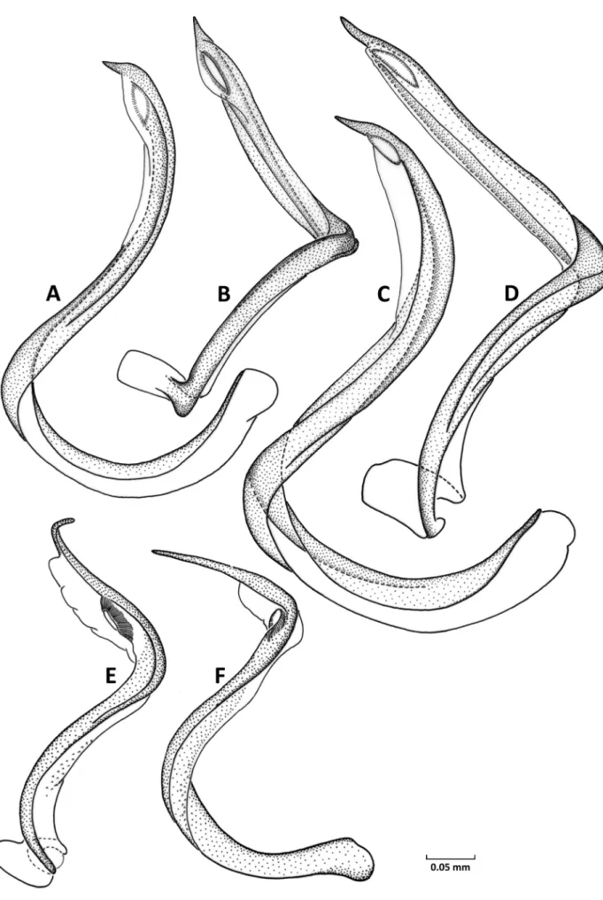
ally curving subapically; pulvilli narrow, reaching one-half length of claw, apically free (Fig. 6J – L); vesica relatively simple, thin, S-shaped, apically terminating with single gradually tapering blade (Figs. 9 – 11, 14A – P); secondary gonopore located at base of apical blade. Species primarily feeding on Artemisia spp. (Asteraceae) and sometimes on halophytes from Amaranthaceae.
According to the original description (reuter 1903), Agraptocoris is most closely related to the genus Pasto
coris Reuter, 1879. Both genera are similar in overall size and body proportions, color pattern of dorsum, long and thin tarsi, and slender claws. However Pastocoris dif
fers from Agraptocoris in many features including ma
cropterous females and very small pulvilli, whereas the structure of vesica in Pastocoris unequivocally suggests its close affinity to the SolenoxyphusBoopidocoris group of genera due to the presence of characteristic step-shaped projection distal to secondary gonopore (Konstantinov
2008b; Konstantinov & Korzeev 2014). The phylogene
tic analysis rendered the monotypic genus Josifovius as a sister clade to Agraptocoris (Fig. 1). Josifovius dimorphus Wagner, 1961, although similar in the general appearance, pretarsal structure, and the form of female genitalia, dif
fers from Agraptocoris spp. in having an entirely diffe
rent vesica structure (Konstantinov 2008c: figs. 18, 20).
Females of Josifovius further differ in having long and thin, distinctly not saltatorial hind femur and only slightly shortened, submacropterous hind wing reaching abdomi
nal tergite XI (Konstantinov 2008c: fig. 6).
Among Palearctic phylines, species of Agraptocoris appear to be most similar to those of Compsidolon Reuter, 1899 in the male body proportions, color pattern of dor
sum frequently composed of dense minute brown spots, vestiture composed of silvery sericeous and dark simp
le setae, claw structure, and the relatively simple vesica with subapical secondary gonopore and single, gradually tapering apical blade. In addition, many Compsidolon species from the subgenus Apsinthophylus Wagner, 1965 utilize Artemisia spp. as hosts and females of one spe
cies from this subgenus, C. hiemale Konstantinov, 2006, are brachypterous (Konstantinov 2006: fig. 38). Species of the genus Compsidolon can be distinguished from Agraptocoris by the presence of several rows of minute dark spinules along entire length of tibia, pulvillus adnate to claw along entire length, and characteristic curvature of the apical blade of vesica typical for Apsinthophylus spp. (linnavuori 1971: figs. 2, 3; Konstantinov 2006:
figs. 8 – 14).
The male genitalia structure, particularly the vesica of Agraptocoris, is most similar to those of Psallomorpha Duwal, Yasunaga & Lee, 2010 known only from Nepal.
However, the latter genus differs from Agraptocoris in many other respects including the intense dark color pat
tern of dorsum, presence of a series of black round spots along the fore margin of hind femur, the darkened bases of tibial spines, and macropterous females. In addition, all 6 species of Psallomorpha are associated with broad- leaved trees in the Fagaceae, Rosaceae, and Theaceae (duWal et al. 2010).
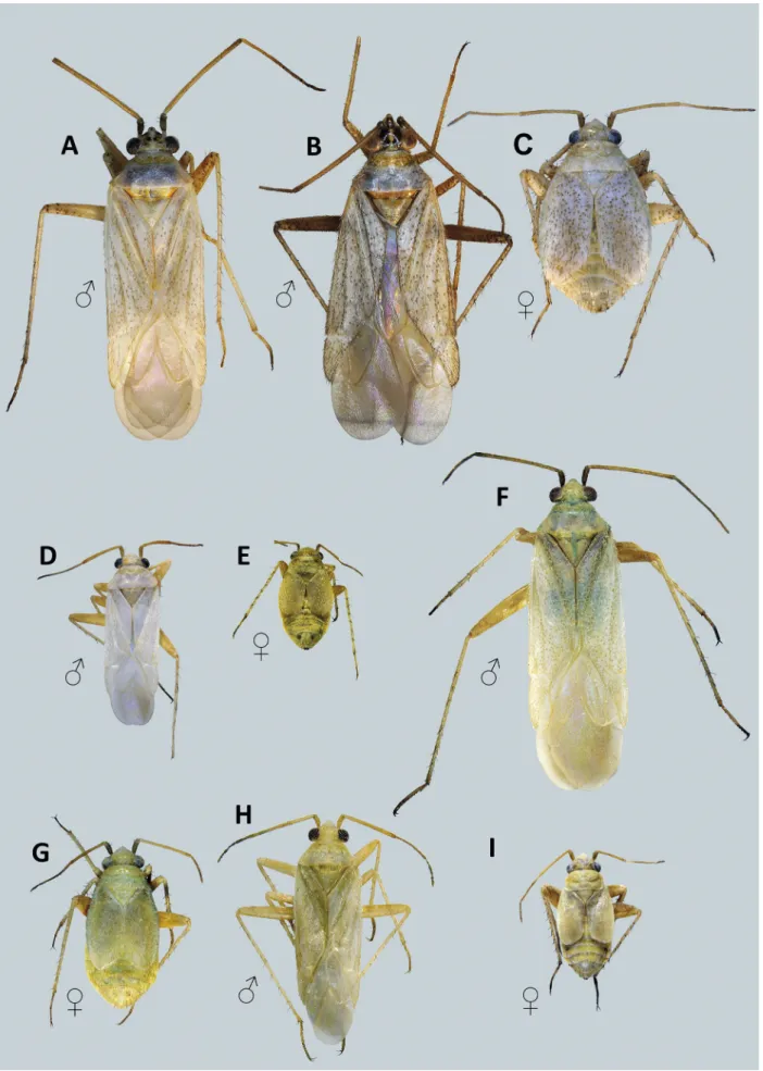
Redescription. Male: Small to medium-sized, total length 3.3 – 6.2, macropterous, body elongate and near
ly parallelsided. Coloration (Figs. 3, 4): Dorsum and venter generally unicolorous, ground-color varying from whitish to dirty-yellow; head usually uniformly pale, so
metimes partly darkened or with two brown spots on ver
tex (Fig. 8), labial segment IV apically or entirely darke
ned; pronotum and scutellum without dark color pattern;
hemelytron frequently with faint, minute, rounded brown spots, sometimes uniformly pale yellow; femora usually with pale brown markings apically; tibiae without dark spots at bases of tibial spines. Surface and vestiture (Figs.
7A – C, 8): Dorsum smooth, moderately shining, clothed with dense, recumbent, weakly woolly, adpressed silvery setae intermixed with comparatively long, semierect to adpressed, dark brown simple setae; dark simple setae of
ten present only on apical half of hemelytron, sometimes covering entire dorsum; antenna and legs clothed with comparatively short, dense, adpressed, silvery simple se
tae; antennomere I with one dark spinelike seta ventrally and 2 – 7 dark spinelike setae dorsomesially; femora with several pairs of dark brown, rarely pale brown spinelike setae apically; hind tibial spines always dark brown, mi
nute black spinules on tibiae scarce, located close to ex
treme apex and not arranged in regular rows. Structure:
Head anterior to eyes roughly triangular in dorsal view (Fig. 8); eyes relatively large, occupying almost entire height of head in lateral view (Fig. 7B), posterolateral
margins of eyes almost contiguous with anterolateral margins of pronotum; vertex weakly convex, clypeus distinctly produced anteriorly and always visible in dor
sal view (Fig. 7A); antennal fossae located well above ventral margin of eye; antennomere I somewhat swollen along entire length, as long as or somewhat longer than width of vertex; segment II linear, 0.9 – 1.5 × as long as basal width of pronotum; labium reaching or slightly surpassing metacoxae. Pronotum trapezoidal, with indis
tinct calli and weakly convex disk, posterior margin ne
arly straight or weakly concave medially, lateral margins straight, posterolateral angles broadly rounded; mesono
tum moderately exposed; metathoracic scent-gland eva
poratory area roughly triangular, peritreme apically roun
ded, raised above pleural surface (Fig. 7D,E). Hind femur elongate, not swollen, tibia cylindrical, second tarsal seg
ment somewhat longer than third, claw (Fig. 6J – L) long and thin, gradually bent in apical part, pulvillus small, not reaching midpoint of claw, apically free; unguitractor with broadly rounded lamellae arranged in three wide
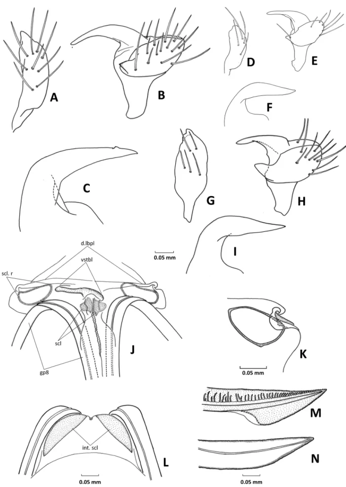
ly spaced columns (Fig. 6J). Genitalia: Genital segment conical, of typical phyline shape and devoid of distincti
ve ornamentation, more or less elongated, 1.1 – 1.4 × as long as width at base; apex of phallotheca narrow, ty
pically L-shaped (Figs. 12G,J,N, 13C,I, 14Q – T); right paramere lanceolate (Figs. 12A,E,I,L, 13A,D,G); left paramere boat-shaped, with apically rounded or pointed hypophysis Figs. 12B,D,F,H,K,M,O, 13B,E,H); vesica S-shaped, typical of Phylini, terminating with single api
cal blade, usually long and bent at midpoint, sometimes short and pointed; secondary gonopore located subapi
cally and surrounded with more or less developed mem
brane (Figs. 9 – 11, 14). — FeMale: Body (Figs. 3B,E,H, 4C,E,I) short, stout, strongly brachypterous, total length 2.5 – 4.1 mm. Coloration: Similar to male but usually with poorly expressed color pattern of dorsum; heme
lytron frequently uniformly pale yellow, without minute pale brown spots. Surface and vestiture: Similar to male.
Structure: Head similar to that of male but with distinctly smaller eyes. Thorax not elevated posteriorly, pronotum and scutellum almost flat in lateral view; suture between mesonotum and scutellum shallow, mesonotum usu
ally entirely covered by pronotum. Hemelytron strong- ly brachypterous, corium and clavus fused, cuneus and membrane absent; lateral margins broadly convex, apex of hemelytron broadly rounded or obliquely truncated, reaching abdominal tergite V – VII. Legs not as elonga
te as in male, with thicker fore and middle femora, hind femur swollen, saltatorial, almost reaching apex of abdo
men. Abdomen broad, elongate-oval, 1.1 – 1.3 × as long as broad, 1.5 – 1.7 × as broad as basal width of pronotum.
Genitalia: Dorsal labiate plate of bursa copulatrix with relatively large, oval to roughly triangular sclerotized rings (Fig. 13K); sclerites encircling vulva symmetric, of typical phyline shape; vestibulum weakly sclerotized, slightly bent leftwards (Fig. 13J); posterior wall with two distinct and symmetrical blade-shaped sclerites at sides (Fig. 13L); gonapophysis 8 gradually tapering, gonapo
physis 9 sagittate (Fig. 13M,N).
Note. Female genitalia of Agraptocoris showed compa
ratively low interspecific variability and did not allow for separation of many species. Some distinctions were observed in the shape of sclerotized rings of the dorsal la
biate plate, roughly triangular in A. concolor and A. sub
concolor, more ovoid in other species.
4.2. Identification key to species
1 Head in male partly darkened or with two brown ma
cula on vertex (Fig. 8C,E,F), rarely uniformly pale yellow; secondary gonopore located close to apex of vesica, apical blade short and straight (Fig. 10A – D).
... 2 1’ Head in male without dark brown pattern, rarely frons
with indistinct pale brown stripes radiating from mid
line (Fig. 8A,D,G – I); secondary gonopore removed from apex of vesica, apical blade smoothly curved, at least 3 × as long as gonopore (Figs. 9, 10E,F, 11). … 3 2 Antennomere I in both sexes with three brown spi
nelike setae on dorsomesial surface (Fig. 8E,F); he
melytron in male with dense minute pale brown spots and two or three pale brown longitudinal lines com
posed of confluent spots, sometimes interrupted and indistinct (Fig. 4A,B); hemelytron in female large, always covering abdominal tergite VII, with minute pale brown spots (Fig. 4C); phallotheca with subapi
cal tooth (Figs. 13C, 14Q); vesica as in Figs. 10C,D, 14I,L. ... A. oncotyloides 2’ Antennomere I in both sexes with 5 – 7 brown spine
like setae on dorsomesial surface (Fig. 8B,C); hemely
tron in male with regular, minute, pale brown spots not forming longitudinal lines (Fig. 3C,D); hemelytron in female at most partly covering abdominal tergite VI, without dark color pattern (Fig. 3E); phallotheca wi
thout subapical tooth (Figs. 12G, 14R); vesica as in Figs. 10A,B, 14H,K. ... A. eugeniae 3 Vesica strongly bent just distal to secondary gonopore,
apical blade located almost at right angle to basal 2/3 of vesica in lateral view (Figs. 10F, 11A,B, 14M,N);
hemelytron in female short, reaching or almost co
vering abdominal tergite V (Figs. 3H, 4E, unknown in A. margaretae). ... 4 3’ Vesica smoothly and gradually curved, without bend
distal to secondary gonopore (Figs. 9A – C, 14A – D, G); hemelytron in female at least partly covering ab
dominal tergite VI (Figs. 3B, 4G,I). ... 6 4 Pronotum, scutellum and hemelytron with small dif
fuse pale brown spots (Fig. 3F); apical blade of vesica without additional processes (Fig. 10E,F) [Kashmir region]. ... A. margaretae 4’ Dorsum immaculate, rarely with diffuse minute spots
on clavus and endocorium (Figs. 3C,D, 4D); apical blade of vesica with small flattened outgrowth distal to secondary gonopore (Figs. 11A – D, 14M – P) [Mon
golia]. ... 5 5 Entire dorsum including head and pronotum with con
spicuous black simple setae in both sexes (Fig. 8D);
Fig. 3. Dorsal habitus of Agraptocoris spp. A,B: A. concolor. C – E: A. eugeniae. F: A. margaretae. G,H: A. nigrisetosus.
Fig. 4. Dorsal habitus of Agraptocoris spp. A – C: A. oncotyloides. D,E: A. pallescens. F,G: A. pamiricus. H,I: A. subconcolor.
antennomere II distinctly longer than posterior width of pronotum in male and head width in female; scle- rotized outgrowth of vesica thornshaped, posteriorly attenuated (Figs. 11C,E, 14O). ... A. nigrisetosus 5’ Dorsum in both sexes without dark setae or with a
few brown setae in apical part of corium (Fig. 4D,E);
antennomere II shorter than or equals to posterior width of pronotum in male and to head width in fe
male; outgrowth of vesica semicircular, weakly scle- rotized (Fig. 11B,F, 14P). ... A. pallescens 6 Antennomere I with four mesial spinelike setae in
both sexes (Fig. 8H); hemelytron with regularly dis
tributed, dense, minute pale brown spots (Fig. 4F);
apical blade of vesica straight, slightly bent only at extreme apex, abruptly narrowing distal to secondary gonopore (Figs. 9A,F, 14J). ... A. pamiricus 6’ Antennomere I with two mesial spinelike setae in
both sexes (Fig. 8A,I); hemelytron immaculate or with irregular brown spots more dense along claval vein and medial fracture of corium (Figs. 3A, 4H);
apical blade of vesica bent at midpoint and gradually narrowing towards apex (Figs. 9B – E, 14A – F). .... 7 7 Larger, body length male 4.8 – 5.6 mm, female
2.8 – 3.6 mm; hemelytron in male usually with dif
fuse pale brown minute spots along claval vein and medial fracture (Fig. 3A); apical blade of vesica lan
cet-shaped in lateral view (Figs. 9C, 14A,B), strongly curved at midpoint (Figs. 9D, 14E). ... A. concolor 7’ Smaller, body length male 3.8 – 4.7 mm, female 2.5 –
3.0 mm; hemelytron in male without pale brown spots or with few minute pale brown spots (Fig. 4H); apical blade of vesica thin and gradually tapering towards apex in lateral view (Figs. 9B, 14C,D), smoothly cur
ved at midpoint (Figs. 9E, 14F). ... A. subconcolor
4.3. Agraptocoris concolor Reuter, 1903 Figs. 3A,B, 7D, 8A, 9C,D, 12A – D, 13J – L, 14A,B,E, 15
Agraptocoris concolor Reuter, 1903: 7 (new species); Kerzhner et al. 1997: 127 (lectotype designation); vinoKurov & KanyuKo
va 1995b: 121 (key).
Diagnosis. Recognized by the following combination of characters: body relatively large, male 4.8 – 5.6 mm, female 2.8 – 3.6 mm; antennomere I with two spinelike dark setae on dorsomesial surface (Fig. 8A); hemelytron in male usually with indistinct pale brown minute spots more dense along medial fracture of corium (Fig. 3A), rarely dorsum uniformly pale; dark setae absent on head and pronotum, located only on distal 2/3 of hemelytron;
vesica S-shaped, gradually curved, without additional outgrowth, secondary gonopore removed from apex;
apical blade of vesica straight, of almost same width through out its length, abruptly tapering at extreme apex (Figs. 9C,D, 14A,B,E).
Most similar to A. subconcolor sp.n. and apparently not always separable from it without careful investigati
on of the vesica. Males of A. subconcolor differ in appea
rance from A. concolor in having smaller size (3.8 – 4.7 mm) and pale dorsum without pale brown mottling along medial fracture. The apical blade of A. concolor is lancet
shaped, comparatively wide, abruptly narrowing at extre
me apex in lateral view (Figs. 9C, 14A,B) and strongly bent at midpoint between secondary gonopore and apex of vesica in ventral view (Figs. 9D, 14E). In contrast, the apical blade of A. subconcolor is distinctly thinner, gra
dually tapering (Figs. 9B, 14C,D) and smoothly curved at midpoint (Figs. 9D, 14F).
Redescription. Male: Coloration (Fig. 3A): Whitish yellow. Head: Pale yellow, without dark markings; an
tenna uniformly pale to dirty yellow, sometimes with somewhat darkened segment IV; labrum dirty yellow;
labium yellow, with darkened apex of segment III and entire segment IV. Thorax: Pronotum, exposed part of mesonotum and scutellum pale yellow, immaculate, posterior part of pronotum and midline of scutellum sometimes with greenish tinge; hemelytron whitish yel
low, with indistinct small pale brown spots along medial fracture on corium, usually with similar spots on exoco
rium and cuneus, rarely hemelytron without any spots;
membrane pale brown, semitransparent; veins whitish.
Femora pale yellow, hind femur with very minute and barely recognizable pale brown spots on apex of dorsal surface and ventrally along posterior margin; tarsi api
cally darkened. Thoracic venter and abdomen uniform
ly pale yellow. Vestiture: Dorsum with dense woolly silvery setae, semierect on vertex and anterior part of pronotum, adpressed elsewhere; dark setae absent on head, pronotum and basal part of hemelytron; apical part of corium and cuneus with dark brown adpressed simple setae; venter, antenna, and legs with compara
tively short silvery adpressed simple setae; antennomere I with two brown spinelike setae on medial surface and one subapical spinelike seta ventrally (Fig. 8A). Struc- ture: Body 4.1 – 4.3 × as long as width of pronotum;
total length 4.8 – 5.6 mm; vertex 1.2 – 1.4 × as wide as eye, 0.8 – 0.9 × as wide as length of antennomere I; an
tennomere II 1.1 – 1.4 × as long as basal width of pro
notum, 1.6 – 2.0 × as long as width of head; pronotum 2.1 – 2.2 × as wide as long. Genitalia: Genital segment conical, distinctly elongated, 1.3 – 1.4 × as long as width at base; phallotheca with finely attenuated apex, without subapical tooth (Fig. 9A), parameres as in Fig. 9A,B,D;
vesica S-shaped, gradually curved along entire length, secondary gonopore removed from apex, apical blade without additional processes, lancet-shaped, curved at midpoint (Figs. 9C,D, 14A,B,E). — FeMale: Colorati- on: Similar to male, uniformly pale yellow, sometimes with greenish tinge, hemelytron immaculate, rarely with diffuse and hardly recognizable pale brown minute spots in middle (Fig. 3B). Vestiture: As in male. Structure:
Body 2.8 – 3.3 × as long as width of pronotum; total length 2.8 – 3.6 mm; vertex 1.9 – 2.1 × as wide as eye, 1.2 – 1.3 × as wide as length of antennomere I; antenno
mere II 1.1 – 1.3 × as long as basal width of pronotum, 1.3 – 1.6 × as long as width of head; pronotum 2.1 – 2.4 ×