Ministry of Health of Ukraine Kharkiv National Medical University
MAIN COMPLAINTS OF PATIENTS WITH RESPIRATORY ORGANS DISEASES. STATIC AND DYNAMIC
INSPECTION OF THE CHEST. PALPATION OF THE CHEST Methodical instructions for students
Рекомендовано Ученым советом ХНМУ Протокол №__от_______2017 г.
Kharkiv KhNMU 2017
Main complaints of patients with respiratory organs diseases.
Static and dynamic inspection of the chest. Palpation of the chest:
Меthod. instr. for students / Authors. Т.V. Ashcheulova, O.M.
Kovalyova, G.V. Demydenko. – Kharkiv: KhNMU, 2017. – 26 с.
Authors: Т.V. Ashcheulova O. M. Kovalyova G.V. Demydenko
INQUIRY
The main complaints of the patients with disease of the respiratory system are: dyspnoea (breathlessness), cough, and chest pain.
Dyspnoea is determined as an abnormally uncomfortable awareness of breathing.
All normal subjects will have experienced dyspnoea on heavy exertion – physiological dyspnoea. Pathological dyspnoea is the same sensation occurring at lower workloads or at rest, and includes a perception that the awarness of breathing is unpleasant and/or inappropriate to the situation. The gradation of dyspnoea may usefully be based on the amount of physical exertion required to produce the sensation.
Dyspnoea in its manifestation can be subjective, objective, and mixed. By subjective dyspnoea is understood the subjective feeling of difficult breathing. Objective dyspnoea is characterized by changes in respiration rate, depth, or rhythm, and also the duration of inspiration and expiration. Respiratory diseases are often accompanied by mixed dyspnoea.
Dyspnoea is possible with normal, rapid breathing (tachypnoea), and slow rate of breathing (bradypnoea).
Three types of dyspnoea quality are differentiated by the prevalent breathing phase: inspiratory dyspnoea (more difficult to breath in than out), expiratory dyspnoea (more difficult to breath out than in), and mixed dyspnoea when both inspiration and expiration phases become difficult.
Of the greatest value in separating out conditions likely to be associated with breathlessness is noting its rate of onset. There are five categories (Tab.1.1).
Dyspnoea may be of dramatic onset (over minutes), acute onset (over hours), subacute onset (over weeks), or chronic onset (over month or years), or it may be intermittent.
Cough is a defensive reflex designed to clear and protect the lower respiratory tract. The cough reflex can be initiated by stimulation of irritant receptors in the larynx, trachea, and major bronchi.
The clinical description of cough relies to its character, its timing, and whether or not there is expectoration.
A cough may fail to produce expectoration; such type of cough is called dry. The cough productive of sputum can be described as moist.
A dry cough with irritative barking quality, short and often repeated, is heard in inflammatory conditions of pharynx, tracheobronchitis, and early pneumonia. With laryngitis the sound is
harsh and hoarse. The long inspiratory sound that gives whooping cough its name is also produced by tracheal and laryngeal inflammation.
Table 1.1. Conditions causing dyspnoea classified by rate of onset
№ Categories Causes
1. Dramatically sudden:
over minutes Pneumothorax
Pulmonary embolism Pulmonary oedema 2. Acute:
over hours Pneumonia
Acute pulmonary infiltrations, e.g.
allergic alveolitis Asthma
Left ventricular failure 3. Subacute:
over weeks Pleural effusion
Bronchogenic carcinoma
Subacute pulmonary infiltrations, e.g.
sarcoidosis 4. Chronic:
over month or years
Chronic airflow obstruction Diffuse fibrosing conditions
Chronic non-pulmonary causes, e.g.
anaemia, hyperthyroidism 5. Intermittent:
Episodic breathlessness Asthma
Left ventricular failure
Cough with expectoration on rising in the morning is characteristic of chronic bronchitis (‘morning cough’), although it may also be reported in asthmatics. Patients with pneumonia and acute bronchitis may complain of cough attacks during the entire day, but attacks may intensify by night (‘evening cough’). ‘Night cough’ is characteristic of tuberculosis, lymphogranulomatosis, or tumor.
Cough may be periodic or permanent. Periodic cough occurs more frequently. Such cough is characteristic of influenza, pneumonia, pulmonary tuberculosis, and chronic bronchitis. Permanent cough occurs in laryngitis, acute bronchitis, bronchogenic tumor of the lungs, and in certain forms of tuberculosis.
If the patient complains of cough with sputum you should try to determine:
o the amount of sputum during one fit and during entire day;
o the time of day when sputum is expectorated;
o posture of the patient at which cough is provoked;
o properties of the sputum (the color, odour, etc.).
In the patients with chronic bronchitis, bronchiectasis, lung abscess, and cavernous tuberculosis of the lungs, the sputum accumulates during the night sleep in the lungs and bronchi. When the patient gets up in the morning, the sputum moves to stimulate the reflex zones of the bronchial mucosa and cause cough and expectoration of sputum. The amount of the morning sputum is two thirds of the entire daily expectoration. The daily amount of sputum may vary from 10-15 ml to 2 liters. In unilateral bronchiectasis, sputum may be better expectorated when the patient lies on the healthy side.
Haemoptysis defined as the expectoration of blood from the respiratory tract.
In the assessment of the haemoptysis it is important to establish first that the blood-stained material has come from the chest and not from the gastrointestinal tract. Haemoptysis is produced with a “cough”
not a “retch”. Gastric contents should be acid, bronchial contents should be alkaline.
The most common site of bleeding is the airways, i.e., tracheobronchial tree, which can be affected by inflammation (acute or chronic bronchitis, bronchiectasis) or by neoplasm (bronchogenic carcinoma, or bronchial carcinoid tumor). Blood originated from the pulmonary parenchyma can be either from a localized source, such as an infection (pneumonia, lung abscess, tuberculosis), or from a process diffusely affecting the parenchyma. Disorders primarily affecting the pulmonary vasculature include pulmonary embolic disease and those conditions associated with elevated pulmonary venous and capillaries pressure, such as mitral stenosis or left ventricular failure.
In tuberculosis frank blood in otherwise mucoid sputum is well recognized. Heamoptysis following the acute onset of pleuritic chest pain and dyspnoea is suggesting the pulmonary embolism. In bronchial neoplasia there may be streaking of the sputum with blood or more substantial bleeding with clots, often observed daily.
In general context, it may necessary to consider thoracic trauma, endometriosis, or a blood coagulation disorder as cause of haemoptysis.
Chest pain. The greater part of the lower respiratory tract is insensitive to pain. However, the parietal pleura, is exquisitely sensitive to painful stimuli and unpleasant sensations can arise from the tracheobronchial tree.
Typical pleural pain has a sharp stabbing and knife-like character in pleurisy and is accentuated by respiratory movement. Hence it is
aggravated by respiration and coughing thus leading to rapid shallow breathing and a suppressed cough. If pleurisy progresses to pleural effusion, the sharp pain largely disappears and is replaced by a dull and more constant ache or heaviness, quantitatively roughly proportional to the amount of fluid.
Most conditions giving rise to pleuritic pain are acute and inflammatory origin: either infective when there is usually associated pneumonia (pleurisy is particularly common in pneumococcal pneumonia) or infarctive as pulmonary embolism.
Pain due to strain or tearing of thoracic muscles can quite sharp, and since it may be caused by coughing and may cause shallow respiration, it can easily be confused with pleurisy. However, there is always local tenderness over affected muscle and none of the ancillary investigations for pleurisy prove positive. Patients with persistent cough or distressing breathlessness, particularly due to asthma, may complain of muscular pain around the lower rib cage.
INSPECTION OF THE CHEST
The patient should be better examined in standing or sitting position with the chest being naked. Illumination of the body should be uniform.
To describe an abnormality on the chest, you need to locate it in two dimensions: along vertical axis and around the circumference of the chest.
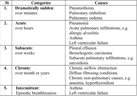
To locate vertically, you must be able to number the ribs and interspaces accurately (Fig.1.1).
Fig. 1.1. Anatomy of the chest wall. Anterior view.
Note that an interspace between two ribs is numbered by the rib above it. As the 1st rib is covered by clavicle, the 1st interspace is below it.
When
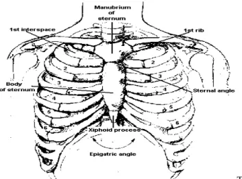
estimating location posteriorly, remember that the inferior angle of the scapula usually lies at the level of the 7th rib or interspace (Fig. 1.2.).
Fig. 1.2. Anatomy of the chest wall. Posterior view.
When a patient flexes the neck forward, the prominent process is usually that of the 7th cervical.
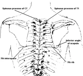
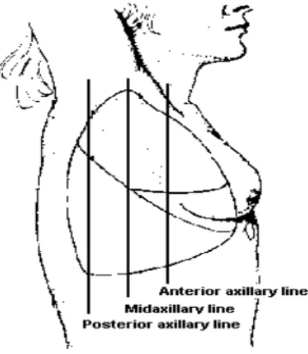
To locate findings around the circumference of the chest, use a topographic vertical lines.
The median (or midsternal) and vertebral lines are precise; the others are estimated. The midclavicular line drops vertically from the midpoint of the clavicle. (Fig. 1.3).
Fig. 1.3. Topographic lines. Anterior view.
The anterior and posterior axillary lines drop vertically from the anterior and posterior axillary folds (the muscles that border the axilla).
The midaxillary line drops from the apex of axilla (Fig. 1.4).
Fig. 1.4. Topographic lines. Right anterior oblique view.
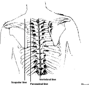
Posteriorly, the vertebral line follows the spinal processes of the vertebra. Paraspinal lines drop along vertebra; each scapular line drops from the inferior angle of the scapular (Fig. 1.5).
Fig. 1.5. Topographic lines. Posterior view.
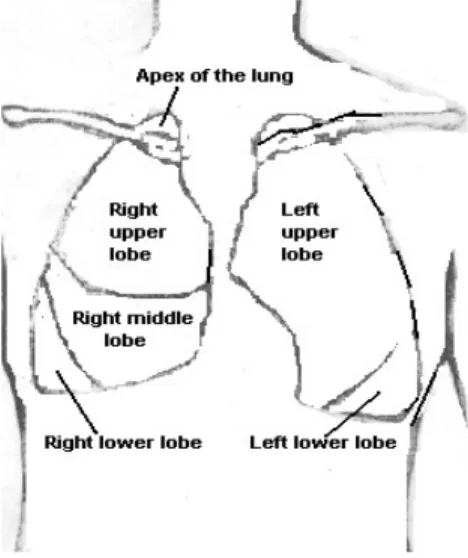
The lungs lobes and fissures can be outlined mentally on the chest wall. Anteriorly, the apex of the each lung rises about 2 cm to 4 cm above the inner third of the clavicle (Fig. 1.8). The lower border of the
lung passes the 6th rib at the midclavicular line and 8th rib at the midaxillary line.
Posteriorly, the lower border of the lung lies at about the level of the 11th thoracic spinous process at the paraspinal line. On inspiration it descends (Fig.1.6).
Fig. 1.6. Projection of the lungs on the chest wall. Anterior view.
You should usually locate your pulmonary findings in external terms, such as these:
Supraclavicular region – above clavicles;
Infraclavicular region – below clavicle;
Suprascapular region – above scapulae;
Interscapular region – between the scapulae;
Infrascapular region – below scapular;
Bases of the lungs – the lowerst points;
Upper, middle, and lower lungs fields.
While examining the chest, note the shape of the chest, its symmetry (static inspection), type of respiration, participation of the chest wall in breathing act, respiration rate, depth and rhythm (dynamic inspection).
Static inspection
The shape of the chest. To estimate form of the chest, pay attention to:
1. Pronunciation of supraclavicular fosses;
2. Pronunciation of infraclavicular fosses;
3. Pronunciation of intercostal fosses;
4. Direction and width of intercostal spaces and ribs;
5. Epigastrical angle;
6. Anterior-posterior and lateral sizes;
7. Position of scapulas.
Normal and pathological shapes of the chest are distinguished. As three types of the constitution are existed, the chest has different shape according to these constitutional types. There are three normal shapes of the chest: normosthenic chest, hypersthenic chest, and asthenic chest.
Normosthenic chest: the shoulders are under the right angle to the neck, supra- and infraclavicular fossae feebly expressed, the ribs are moderately inclined, the interspaces are visible, but moderate expressed, epigastric angle is near 90˚, the lateral diameter is larger than anteroposterior, shoulder blades closely fits to the chest and are on the same level. The thorax is about the same height as abdominal part of the trunk.
Hypersthenic chest: the shoulders are wide and the neck is short, supra- and infraclavicular fossae are absent (level with the chest), direction of the ribs are nearly horizontal, the interspaces are narrow and slightly expressed, epigastric angle exceeds 90˚, the lateral diameter is about the same as anteroposterior, the chest has form of a cylinder, the shoulder blades closely fit to the chest. The thorax is smaller than abdominal part of the trunk.
Asthenic chest: the shoulders are sloping and are under the dull angle to the neck, clavicles are well visible, supra- and infraclavicular fossae are distinctly pronounced, the ribs direct downward abruptly, more vertical at sides, the interspaces are wide and pronounced, epigastric angle is less than 90˚, both lateral and anteroposterior diameter are smaller than normal, the chest narrow and elongated, the shoulder blades are separate from the chest (scapula alatae) and their angles are well visible. The muscles of the shoulder girdle are underdeveloped. The thorax is longer than abdominal part of the trun.
Pathological shapes of the chest can be caused either by chronic diseases of the lungs and pleura (emphysematous, paralytic chest), or by pathology of the thorax costal skeleton (rachitic, funnel, and foveated chest), or by various deformities of the spine (scoliosis, kyphosis, lordosis, kyphoscoliosis). Pathological shapes of the chest caused by chronic pulmonary diseases.
Emphysematous or barrel chest resembles hypersthenic in its shape. Supra- and infraclavicular fossae are absent or even protruded, the ribs are horizontal, the interspaces are enlarged, epigastric angle is more than 90˚, the chest wall is prominent, chest has an increased anteroposterior diameter and that is why chest has a barrel-like configuration.
Emphysematous chest observes in emphysema of the lungs. The lungs seem to be as if at the inspiration phase due to decreased elasticity of the lungs. The volume of the lungs increases, the natural expiration become difficult (expiratory dyspnoea). Accessory respiratory muscles active participate in the breathing act, especially m.
sternocleidomastoideus and m. trapezius. Elevation and lowering of the entire chest during breathing become evident. A barrel shape is normal during infancy, and often accompanies normal aging.
Paralytic chest. The same signs that peculiar to the asthenic chest but more pronounced characterize paralytic chest. The shoulders are sloping, clavicles are asymmetrical and pronounced, supra- and infraclavicular fossea depresses, the ribs are vertical, the interspaces are wide and depressed, marked atrophy of the chest muscles, epigastric angle is less than 90˚, the shoulder blades are not on the same level, and their movement during breathing are asynchronous.
Paralytic chest is found in emaciated patients, in general asthenia, constitutional underdevelopment and in the patients with long-standing pulmonary pleural diseases, such as pulmonary tuberculosis and pneumosclerosis. Growing of the connective tissue contracts the lungs and diminished their size due to progressive chronic inflammation.
o Pathological shapes of the chest caused by pathology of the thorax costal skeleton.
Rachitic or pigeon chest (Pectus Carinatum) is a result of abnormal skeleton formation in childhood in the patients with rachitis.
In a pigeon chest, the sternum is displaced anteriorly, increasing anteroposterior diameter, resembling the keel of the boat. The ribs meet at an acute angle at the sternum, the costal cartilages thicken like beads at points of their transition to bones (rachitic beads).
Funnel chest (Pectus Excavatum) is characterized by a depression in the lower portion of the sternum near the xiphoid process.
This deformity can be the result of abnormal development of the sternum or prolonged compressing effect. But exact cause is now unknown. In older times such shape of the chest was found in shoemaker adolescents, and was explained by permanent pressure of the chest against the shoe. Therefore, the funnel chest is also called ‘cobbler chest’. Compression of the heart and great vessels may cause murmurs.
Foveated chest is characterized by vertical depression on the upper and middle parts of the anterior surface of the chest. This deformity arises in syringomyelia, a rare disease of the spinal cord.
o Pathological shapes of the chest caused by various deformities of the spin as a result of injuries, tuberculosis of the spine, rheumatoid arthritis, etc.
Four types of spine deformities are distinguished:
Scoliosis – lateral curvature of the spine, is most common. It develops in schoolchildren due to bad habitual posture.
Kyphosis – backward curvature of the spine with formation of the gibbus, occurs less frequently.
Lordosis – forward curvature of the spine, generally in the lumber region, occurs in rare cases.
Kyphoscoliosis – combination of the lateral and backward curvature of the spine.
Kyphosis, lordosis, and kyphoscoliosis change physiological position of thoracic organs and thus interfere with their normal function.
The symmetry of the chest. The right and left sides of the normal chest are symmetrical, the clavicles and shoulder blades are on the same level, the supra- and infraclavicular fossae, and interspaces equally pronounced on both sides. One-sided enlargement or diminutions of the chest (asymmetry) due to the pulmonary and pleural diseases are of great diagnostic value. These changes can transient or permanent.
Enlarged volume of one half of the chest observes in accumulation of considerable amount of fluid (exudates, transudate, blood, pus) in the pleural cavity, or in penetration of air inside the chest in injuries (pneumothorax).
During examination of the enlarged part of the chest you can see asymmetry of the clavicles; leveling or protrusion of the interspaces, and they more wide; the distance between nipple and median line, and from inside edge of scapula to the spine on affected side is larger than on healthy one. Enlarged part of the chest lags in the breathing act. The patient slightly bends to the affected side of the chest. The chest assumes normal symmetrical shape after the fluid or air is removed from the pleural cavity.
Decreased volume of the one part of the chest observes in:
contraction of a considerable portion of the lung due to the growth of connective tissue – pneumosclerosis, after acute or chronic inflammatory processes, such as acute pneumonia (with subsequent carnification of the lung), lung infarction, pulmonary abscess, tuberculosis;
pleural adhesion or contraction of the pleural membranes after resorption of fluid;
obstructive athelectasis;
resection of a part or the entire lung.
During examination of the decreased part of the chest you can see that the shoulder and clavicle lowers, supra- and infraclavicular fossae are more depressed, the interspaces are decreased in size or invisible, the nipple is nearer to the sternum as compared with healthy side, and the scapula lowers. The respiratory movement of clavicle and scapula become slower and limited on affected side.
Dynamic inspection
In dynamic inspection of the chest the pattern of breathing (type of respiration, participation of the chest wall in breathing act, respiration rate, depth and rhythm) must be observed.
Breathing in and out (inspiration and expiration, together called respiration or external respiration) is essential for taking O2 and getting rid of CO2. Respiration is largely an automatic act, controlled in the brain system and mediated by the muscles of respiration. The main respiratory muscles - diaphragm, intercostals muscles, and partly the abdominal wall muscles are normally used for this propose. The accessory muscles – mm. sternocleidomastoideus, trapezius, pectoralis
major et minor, etc., join the respiratory effort in pathological conditions, associated with difficult breathing.
Respiration type. Respiration can be thoracic, abdominal or mixed type.
Thoracic (costal) respiration. Mainly the intercostals muscles carry out respiratory movements. In inspiration the intercostals muscles contract and elevate the ribs, these movements increase the internal capacity of the lungs. As the thoracic wall expands, the lungs also expand and draw in air. In expiration, the thoracic capacity decreases as the inspiratory muscles relax – the lungs then shrink by their own elasticity. This type of breathing is known as costal and is mostly characteristic of women (Fig. 2.9).
Abdominal respiration. The diaphragm is the primary muscle in this type of respiration. In inspiration the diaphragm contracts, descends in the chest and enlarges the thoracic cavity. The thoracic enlargement decreases intrathoracic pressure, draws air through the tracheobronchial tree into the alveoli, and expands the lungs. At the same time it compresses the abdominal contents, pushing the abdominal wall outward. In expiration the chest wall and lungs recoil, the diaphragm rises passively, air flows outward, and the chest and abdomen return to their initial position (Fig. 2.9). This type of breathing is also called diaphragmatic and is mostly characteristic of men.
Mixed respiration. The diaphragm and the intercostals muscles simultaneously carry out respiratory movements. This type of respiration observes in the aged persons and some pulmonary and digestive diseases.
In women mixed respiration occurs in dry pleurisy, pleural adhesion, myosytis, thoracic radiculitis, and lung emphysema.
In men mixed respiration occurs in persons with underdeveloped diaphragmatic muscle, diaphragmatitis, acute cholecystitis, perforating ulcer.
Participation of the chest wall in breathing act. In observing respiratory movement, particular attention must be paid to expansion.
Poor movement of the chest on one side only always indicates pathology on that side. One part of the chest lags in the breathing act in inflammatory infiltration of extensive part of the lung, dry pleurisy, hydrothorax, pneumothorax, ribs fractures, intercostals neuralgia, and myositis. In paralysis or paresis respiratory excursion on the corresponding part is limited.
Respiration rate. In order to determine respiratory rate count the movement of the chest or abdominal wall, with patient being unaware of the procedure.
The repeated cycles of inspiration followed by expiration (respiratory cycle) occur in adults at rest about 16-20 times per minute (the respiratory rate), with inspiration lasting approximately 2 seconds and expiration 3 seconds (Fig. 1.7).
Fig 1.7. Normal breathing.
The respiration rate in newborn is 40-45 per minute, this rate gradually decreasing with age. During night sleep respiratory rate decreases to 12-14 per minute, and increases in physical and emotional exertion, and after heavy meals.
Pathological rapid breathing above 20 per minute is called tachypnea (Fig.1.8).
Fig. 1.8. Tachypnea – rapid shallow breathing.
Tachypnea has a number of causes:
conditions associated with decreased respiratory surface of the lungs: inflammation, tuberculosis, compressive atelectasis (hydrothorax, pneumothorax, mediastinal tumor), obstructive atelectesis, pulmonary emphysema, and pulmonary edema;
narrowing of the small bronchi due to spasm or diffuse inflammation of their mucosa (bronchiolitis), which interfere normal air passage into alveoli;
shallow respiration as a result of difficult contractions of the respiratory muscles in acute pain (dry pleurisy, acute myositis,
intercostals neuralgia, rib fracture) and in elevated abdominal pressure and hight diaphragm level (ascitis, meteorism, late pregnancy).
Pathological slow breathing below 16 per minute is called bradypnea (Fig. 1.9).
Fig. 1.9. Bradypnea – slow breathing.
Bradypnea may be secondary to such causes as increased intracranial pressure (cerebral tumor, hemorrhage, meningitis, brain edema) due to inhibition of the respiratory center, and also due to the toxic effect on respiratory center in uraemia, diabetic or hepatic coma, and drug-induced respiratory depression.
Respiration depth. The volume of the inhaled and exhaled air at rest in adults varies from 300 to 900 ml (500 ml on the average).
Depending on depth, breathing can be shallow or deep.
Shallow respiration is characterized by short inspiratory and expiratory phases. Shallow breathing is usually rapid (Fig. 2.11). In some cases, however, shallow respiration can be slow due to inhibition of the respiratory center, pronounced pulmonary emphysema, and sharp narrowing of the vocal slit or trachea.
Deep respiration is characterized by elongation of the inspiratory and expiratory phases. As a rule, deep respiration is slow (Fig. 2. 12).
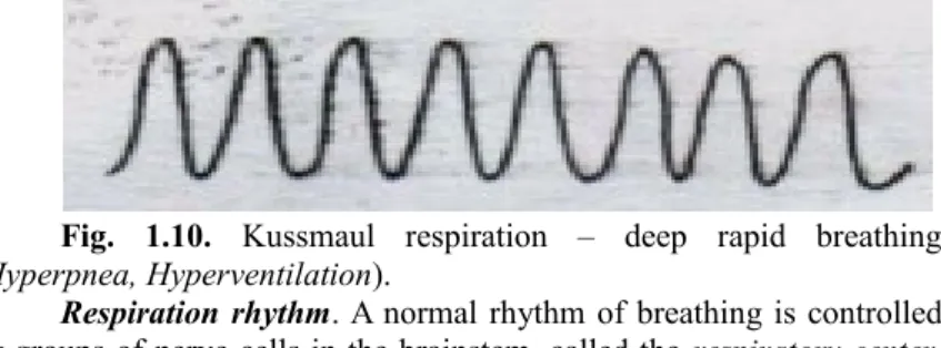
Rapid deep breathing has several causes, including exercise, anxiety, fever, anemia, and metabolic acidosis. Deep rapid breathing, with marked respiratory movements, accompanied by noisy sound is called Kussmaul respiration (Fig. 1.10). This type of breathing observes in the comatose patients due to metabolic acidosis.
Fig. 1.10. Kussmaul respiration – deep rapid breathing (Hyperpnea, Hyperventilation).
Respiration rhythm. A normal rhythm of breathing is controlled by groups of nerve cells in the brainstem, called the respiratory center.
These nerve cells send impulses down the spinal cord to act on the spinal nerve fibers that supply the diaphragm and intercostals muscles.
Respiration of a healthy person is rhythmic, and characterized by uniform depth, and approximately equal duration of inspiratory and expiratory phases (Fig. 2.10). In depression of the respiratory center breathing becomes arrhythmic. Periods of breathing alternate with readily detectable elongation of respiratory pause from few seconds to a minute or with apnea (temporary arrest of breathing) and also respiration may be of different depth. Such type of respiration is called periodic and includes Cheyne-Stokes respiration, Grocco’s respiration, and Biot’s respiration.
Cheyne-Stokes respiration (Fig. 1.11). Noiseless shallow respiration quickly deepens, becomes noisy to attain its maximum at the 5-7 inspirations and slows down gradually. Such periods alternate with periods of apnea (from few second to a minute), during which patient can loses orientation in surroundings or even faints to recover from unconsciousness after respiration restores.
Fig. 1.11. Cheyne-Stokes respiration.
Children and aged people normally may show Cheyne-Stokes respiration in sleep. Other causes include heart failure, uremia, drug- induced respiratory depression, and brain damage (acute or chronic failure of the cerebral circulation, cerebral hypoxia, meningitis).
Grocco’s respiration resembles Cheyne-Stokes respiration except that shallow respiration occurs instead of apnea (Fig. 1.12).
Fig. 1.12. Crocco’s respiration.
Crocco’s respiration caused probably by early stages of the same conditions as Cheyne-Stokes respiration.
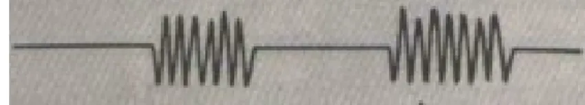
Biot’s respiration (Fig. 1.13). In this type of breathing deep rhythmic respiration alternate with apnea (from few seconds to half minute). Causes include respiratory depression and brain damage (meningitis, agony with disorders of cerebral cerculation).
Fig. 1.13. Biot’s respiration.
PALPATION OF THE CHEST Palpation of the chest has four potential uses:
1. identification of the epigastric angle;
2. identification of the tender areas;
3. assessment of elasticity of the chest;
4. assessment of tactile fremitus.
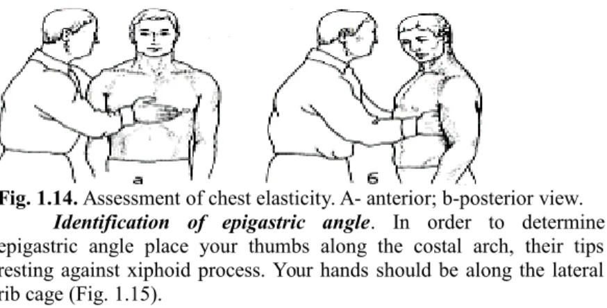
Assessment of elasticity of the chest. Fig. 1.14 Assess elasticity by pressing the chest in anteroposterior and lateral directions. Place your hands parallel: right on the middle of the sternum, left – on the spine and press the chest. Then by both hands press the chest in lateral direction.
The chest of a healthy person is elastic, and yields under the pressure. Rigidity of the chest indicates presence of fluid inn the pleural cavity or pleural tumor, and pulmonary emphysema. In aged persons the chest become rigid due to ossification of the costal cartilages.
Fig. 1.14. Assessment of chest elasticity. A- anterior; b-posterior view.
Identification of epigastric angle. In order to determine epigastric angle place your thumbs along the costal arch, their tips resting against xiphoid process. Your hands should be along the lateral rib cage (Fig. 1.15).
Fig. 1. 15. Estimation of epigastric angle
Identification of tender areas. Carefully palpate from the 1st interspaces on the anterior chest (5 steps), then along midaxillary lines (3 steps), and along the spine on the posterior chest (10 steps).
In rib fracture pain is localized over a limited area, careful displacement of bone fractures attends by a specific sound (crunch).
Intercostals tenderness commonly has musculoskeletal origin. Such pain is called superficial, it intensifies during deep breathing, and when the patient bends or lies on the affected side.
Assessment of tactile fremitus (vocal fremitus pectoralis).
Fremitus refers to the palpable vibrations transmitted through the bronchopulmonary tree to the chest wall when the patient speaks. Ask the patient to repeat the words “ninety-nine” or “one-one-one”. If fremitus is faint, ask the patient to speak more loudly or in a deeper voice.
Palpate and compare symmetrical areas of the chest, using the palms of your both hands simultaneously.
Firstly, place your hands on the patient’s shoulder over the lungs apices projection, then in the infrascapular regions, and axillary regions, using the vibratory sensitivity of your hands to detect fremitus (Fig 1.16).
Fig. 1.16. Assessment of tactile fremitus. Anterior view.
Posteriorly, you should assess vocal fremitus in the supra-, inter-, and infrascapular regions (Fig. 1.17).
Fig. 1.17. Assessment of vocal fremitus. Posterior view.
Identify and locate any areas of increased, decreased or absent fremitus.
Vocal fremitus is typically more prominent in men than inn women and children; in the upper lung fields than in the lower one; and
more prominent on the right side (voice transmission is better through the shorter right main bronchus) than on the left.
Vocal fremitus is increased in consolidation of the pulmonary tissue (lobar pneumonia, lungs infarction, pulmonary tumor, tuberculosis, compressive atelectasis), and also in the presence of an air cavity communicated with bronchus.
Vocal fremitus is decreased when the voice is soft in emaciated patients or when the conduction of vibrations from the larynx to the surface of the chest is impeded. Causes include separation of the lung by moderate amount of fluid (pleural effusion) or air (pneumothorax), by fibrosis (pleural thickening); obstructive atelectasis; and also a very thick chest wall (edema, subcutaneous fat).
Vocal fremitus can be absent when significant amount of fluid or air are accumulated in the pleural cavity.
Tests
1. Paralitic chest shape is observed in:
A. Acute bronchitis
B. Pneumonia
C. Bronchopneumo
nia
D. Lungs tumor
E. Exudation
pleurisy
2. A Boat-shaped chest is observed in:
A. rachitis B. scoliosis C. syringomyelia D. tuberculosis E. bronchitis
3. Barrel-shaped chest is typical for:
A. pulmonary tuberculosis B. emphysema of the lungs C. exudation pleurisy D. pneumothorax E. acute bronchitis
4. Enlargement of one part of the chest is observed in:
A. hydrothorax B. pneumosclerosis
C. obstructive atelectasis of the lung
D. bronchopneumonia E. bronchitis
5. Diminished one part of the chest is observed in:
A. Exudation pleurisy B. pneumothorax C. bronchopneumonia D. pneumosclerosis E. pulmonary emphysema 6. Kussmaul respiration is observed in:
A. diabetic coma B. stroke C. heart failure D. lung failure
E. pulmonary tuberculosis 7. Lateral curvature of the spine is observed in:
A. lordosis B. scoliosis C. kyphosis D. rachitis E. kyphoscoliosis
8. Cheyne-Stocks respiration is typical for:
A. acute insufficiency of the brain circulation
B. pulmonary emphysema C. pneumothorax
D. bronchial asthma E. hydrothorax
9. Increased voice resonance is observed in:
A. hydrothorax
B. compression atelectasis C. pulmonary emphysema D. pneumothorax
E. pneumothorax
10. Decreased voice resonance is observed in
A. hydrothorax B. exudation pleurisy C. pulmonary emphysema D. pneumothorax
E. Acute pneumothorax 11. The patient has an attack of dyspnea. His position is forced; he is sitting resting his hands on the edge of the bed. The voice resonance over the lungs is weak.
What diagnosis can be supposed?
A. Pulmonary emphysema B. Bronchial asthma C. Kussmaul respiration D. atelectasis
E. Cheyne-Stokes respiration
12. The examination has revealed delay in the act of respiration of the right part of the thorax. What diagnosis can be supposed?
A. pneumothorax B. hydrothorax
C. pulmonary emphysema D. obturation atelectasis E. pneumonia
13. The right part of the thorax is protruding, delays in the act of respiration, the voice resonance is not observed. The respiration is superficial. The respiratory rate is 32 per min. What diagnosis can be supposed?
A. Pneumonia B. Hydrothorax,
pneumothorax
C. Pulmonary emphysema D. atelectasis
E. bronchial asthma
14. The chest is asymmetrical, its right half protrudes. The voice resonance downward the middle of the scapula is weak. What diagnosis can be supposed?
A. Hydrothorax, pneumothorax
B. Pulmonary emphysema C. atelectasis
D. pneumonia E. cavity in the lung
15. The chest is ball-shaped. The ratio of anterior-posterior size to transverse size is 8.0. The area of the costal cartilages is thickened.
What diagnosis can be supposed?
A. Asthenic chest B. Rachitic chest C. Hypersthenic chest D. Emphysema chest E. Paralytic chest
16. The right part of the thorax protrudes, delays in the act of respiration, the voice resonance is not heard. The respiration is superficial, 32 per min. What diagnosis can be supposed?
A. Hydrothorax, pneumothorax
B. Pulmonary emphysema C. Pneumonia
D. Atelectasis E. Cavity in the lung
17. Respiratory movements are interrupted with pauses lasting up to 30 seconds. What diagnosis can be supposed?
A. Biots respiration B. Cheyne-Stocks
respiration C. Bronchial asthma D. pneumothorax E. Kussmaul respiration 18. The left part of the thorax delays in the act of respiration, the voice resonance is increased along the paravertebral, scapular, posterior-, mid-, and anterior axillary lines downwards the fifth interspace. What diagnosis can be supposed?
A. atelectasis B. hydrothorax C. pneumothorax D. Cavity in the lung 19. The patient has dyspnea and cyanosis. The right half of the chest protrudes, delays in the act of respiration. The voice resonance is decreased downward the middle of the scapula. What diagnosis can be supposed?
A. Hydrothorax, pneumothorax B. Cavity in the lung C. Pulmonary emphysema D. Atelectasis
E. Pneumonia
Tests: 1D, 2A, 3B, 4A, 5D, 6A, 7B, 8A, 9B, 10E, 11B, 12D, 13B, 14A, 15B, 16A, 17A, 18B, 19A.
Methodical instructions
MAIN COMPLAINTS OF PATIENTS WITH RESPIRATORY ORGANS DISEASES. STATIC AND DYNAMIC
INSPECTION OF THE CHEST. PALPATION OF THE CHEST Methodical instructions for students
Authors: Т.V. Ashcheulova O. M. Kovalyova G.V. Demydenko Chief Editor Ashcheulova Т.V.
Редактор____________
Корректор____________
Компьютерная верстка_____________
Пр. Ленина, г. Харьков, 4, ХНМУ, 61022 Редакционно-издательский отдел